
Article Figures & Data
Figures
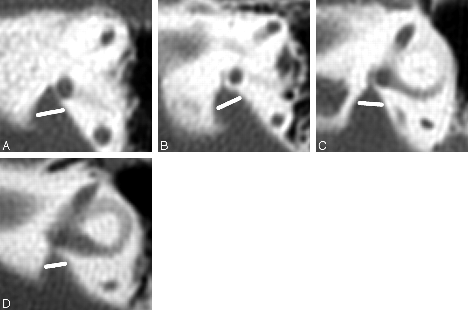
- Fig 1.
Axial CT images showing the coronal planes used to define the VA midpoint plane in 4 temporal bones. The vestibular plane (V) lies at the level of the posterior wall of the vestibule or crus commune. The opercular plane is at the level of the VA operculum (O) or adjacent posterior wall of the petrous bone. The midpoint plane (M) is equidistant from the vestibular and opercular planes. Magnification of all images is the same. Enlarged rather than normal VAs are used for illustration so that the relationships of the VAs to the planes are easier to see.
A, The posterior wall of the vestibule defines the vestibular plane, and the edge of the operculum defines the opercular plane.
B, The transition between the posterior wall of the vestibule and crus commune defines the vestibular plane, and the edge of the operculum defines the opercular plane.
C, The posterior wall of the crus commune defines the vestibular plane, and the edge of the operculum defines the opercular plane.
D, The posterior wall of the vestibule defines the vestibular plane, and the posterior wall of the temporal bone just above the operculum defines the opercular plane.
- Fig 2.
The same axial CT images seen in Fig 1 with the VA midpoint measurement lines drawn. Magnification of all images is the same.
A, The VA midpoint width is 2.0 mm.
B, The VA midpoint width is 1.4 mm.
C, The VA midpoint width is 1.0 mm.
D, The VA midpoint width is 0.8 mm.
- Fig 3.
Axial CT images showing VA midpoint measurements in 4 additional children with large midpoints. Magnification of all images is the same. Enlarged rather than normal VAs are used for illustration so that the relationships of the VAs to the planes are easier to see.
A, VA midpoint measures 2.8 mm.
B, VA midpoint measures 2.6 mm.
C, VA midpoint measures 2.5 mm. D, VA midpoint measures 2.3 mm.
- Fig 4.
Technique of measuring the width of the VA at the operculum. Asterisks (*) mark the opercular margins of the VAs. The widths (white lines) are measured from the opercular margins to the spots on the posterior temporal bone walls whose surface is perpendicular to the measurement lines. Tangents (black lines) to these spots are shown to illustrate these right-angle relationships. The magnification of all images is the same.
A, VA opercular width is 5.3 mm.
B, VA opercular width is 4.0 mm.
C, VA opercular width is 4.0 mm.
D, VA opercular width is 3.7 mm.
- Fig 5.
Normal opercular and midpoint VA measurements. The width of the VA at the operculum (*) is 1.4 mm (long white line), and the width at the midpoint (short white line) is 0.4 mm in this temporal bone. The crus commune (CC) is in the axial plane of this VA.
- Fig 6.
Correlation structure of the midpoint and the operculum measurements of 146 ears without regard to right or left side.
- Fig 7.
Scatter and box plot of midpoint measurement compared with age of subject. The dots represent the scatter plot of midpoint measurements (mm) by age of subject in years. The bar graphs represent the bottom offset axis for the median midpoint measurements (mm) by the 4 age categories (0–3.9 years, 4–6.9 years, 7–9.9 years, and >10 years).
- Fig 8.
Scatter and box plot of opercular measurement in comparison to age of subject The dots represent the scatter plot of operculum measurements (mm) by age of subject in years. The bar graphs represent the bottom offset axis for the median operculum measurements (mm) by the 4 age categories (0–3.9 years, 4–6.9 years, 7–9.9 years, and >10 years). To the eye, the boxes look like a trend toward increasing size with increasing patient age, but this is insignificant.


Tables
The upper ranges of normal vestibular aqueduct widths
Plane and Percentile Size (mm) All Ears(n = 146) Left Side(n = 73) Right Side(n = 73) Opercular Maximum 3.4 3.4 2.7 99th 2.7 3.4 2.7 97.5th 2.3 2.3 2.4 95th 1.9 2.0 1.9 90th 1.7 1.7 1.5 75th 1.2 1.2 1.1 50th 0.9 0.9 0.8 Midpoint Maximum 1.8 1.8 1.3 99th 1.3 1.8 1.3 97.5th 1.2 1.2 1.2 95th 0.9 0.8 1.0 90th 0.7 0.7 0.7 75th 0.4 0.4 0.4 50th 0.2 0.2 0.2


