
Article Figures & Data
Figures
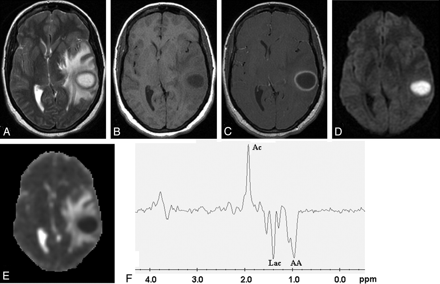
- Fig 1.
Pyogenic abscess in the left temporal lobe of a 31-year-old woman.
Axial T2-weighted image (A) shows a well-defined hyperintense lesion with hypointense wall that appears hypointense on axial T1-weighted image (B) with isointense wall. On postcontrast T1-weighted image (C), it shows ring enhancement. Diffusion-weighted image shows homogeneous hyperintensity in the cavity (D) with low ADC (0.63 × 10⁻³ mm2/s) (E). PMRS from the center of the lesion with a voxel size of 2.4 mL shows amino acids (AA, 0.9 ppm), lactate (Lac, 1.3 ppm), and acetate (Ac, 1.9 ppm) (F). Culture from pus grew Bacteroides species.
- Fig 2.
Tubercular abscess in the right basal ganglia of a 25-year-old woman.
A well-defined hyperintense lesion is seen with a hypointense wall on axial T2-weighted image (A), which shows ring enhancement on postcontrast T1-weighted image (B). Diffusion-weighted image shows homogeneous hyperintensity in the cavity (C) with low ADC (0.54 × 10⁻³ mm2/s) (D). PMRS from the center of the lesion with a voxel size of 2 mL shows predominant lipid peak (Lip, 1.3 ppm) (E). Culture from pus shows the presence of Mycobacterium tuberculosis.
- Fig 3.
Fungal abscess in the frontal lobe of a 39-year-old man with non-Hodgkin lymphoma on treatment.
Axial T2-weighted image (A) shows a well-defined heterointense lesion in the right frontal lobe with an irregular hypointense wall. Hypointense projections attached to the wall are well demonstrated. White arrows show the crenated margin of the fungal wall. Axial T1-weighted image (B) shows a hypointense core with isointense intracavitary projections. Postcontrast axial T1-weighted image (C) shows peripheral enhancement of the wall with no enhancement of intracavitary projections. Note the presence of 2 more enhancing lesions in the right thalamus and right occipital regions, which are better seen in adjoining sections. Diffusion-weighted image (D) shows hyperintensity in the projections with hypointensity in the cavity. On an ADC map (E), intracavitary projections show low ADC (0.46 × 10⁻³ mm2/s) with high ADC in the cavity (2.22 × 10⁻³ mm2/s). PMRS (F) obtained with a voxel size of 2 mL shows lactate along with lipid (Lac/Lip, 1.3 ppm). Culture from pus grew Aspergillus flavus.
- Fig 4.
Fungal abscess in the left frontoparietal region of a 46-year-old man who was immunocompetent.
The lesion appears as a well-defined hyperintense mass with an irregular wall and intracavitary hypointense projections on axial T2-weighted image (A). Axial T1-weighted image (B) shows a hypointense core with isointense intracavitary projections. Postcontrast axial T1-weighted image (C) shows peripheral enhancement of the wall with no enhancement of intracavitary projections. Hyperintense projections with hypointense cavity are seen on diffusion-weighted image (D). ADC map (E) shows low ADC value (0.53 × 10⁻³ mm2/s) in the intracavitary projections with high ADC (2.17 × 10⁻³ mm2/s) in the cavity. PMRS (F) shows amino acids (AA, 0.9 ppm) and lactate (Lac, 1.3 ppm) with multiple peaks at 3.6 and 3.8 ppm. Stereomicroscopic view (G) of the wall of the abscess shows it to be composed of fibrocollagenous tissue with inflammation and neovascularization. The lumen is lined by frayed necrotic material (H&E × 4). Inset high-power view shows branching slender fungal hyphae in a background of necrotic material (GMS × 200). Culture from pus grew Aspergillus fumigatus.


Tables
- Table 1:
Summary of the mean and SD of ADC values in the walls and cavities of the 3 different types of brain abscess
Type of Abscess Apparent Diffusion Coefficient (mean ± SD) × 10−3 mm2/s* Wall Restricted Portion of Cavity Nonrestricted Portion of Cavity Pyogenic (n = 91) 0.79 ± 0.19 0.73 ± 0.18 1.32 ± 0.41 Tubercular (n = 11) 0.83 ± 0.34 0.66 ± 0.23 1.61 ± 0.42 Fungal (n = 8) 0.81 ± 0.22 0.50 ± 0.05 1.50 ± 0.42 P values P = 0.35‡§ P = 0.23‡§ P = 0.90‡§ P = 0.77†§ P = 0.01†§ P = 0.41†§ P = 0.36†‡ P = 0.51†‡ P = 0.39†‡ * The mean ADC of normal-appearing white matter in our study was 0.74 ± 0.07 × 10⁻³ mm2/s.
† Pyogenic abscess.
‡ Tubercular abscess.
§ Fungal abscess.
Type of Abscess Metabolite* Amino Acid (0.9 ppm) Lipid (1.3 ppm) Lactate (1.3 ppm) Lipid + Lactate Acetate (1.9 ppm) Succinate (2.4 ppm) (3.6–3.8 ppm)† Pyogenic 89 – 46 45 25 18 – Tubercular – 9 – 2 – – – Fungal 4 1 4 3 – – 5 * The figures indicate the number of abscesses that showed the presence of a particular metabolite.
† Multiple peaks between 3.6 and 3.8 were assigned to trehalose, as reported in the literature.12
In this issue


Jump to section
Related Articles
Cited By...
- Multiparametric imaging in the evaluation of intracerebral abscesses
- Multiparametric imaging in the evaluation of intracerebral abscesses
- Fungal brain abscess in a post COVID-19 patient
- Clinicoradiological improvement of intracranial tubercular abscess with medical management alone
- Migration: A Notable Feature of Cerebral Sparganosis on Follow-Up MR Imaging
- Persistent Diffusion-Restricted Lesions in Bevacizumab-Treated Malignant Gliomas Are Associated with Improved Survival Compared with Matched Controls
- In Vivo Proton MR Spectroscopy Evaluation of Pyogenic Brain Abscesses: A Report of 194 Cases