Advertisement

Main menu

  • Home
  • Content
    • Current Issue
    • Accepted Manuscripts
    • Article Preview
    • Past Issue Archive
    • Video Articles
    • AJNR Case Collection
    • Case of the Week Archive
    • Case of the Month Archive
    • Classic Case Archive
  • Special Collections
    • AJNR Awards
    • Low-Field MRI
    • Alzheimer Disease
    • ASNR Foundation Special Collection
    • Photon-Counting CT
    • View All
  • Multimedia
    • AJNR Podcasts
    • AJNR SCANtastic
    • Trainee Corner
    • MRI Safety Corner
    • Imaging Protocols
  • For Authors
    • Submit a Manuscript
    • Submit a Video Article
    • Submit an eLetter to the Editor/Response
    • Manuscript Submission Guidelines
    • Statistical Tips
    • Fast Publishing of Accepted Manuscripts
    • Graphical Abstract Preparation
    • Imaging Protocol Submission
    • Author Policies
  • About Us
    • About AJNR
    • Editorial Board
    • Editorial Board Alumni
  • More
    • Become a Reviewer/Academy of Reviewers
    • Subscribers
    • Permissions
    • Alerts
    • Feedback
    • Advertisers
    • ASNR Home

User menu

  • Alerts
  • Log in
  • Log out

Search

  • Advanced search
American Journal of Neuroradiology

American Journal of Neuroradiology

ASHNR American Society of Functional Neuroradiology ASHNR American Society of Pediatric Neuroradiology ASSR
  • Alerts
  • Log in
  • Log out

Advanced Search

  • Home
  • Content
    • Current Issue
    • Accepted Manuscripts
    • Article Preview
    • Past Issue Archive
    • Video Articles
    • AJNR Case Collection
    • Case of the Week Archive
    • Case of the Month Archive
    • Classic Case Archive
  • Special Collections
    • AJNR Awards
    • Low-Field MRI
    • Alzheimer Disease
    • ASNR Foundation Special Collection
    • Photon-Counting CT
    • View All
  • Multimedia
    • AJNR Podcasts
    • AJNR SCANtastic
    • Trainee Corner
    • MRI Safety Corner
    • Imaging Protocols
  • For Authors
    • Submit a Manuscript
    • Submit a Video Article
    • Submit an eLetter to the Editor/Response
    • Manuscript Submission Guidelines
    • Statistical Tips
    • Fast Publishing of Accepted Manuscripts
    • Graphical Abstract Preparation
    • Imaging Protocol Submission
    • Author Policies
  • About Us
    • About AJNR
    • Editorial Board
    • Editorial Board Alumni
  • More
    • Become a Reviewer/Academy of Reviewers
    • Subscribers
    • Permissions
    • Alerts
    • Feedback
    • Advertisers
    • ASNR Home
  • Follow AJNR on Twitter
  • Visit AJNR on Facebook
  • Follow AJNR on Instagram
  • Join AJNR on LinkedIn
  • RSS Feeds

AJNR Awards, New Junior Editors, and more. Read the latest AJNR updates

OtherHead and Neck Imaging

False-Positive Positron-Emission Tomography-CT of a Teflon Granuloma in the Parapharyngeal Space Occurring after Treatment for a Patulous Eustachian Tube

C.F.E. Kirsch, J.D. Suh, R.B. Lufkin and R.F. Canalis
American Journal of Neuroradiology August 2007, 28 (7) 1371-1372; DOI: https://doi.org/10.3174/ajnr.A0554
C.F.E. Kirsch
  • Find this author on Google Scholar
  • Find this author on PubMed
  • Search for this author on this site
J.D. Suh
  • Find this author on Google Scholar
  • Find this author on PubMed
  • Search for this author on this site
R.B. Lufkin
  • Find this author on Google Scholar
  • Find this author on PubMed
  • Search for this author on this site
R.F. Canalis
  • Find this author on Google Scholar
  • Find this author on PubMed
  • Search for this author on this site
  • Article
  • Figures & Data
  • Info & Metrics
  • Responses
  • References
  • PDF
Loading

Abstract

SUMMARY: This report presents a 55-year-old woman who underwent 2 Teflon injections in 1971 for a patulous eustachian tube. The patient returned in 2006 with a bloody left otorrhea. A positron-emission tomography-CT scan demonstrated a 2-cm hypermetabolic parapharyngeal mass, initially interpreted as a skull base tumor. Repeat neck CT confirmed a 2-cm hyperattenuated left parapharyngeal granulomatous mass. This is the first reported case of a Teflon granuloma presenting as a false-positive parapharyngeal mass.

Positron-emission tomography (PET) with [18F]fluorodeoxyglucose is a useful clinical tool for diagnosing and staging head and neck malignancies. Although sensitive for assessing neoplasms, increased uptake may be seen in nondiseased muscle, fat, glands, or tissues involved with infection and inflammatory granulomatous disease. This report presents a case of a 55-year-old woman who underwent 2 Teflon injections for treatment of a patulous eustachian tube (pET) in 1971 with a resultant false-positive PET-CT in 2006. In the 1970s, Teflon (polytetrafluoroethylene) was used to treat pET via a Bruning syringe for injection of Teflon into the eustachian tube orifice to create mass effect. This patient underwent 2 Teflon injections for pET treatment in 1971 for discomfort experienced while singing. The patient returned in 2006 for evaluation of left-sided bloody otorrhea. Her work-up included a PET-CT scan demonstrating a hypermetabolic parapharyngeal mass below the skull base measuring 2 cm in diameter, initially interpreted as a skull base tumor at an outside institution. Repeat CT, MR imaging, and biopsy confirmed the presence of a 2-cm hyperattenuated mass in the left parapharyngeal space with no evidence of neoplasm. Combined PET-CT is useful in correlating anatomic and physiologic findings; however, care should be taken when evaluating patients who have had Teflon injections for treatment of head and neck disorders.

Case Report

A 55-year-old woman with history of a left pET presented to her otolaryngologist with an episode of painless left-sided otorrhea. Thirty-five years before, the patient underwent 2 Teflon injections via a Bruning syringe into the eustachian tube (ET) orifice for aural fullness experienced while singing (Fig 1). The patient experienced relief after the 2 injections and requested a third injection from her physician, who declined. Although she experienced mild relief, she subsequently required treatment with 6 pressure equalization tubes in her left ear for recurrent effusions. Physical examination revealed a small left tympanic membrane perforation with thick brown otorrhea. Flexible nasopharyngoscopy demonstrated no masses or obstruction at the torus tubarius. The patient underwent a PET-CT scan in April 2006 demonstrating a 2-cm markedly hypermetabolic mass in the left parapharyngeal space, which was initially interpreted at an outside institution as suspicious for a “skull base tumor” (Fig 2). On CT scans, hyperattenuated material was located just inferior and medial to the left foramen ovale (Fig 3). On MR imaging, the mass was isointense to muscle on both T1- and T2-weighted imaging and demonstrated only rim enhancement (Fig 4A and B). Biopsy confirmed no tumor cells present and that the hypermetabolic mass within the left parapharyngeal space on PET-CT and MR imaging was consistent with a Teflon granuloma.

Fig 1.
  • Download figure
  • Open in new tab
  • Download powerpoint
Fig 1.

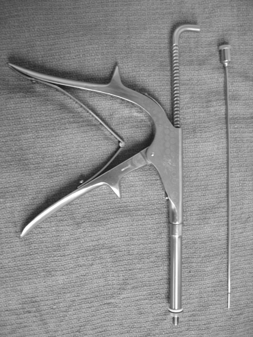
Bruning syringe for Teflon injection.

Fig 2.
  • Download figure
  • Open in new tab
  • Download powerpoint
Fig 2.

PET-CT with 2-cm hypermetabolic mass in the left skull base.

Fig 3.
  • Download figure
  • Open in new tab
  • Download powerpoint
Fig 3.

CT scan demonstrating hyperattenuated material within the left parapharyngeal fat.

Fig 4.
  • Download figure
  • Open in new tab
  • Download powerpoint
Fig 4.

T1 pregadolinium (A) and postgadolinium (B) with fat saturation demonstrating a mass isointense to muscle in the left parapharyngeal fat with peripheral rim enhancement.

Discussion

The pET first described by Schwartze in 18641 is a rare disorder where the ET remains abnormally patent, creating aural fullness, tinnitus, audible respiratory sounds, and autophony or the sensation of hearing one's own physiologic sounds, speech, respiration, and occasionally pulse. In some patients, the symptoms are so disruptive and disturbing that they lead to psychosis or depression.2 The pET is associated with depletion of soft tissue surrounding the cartilaginous medial two thirds of the ET.2 On otoscopic examination, the tympanic membrane moves freely in and out with respiration, enhanced by obstructing a nostril. Patients often have a hyponasal voice from subconsciously trying to close the ET by closing their velum.2 Recent literature notes that the pET is caused by an incompetent tubal valve from a longitudinal scaphoid defect in the anterior lateral cartilaginous lumen, which does not allow the valve to close completely. This defect can be observed endoscopically and confirmed with CT imaging.1 Although many medical and surgical treatments are described in the literature, no definitive treatment is noted.1,2 In this patient, Teflon was injected into the ET via a Bruning syringe to create mass effect and obstruct the ET.

Teflon

Polytetrafluoroethylene was discovered in 1938 as an inert polymer.3 It was used in the 1960s for vocal cord dysphonia.4 Reports from that time noted foreign body granuloma formation at the sites of injection and of local and distant migration.5,6 Teflon causes a marked inflammatory reaction with formation of giant cells and active phagocytosis. After 2 months, the acute inflammatory reaction may subside, whereas giant cells may persist, actively phagocytosing the Teflon particles. An encapsulating granuloma may start to form at 3–6 months.7 This foreign-body reaction with granulomatous formation and fibrosis may lead to a markedly false-positive PET-CT scan secondary to the propensity of FDG accumulation in both lymphocytes and macrophages.7,8 Although Teflon granulomas in the true cords and nasopharynx occurring secondary to treatment of vocal cord and velopharyngeal insufficiency are reported, this is the first case to our knowledge of a Teflon granuloma in the parapharyngeal space occurring after treatment for a pET. Because PET-CT is now increasingly used in the evaluation of head and neck malignancies, awareness of clinical history of Teflon injections is necessary to avoid a potential false-positive misinterpretation.

Footnotes

  • Paper previously presented at: Annual Meeting of the American Society of Head and Neck Radiology, September 27–October 1, 2006; Phoenix, Ariz.

References

  1. ↵
    Grimmer JF, Poe DS. Update on eustachian tube dysfunction and the patulous Eustachian tube. Curr Opin Otolaryngol Head Neck Surg 2005;13:277–82
    CrossRefPubMed
  2. ↵
    Doherty JK, Slattery WH III. Autologous fat grafting for the refractory patulous eustachian tube. Otolaryngol Head Neck Surg 2003;128:88–91
    Abstract/FREE Full Text
  3. ↵
    Ward PH. Uses of injectable Teflon in otolaryngology. Ach Otolaryngol 1968;87:637–43
  4. ↵
    Ward PH, Hanson DG, Abemayor E. Transcutaneous Teflon injection of the paralyzed vocal cord: a new technique: Laryngoscope 1985;95:644–49
    PubMed
  5. ↵
    Rubin HJ. Misadventures with injectable polytef (Teflon). Arch Otolaryngol 1975;101:114–16
    CrossRefPubMed
  6. ↵
    Malizia AA Jr, Reiman HM, Myers RP, et al. Migration and granulomatous reaction after periurethral injection of polytef (Teflon). JAMA 1984;251:3277–81
    CrossRefPubMed
  7. ↵
    Yeretsian RA, Blodgett TM, Branstetter BF 4th, et al. Teflon-induced granuloma: a false-positive finding with PET resolved with combined PET and CT. AJNR Am J Neuroradiol 2003;24:1164–66
    Abstract/FREE Full Text
  8. ↵
    Harrigal C, Branstetter BF 4th, Snyderman CH, et al. Teflon granuloma in the nasopharynx: a potentially false-positive PET/CT finding. AJNR Am J Neuroradiol 2005;26:417–20
    Abstract/FREE Full Text
  • Received October 12, 2006.
  • Accepted after revision December 1, 2006.
  • Copyright © American Society of Neuroradiology
View Abstract
PreviousNext
Back to top

In this issue

American Journal of Neuroradiology: 28 (7)
American Journal of Neuroradiology
Vol. 28, Issue 7
August 2007
  • Table of Contents
  • Index by author
Advertisement
Print
Download PDF
Email Article

Thank you for your interest in spreading the word on American Journal of Neuroradiology.

NOTE: We only request your email address so that the person you are recommending the page to knows that you wanted them to see it, and that it is not junk mail. We do not capture any email address.

Enter multiple addresses on separate lines or separate them with commas.
False-Positive Positron-Emission Tomography-CT of a Teflon Granuloma in the Parapharyngeal Space Occurring after Treatment for a Patulous Eustachian Tube
(Your Name) has sent you a message from American Journal of Neuroradiology
(Your Name) thought you would like to see the American Journal of Neuroradiology web site.
CAPTCHA
This question is for testing whether or not you are a human visitor and to prevent automated spam submissions.
Cite this article
C.F.E. Kirsch, J.D. Suh, R.B. Lufkin, R.F. Canalis
False-Positive Positron-Emission Tomography-CT of a Teflon Granuloma in the Parapharyngeal Space Occurring after Treatment for a Patulous Eustachian Tube
American Journal of Neuroradiology Aug 2007, 28 (7) 1371-1372; DOI: 10.3174/ajnr.A0554

Citation Manager Formats

  • BibTeX
  • Bookends
  • EasyBib
  • EndNote (tagged)
  • EndNote 8 (xml)
  • Medlars
  • Mendeley
  • Papers
  • RefWorks Tagged
  • Ref Manager
  • RIS
  • Zotero
0 Responses
Respond to this article
Share
Bookmark this article
False-Positive Positron-Emission Tomography-CT of a Teflon Granuloma in the Parapharyngeal Space Occurring after Treatment for a Patulous Eustachian Tube
C.F.E. Kirsch, J.D. Suh, R.B. Lufkin, R.F. Canalis
American Journal of Neuroradiology Aug 2007, 28 (7) 1371-1372; DOI: 10.3174/ajnr.A0554
del.icio.us logo Twitter logo Facebook logo Mendeley logo
  • Tweet Widget
  • Facebook Like
  • Google Plus One
Purchase

Jump to section

  • Article
    • Abstract
    • Case Report
    • Discussion
    • Footnotes
    • References
  • Figures & Data
  • Info & Metrics
  • Responses
  • References
  • PDF

Related Articles

  • No related articles found.
  • PubMed
  • Google Scholar

Cited By...

  • No citing articles found.
  • Crossref
  • Google Scholar

This article has not yet been cited by articles in journals that are participating in Crossref Cited-by Linking.

More in this TOC Section

  • Hydrops Herniation into the Semicircular Canals
  • ASL Sensitivity for Head and Neck Paraganglioma
  • Post SRS Peritumoral Hyperintense Signal of VSs
Show more HEAD AND NECK IMAGING

Similar Articles

Advertisement

Indexed Content

  • Current Issue
  • Accepted Manuscripts
  • Article Preview
  • Past Issues
  • Editorials
  • Editor's Choice
  • Fellows' Journal Club
  • Letters to the Editor
  • Video Articles

Cases

  • Case Collection
  • Archive - Case of the Week
  • Archive - Case of the Month
  • Archive - Classic Case

More from AJNR

  • Trainee Corner
  • Imaging Protocols
  • MRI Safety Corner
  • Book Reviews

Multimedia

  • AJNR Podcasts
  • AJNR Scantastics

Resources

  • Turnaround Time
  • Submit a Manuscript
  • Submit a Video Article
  • Submit an eLetter to the Editor/Response
  • Manuscript Submission Guidelines
  • Statistical Tips
  • Fast Publishing of Accepted Manuscripts
  • Graphical Abstract Preparation
  • Imaging Protocol Submission
  • Evidence-Based Medicine Level Guide
  • Publishing Checklists
  • Author Policies
  • Become a Reviewer/Academy of Reviewers
  • News and Updates

About Us

  • About AJNR
  • Editorial Board
  • Editorial Board Alumni
  • Alerts
  • Permissions
  • Not an AJNR Subscriber? Join Now
  • Advertise with Us
  • Librarian Resources
  • Feedback
  • Terms and Conditions
  • AJNR Editorial Board Alumni

American Society of Neuroradiology

  • Not an ASNR Member? Join Now

© 2025 by the American Society of Neuroradiology All rights, including for text and data mining, AI training, and similar technologies, are reserved.
Print ISSN: 0195-6108 Online ISSN: 1936-959X

Powered by HighWire