Advertisement

Main menu

  • Home
  • Content
    • Current Issue
    • Accepted Manuscripts
    • Article Preview
    • Past Issue Archive
    • Video Articles
    • AJNR Case Collection
    • Case of the Week Archive
    • Case of the Month Archive
    • Classic Case Archive
  • Special Collections
    • AJNR Awards
    • Low-Field MRI
    • Alzheimer Disease
    • ASNR Foundation Special Collection
    • Photon-Counting CT
    • View All
  • Multimedia
    • AJNR Podcasts
    • AJNR SCANtastic
    • Trainee Corner
    • MRI Safety Corner
    • Imaging Protocols
  • For Authors
    • Submit a Manuscript
    • Submit a Video Article
    • Submit an eLetter to the Editor/Response
    • Manuscript Submission Guidelines
    • Statistical Tips
    • Fast Publishing of Accepted Manuscripts
    • Graphical Abstract Preparation
    • Imaging Protocol Submission
    • Author Policies
  • About Us
    • About AJNR
    • Editorial Board
    • Editorial Board Alumni
  • More
    • Become a Reviewer/Academy of Reviewers
    • Subscribers
    • Permissions
    • Alerts
    • Feedback
    • Advertisers
    • ASNR Home

User menu

  • Alerts
  • Log in

Search

  • Advanced search
American Journal of Neuroradiology

American Journal of Neuroradiology

ASHNR American Society of Functional Neuroradiology ASHNR American Society of Pediatric Neuroradiology ASSR
  • Alerts
  • Log in

Advanced Search

  • Home
  • Content
    • Current Issue
    • Accepted Manuscripts
    • Article Preview
    • Past Issue Archive
    • Video Articles
    • AJNR Case Collection
    • Case of the Week Archive
    • Case of the Month Archive
    • Classic Case Archive
  • Special Collections
    • AJNR Awards
    • Low-Field MRI
    • Alzheimer Disease
    • ASNR Foundation Special Collection
    • Photon-Counting CT
    • View All
  • Multimedia
    • AJNR Podcasts
    • AJNR SCANtastic
    • Trainee Corner
    • MRI Safety Corner
    • Imaging Protocols
  • For Authors
    • Submit a Manuscript
    • Submit a Video Article
    • Submit an eLetter to the Editor/Response
    • Manuscript Submission Guidelines
    • Statistical Tips
    • Fast Publishing of Accepted Manuscripts
    • Graphical Abstract Preparation
    • Imaging Protocol Submission
    • Author Policies
  • About Us
    • About AJNR
    • Editorial Board
    • Editorial Board Alumni
  • More
    • Become a Reviewer/Academy of Reviewers
    • Subscribers
    • Permissions
    • Alerts
    • Feedback
    • Advertisers
    • ASNR Home
  • Follow AJNR on Twitter
  • Visit AJNR on Facebook
  • Follow AJNR on Instagram
  • Join AJNR on LinkedIn
  • RSS Feeds

AJNR Awards, New Junior Editors, and more. Read the latest AJNR updates

Research ArticleNeurointervention

Embolization of High-Flow Craniofacial Vascular Malformations with Onyx

A. Arat, B.E. Cil, I. Vargel, B. Turkbey, M. Canyigit, B. Peynircioglu and Y.O. Arat
American Journal of Neuroradiology August 2007, 28 (7) 1409-1414; DOI: https://doi.org/10.3174/ajnr.A0547
A. Arat
  • Find this author on Google Scholar
  • Find this author on PubMed
  • Search for this author on this site
B.E. Cil
  • Find this author on Google Scholar
  • Find this author on PubMed
  • Search for this author on this site
I. Vargel
  • Find this author on Google Scholar
  • Find this author on PubMed
  • Search for this author on this site
B. Turkbey
  • Find this author on Google Scholar
  • Find this author on PubMed
  • Search for this author on this site
M. Canyigit
  • Find this author on Google Scholar
  • Find this author on PubMed
  • Search for this author on this site
B. Peynircioglu
  • Find this author on Google Scholar
  • Find this author on PubMed
  • Search for this author on this site
Y.O. Arat
  • Find this author on Google Scholar
  • Find this author on PubMed
  • Search for this author on this site
  • Article
  • Figures & Data
  • Info & Metrics
  • Responses
  • References
  • PDF
Loading

Article Figures & Data

Figures

  • Tables
  • Fig 1.
    • Download figure
    • Open in new tab
    • Download powerpoint
    Fig 1.

    Patient 1.

    A, Lateral projection of the left external carotid arteriogram demonstrates a periorbital arteriovenous malformation supplied by the branches of the superficial temporal (black dotted arrow) and internal maxillary arteries (white dotted arrow, middle meningeal branch: white arrow).

    B, Lateral projection of the left internal carotid arteriogram showing ophthalmic artery contribution to the supply of the arteriovenous malformation.

    C, Lateral projection of the left internal carotid arteriogram during second session embolization, after completion of the embolization of the external carotid supply discloses the drainage of the ophthalmic supply (marked by the tip of the hemostatic clamp). This drainage vein was selectively punctured and embolized.

    D, Native image of the venous phase of a postembolization left common carotid arteriogram showing the embolic cast. The defect within the embolic cast denoted with the solid white arrow is the point of balloon inflation. Embolic agent is also noted in the vein draining the ophthalmic supply (white dotted arrow).

    E, Left common arteriogram after the final session of embolization shows almost total obliteration of the lesion angiographically.
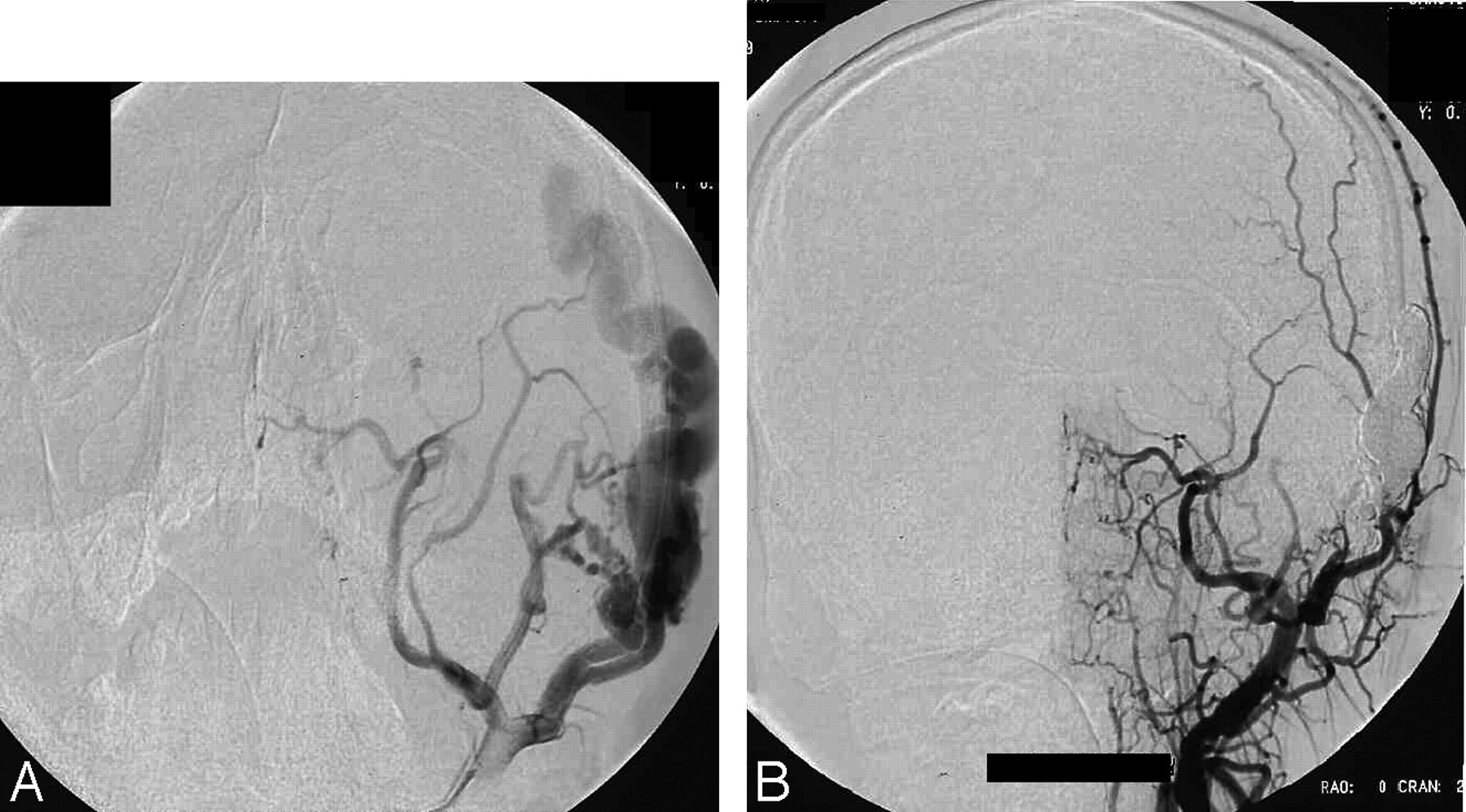
  • Fig 2.
    • Download figure
    • Open in new tab
    • Download powerpoint
    Fig 2.

    Patient 6.

    A, Native radiogram of right external carotid arteriogram in lateral projection reveals a fistulous CHVM supplied by the superficial temporal artery and collateral supply by ipsilateral middle meningeal and occipital arteries.

    B, The inflated HyperForm balloon is visible (white arrows) on the roadmap capture in lateral projection obtained during embolization; note the retrograde filling of the middle meningeal and occipital collateral supply.

    C, Postembolization ipsilateral external carotid arteriogram in the same projection demonstrates no evidence of residual arteriovenous shunt surgery.

  • Fig 3.
    • Download figure
    • Open in new tab
    • Download powerpoint
    Fig 3.

    Patient 8.

    A, Anteroposterior projection of a left external carotid arteriogram in Waters projection depicts a fistulous CHVM supplied by the superficial temporal, anterior deep temporal, and middle meningeal arteries.

    B, Postembolization external carotid arteriogram in anteroposterior projection reveals total obliteration of the lesion.

  • Fig 4.
    • Download figure
    • Open in new tab
    • Download powerpoint
    Fig 4.

    Patient 9.

    A, Lateral projection of the left superficial temporal arteriogram demonstrates a fistulous CHVM; there is attenuated opacification of the draining vein, which projects in between the 2 branches of the artery.

    B, Floroscopic image captured during percutaneous embolization shows impediment of venous filling by the external compression over the vein with a hemostatic clamp; retrograde filling in numerous arterial pedicles is also noted.

    C, Postprocedure external carotid injection in lateral projection demonstrated no evidence of a residual lesion.

    D, Postembolization photograph demonstrates blackish discoloration at the injection site.

    E, Photograph after resection of the cast on postoperative day 5 shows residual discoloration at the margins of the incision.

Tables

  • Figures
  • Clinical and angiographic characteristics of patients

    Patient No. (Figure No.)Age/SexLocationMain SymptomsTypeAccessReason to Use OnyxAssist TechniqueOutcomeNotes
    1 (Figure 1)21/ML periorbitalPainful pulsatile mass totally obstructing visionvmIA × 3 PCAnticipation of better penetration into the nidus of the malformation with IA injection and direct punctureBalloon, direct puncture of draining vein remote to fistulaTotal removal after 2 series of embolizations and surgeriesCatheter tip retained and removed later during surgery, difficulty in balloon retrieval, supplemented with glue for cost containment purposes and also for direct puncture of draining vein
    224/FL cheekPainful mass, cosmetic concernsvmIA × 1Anticipation of better penetration into the nidus of the malformation with IA injectionBalloonResidual AVMIncomplete treatment, patient unwilling to pursue further treatment
    310/MAt mentum of mandibleMass with intermittent bleedingvmIA × 2Anticipation of better penetration into the nidus of the malformation with IA injectionTotal elimination of malformationSmall AVM, bilateral facial approach, no surgery, catheter tip retained on one side
    47/ML preauricularEnlarging PMfsIA × 1Plan to use external compression to limit embolization to the exact point of fistula under controlled injection to avoid excessive venous filling & arterial refluxEC on both arterial and venous side of AVFTotal elimination of shunt
    59/MR angle of mandibleEnlarging PMfsIA × 1 TV in session 1, PC for 2nd sessionPlan to use balloon assistBalloon for session 1, venous EC for both sessionsTotal elimination of shunt after 2 sessions of embolizationSignificant filling of lesion through collaterals after session 1, glue used during second session to fill in large venous pouch
    6 (Figure 2)25/FR zygomaBruit, PMfsIA × 1Plan to use balloon assistBalloonTotal elimination of shunt
    726/ML templefsPCAnticipation of retrograde filling of feedersVenous ECTotal elimination of shuntSurgical excision of cast
    8 (Figure 3)33/ML occipitalPainful large scalp massfsPCAnticipation of retrograde filling of feedersVenous ECTotal elimination of shuntSurgical excision of cast
    9 (Figure 4)24/ML foreheadCosmetic concernsfsPCAnticipation of retrograde filling of feedersVenous ECTotal elimination of shuntSurgical excision of cast, second surgery for blackish discoloration at injection site
    • Note:—M indicates male; F, female; L, left; R, right; PM, pulsatile mass; vm or AVM, arteriovenous malformation; AVF, arteriovenous fistula; fs, fistulous lesion; IA, intra-arterial; PC, percutaneous; TV, transvenous; EC, external compression.

PreviousNext
Back to top

In this issue

American Journal of Neuroradiology: 28 (7)
American Journal of Neuroradiology
Vol. 28, Issue 7
August 2007
  • Table of Contents
  • Index by author
Advertisement
Print
Download PDF
Email Article

Thank you for your interest in spreading the word on American Journal of Neuroradiology.

NOTE: We only request your email address so that the person you are recommending the page to knows that you wanted them to see it, and that it is not junk mail. We do not capture any email address.

Enter multiple addresses on separate lines or separate them with commas.
Embolization of High-Flow Craniofacial Vascular Malformations with Onyx
(Your Name) has sent you a message from American Journal of Neuroradiology
(Your Name) thought you would like to see the American Journal of Neuroradiology web site.
CAPTCHA
This question is for testing whether or not you are a human visitor and to prevent automated spam submissions.
Cite this article
A. Arat, B.E. Cil, I. Vargel, B. Turkbey, M. Canyigit, B. Peynircioglu, Y.O. Arat
Embolization of High-Flow Craniofacial Vascular Malformations with Onyx
American Journal of Neuroradiology Aug 2007, 28 (7) 1409-1414; DOI: 10.3174/ajnr.A0547

Citation Manager Formats

  • BibTeX
  • Bookends
  • EasyBib
  • EndNote (tagged)
  • EndNote 8 (xml)
  • Medlars
  • Mendeley
  • Papers
  • RefWorks Tagged
  • Ref Manager
  • RIS
  • Zotero
0 Responses
Respond to this article
Share
Bookmark this article
Embolization of High-Flow Craniofacial Vascular Malformations with Onyx
A. Arat, B.E. Cil, I. Vargel, B. Turkbey, M. Canyigit, B. Peynircioglu, Y.O. Arat
American Journal of Neuroradiology Aug 2007, 28 (7) 1409-1414; DOI: 10.3174/ajnr.A0547
del.icio.us logo Twitter logo Facebook logo Mendeley logo
  • Tweet Widget
  • Facebook Like
  • Google Plus One
Purchase

Jump to section

  • Article
    • Abstract
    • Materials and Methods
    • Results
    • Discussion
    • Conclusion
    • Footnotes
    • References
  • Figures & Data
  • Info & Metrics
  • Responses
  • References
  • PDF

Related Articles

  • No related articles found.
  • PubMed
  • Google Scholar

Cited By...

  • Embolization of palpebral and orbito-frontal fistulas: technical and anatomical considerations in treating high-flow superficial skin lesions with liquid embolics
  • Periorbital arteriovenous malformations: a word of caution
  • Interventional management of high-flow craniofacial vascular malformations: a database analysis and review of the literature
  • Balloon augmented Onyx embolization utilizing a dual lumen balloon catheter: utility in the treatment of a variety of head and neck lesions
  • Endovascular treatment for traumatic scalp arteriovenous fistulas: results with Onyx embolization
  • Flow control techniques for Onyx embolization of intracranial dural arteriovenous fistulae
  • Onyx embolization of an extensive mandibular arteriovenous malformation via a dual lumen balloon catheter: a technical case report
  • Balloon-augmented Onyx embolization of a dural arteriovenous fistula arising from the neuromeningeal trunk of the ascending pharyngeal artery: technical report
  • Safety and Clinical Efficacy of Onyx for Embolization of Extracranial Head and Neck Vascular Anomalies
  • Crossref
  • Google Scholar

This article has not yet been cited by articles in journals that are participating in Crossref Cited-by Linking.

More in this TOC Section

  • A Retrospective Study in Tentorial DAVFs
  • Proximal Protection Devices for Carotid Stenting
  • Guided vs Conventional Angiography Systems
Show more NEUROINTERVENTION

Similar Articles

Advertisement

Indexed Content

  • Current Issue
  • Accepted Manuscripts
  • Article Preview
  • Past Issues
  • Editorials
  • Editor's Choice
  • Fellows' Journal Club
  • Letters to the Editor
  • Video Articles

Cases

  • Case Collection
  • Archive - Case of the Week
  • Archive - Case of the Month
  • Archive - Classic Case

More from AJNR

  • Trainee Corner
  • Imaging Protocols
  • MRI Safety Corner
  • Book Reviews

Multimedia

  • AJNR Podcasts
  • AJNR Scantastics

Resources

  • Turnaround Time
  • Submit a Manuscript
  • Submit a Video Article
  • Submit an eLetter to the Editor/Response
  • Manuscript Submission Guidelines
  • Statistical Tips
  • Fast Publishing of Accepted Manuscripts
  • Graphical Abstract Preparation
  • Imaging Protocol Submission
  • Evidence-Based Medicine Level Guide
  • Publishing Checklists
  • Author Policies
  • Become a Reviewer/Academy of Reviewers
  • News and Updates

About Us

  • About AJNR
  • Editorial Board
  • Editorial Board Alumni
  • Alerts
  • Permissions
  • Not an AJNR Subscriber? Join Now
  • Advertise with Us
  • Librarian Resources
  • Feedback
  • Terms and Conditions
  • AJNR Editorial Board Alumni

American Society of Neuroradiology

  • Not an ASNR Member? Join Now

© 2025 by the American Society of Neuroradiology All rights, including for text and data mining, AI training, and similar technologies, are reserved.
Print ISSN: 0195-6108 Online ISSN: 1936-959X

Powered by HighWire