
Article Figures & Data
Figures
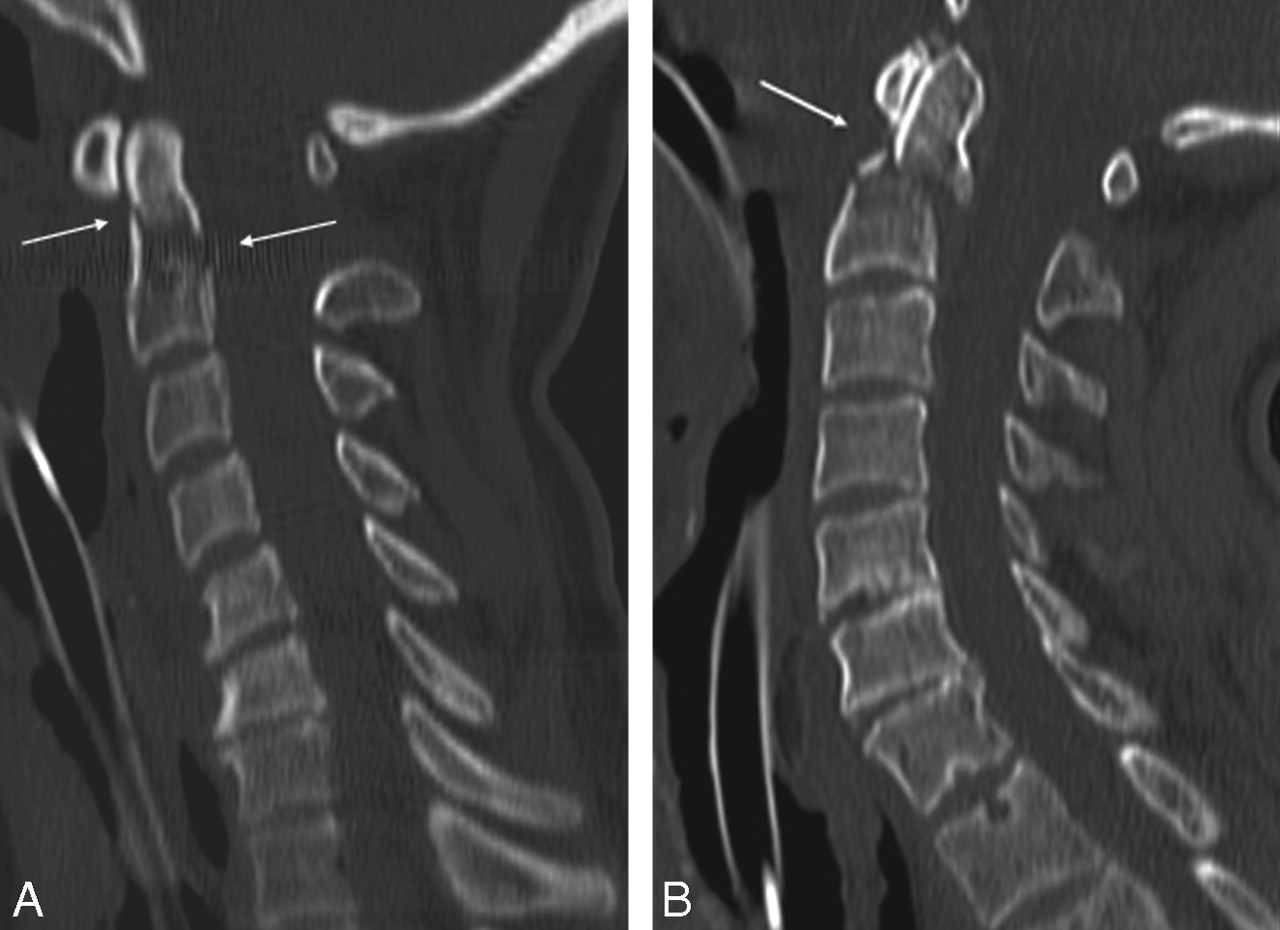
- Fig 1.
A, A 53-year-old woman after polytrauma. Sagittal 2.50 mm image of standard-dose 16-MDCT examination (120 kV and fixed 250 mAs) shows fracture at the base of the dens of C2 (arrow). Calculated effective at examination is 3.9 mSv. B, A 62-year-old man after polytrauma. Sagittal 2.5-mm image of low-dose 16-MDCT examination (100 kV and 141 mAs, after tube current modulation) shows fracture at the base of the dens of C2 (arrow). Calculated effective dose is 1.4 mSv.
- Fig 2.
A, A 53-year-old-man after a high-velocity motor vehicle crash with head injury. Axial 2.5-mm image shows double fracture (arrows) at the transition of the body with each posterior arc of C2. Calculated effective dose of low-dose 16-MDCT examination (100 kV and 96 mAs) is 0.98 mSv. B, Coronal 2.5-mm image shows bilateral fractures at the base of each lateral mass of C2 (arrows). C, Sagittal 2.5-mm image shows fracture at the left arc of C2 (arrow) and at the left arc of C1 (open arrow). D, Sagittal 2.5-mm image shows avulsion fracture of the posterior margin of the body of C2 (open arrow) and posterior subluxation of both posterior arches of C1 and C2 (arrows).
- Fig 3.
A, A 50-year-old woman with history of lung carcinoma with heavy neck pain after minor blunt cervical trauma. Axial 2.5-mm image shows bilateral osteolytic lesions in each lateral mass of the atlas (C1) (open arrows) and midline fracture (arrow) of the posterior arc of C1. Calculated effective dose of low-dose 6-MDCT examination (130 kV and 89 mAs) is 1.48 mSv. B, Coronal 2.5-mm image confirms bilateral osteolytic lesions in each lateral mass of C1 (open arrows) and transverse fracture (arrows) trough the right lateral mass.
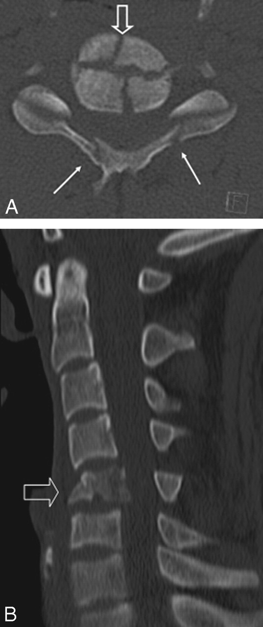
- Fig 4.
A, A 25-year-old man with tetraplegia after a diving accident. Axial 2.5-mm image shows cross-like burst fracture of the body of C5 (open arrow) with posterior displacement and bilateral fractures (arrows) of the posterior arcs. Calculated effective dose of low-dose 16-MDCT examination (120 kV and 160 mAs) is 2.6 mSv. B, Sagittal 2.5-mm image shows burst fracture of the body of C5 (open arrow) with posterior displacement and spinal cord compression.
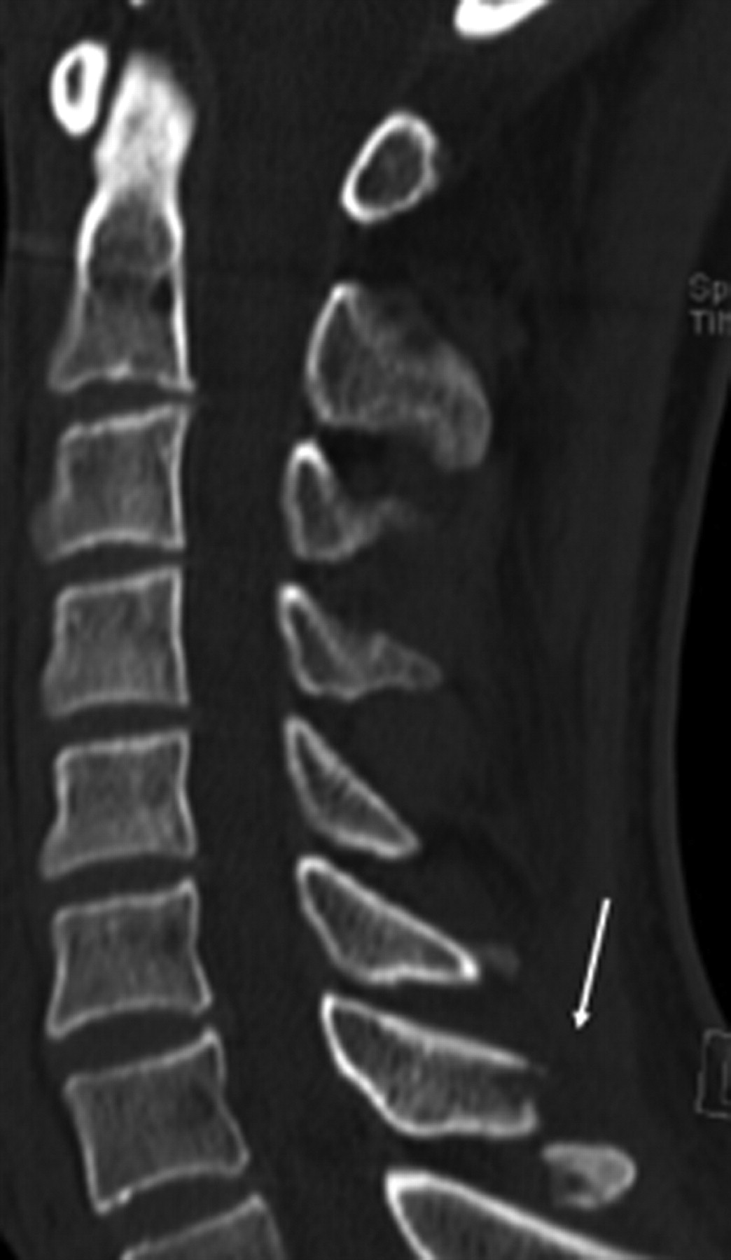
- Fig 5.
A 39-year-old man with hyperextension trauma of the cervical spine. Sagittal 2.5-mm image shows fracture of the spinous process of C7 (arrow). Calculated effective dose of low-dose 6-MDCT examination (110 kV and 56 mAs) is 0.78 mSv.
- Fig 6.
A, An 82-year-old woman with acute neck pain after a motor vehicle crash. Standard radiography, lateral view, was interpreted as negative, but additional CT was proposed because of technical failure to view the lower cervical segment C7 (superposition of the shoulders). B, Standard radiograph with odontoid view was interpreted as negative for fracture in the region of the cranio-cervical junction. C, Sagittal 2.5-mm image of low-dose 16-MDCT-examination (100 kV and 165 mAs) clearly depicts fracture (arrows) at the base of the axis (C2). D, Axial 2.5-mm image of the same low-dose CT examination shows more complex fracture of the body of C2, bilaterally extending in the lateral masses (arrows). Calculated effective dose of MDCT examination is 1.3 mSv.


Tables
Data of standard- and low-dose cervical spine trauma MDCT examinations
Variable Standard Dose Low Dose: High Tube Voltage Low Dose: Low Tube Voltage Tube type Fixed tube current Tube current modulation Tube current modulation No. of patients 51 70 70 Effective dose, mSv* 3.75 ± 0.25 1.57 ± 0.38 1.08 ± 0.28 Image noise, HU* 14.82 ± 5.46 17.46 ± 6.50 19.72 ± 6.17 Image quality scores (0–4)* Reviewer 1 3.02 ± 0.62 3.04 ± 0.51 2.95 ± 0.65 Reviewer 2 3.02 ± 0.47 3.06 ± 0.52 2.94 ± 0.56 Reviewer 3 2.92 ± 0.64 2.99 ± 0.63 2.81 ± 0.62 Reviewer 4 2.82 ± 0.65 2.90 ± 0.71 2.70 ± 0.67 Note:—Reviewers 1 and 2 are experienced radiologists; reviewers 3 and 4 are a second-year and a first-year resident, respectively.
* Data are mean ± SD.
In this issue


Jump to section
Related Articles
Cited By...
- Systematic Radiation Dose Reduction in Cervical Spine CT of Human Cadaveric Specimens: How Low Can We Go?
- Comparison of MRI with radiography for detecting structural lesions of the sacroiliac joint using CT as standard of reference: results from the SIMACT study
- Raise the Bar and Lower the Dose: Current and Future Strategies for Radiation Dose Reduction in Head and Neck Imaging
- Low Kilovoltage CT of the Neck with 70 kVp: Comparison with a Standard Protocol
- Implementation of Automated Tube Current Modulation in PET/CT: Prospective Selection of a Noise Index and Retrospective Patient Analysis to Ensure Image Quality
- Comparison of Image Quality and Radiation Dose between Fixed Tube Current and Combined Automatic Tube Current Modulation in Craniocervical CT Angiography