
Article Figures & Data
Figures
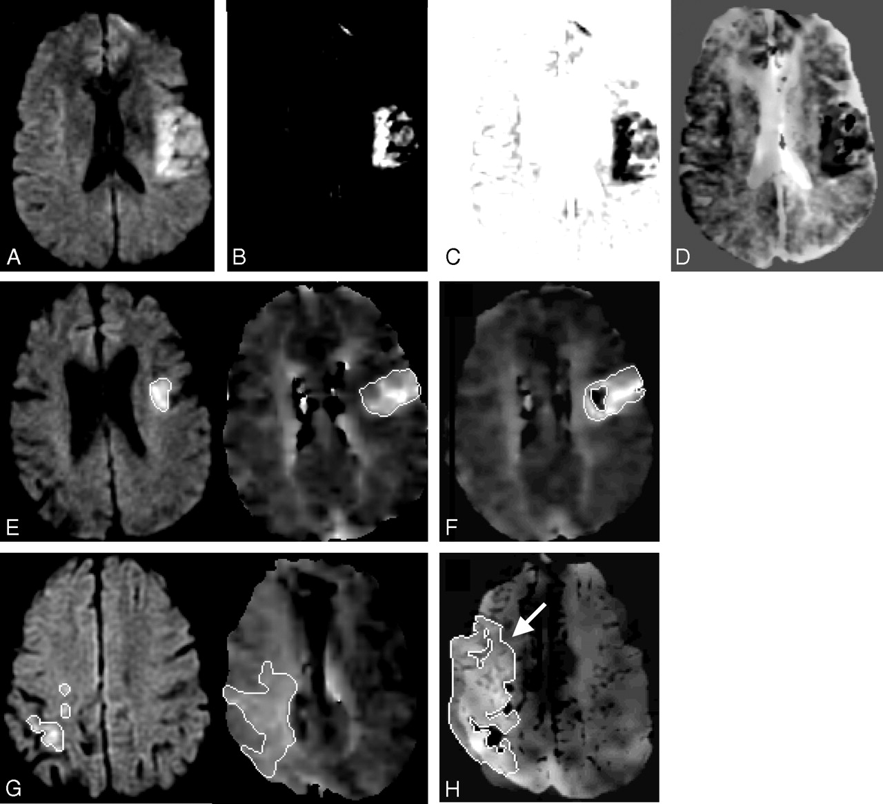
- Fig 1.
Mismatch Method 2. DWI sequence (A) is contrasted to maximize the lesion conspicuity (B) and its intensity values are inverted (C) and then blended with the registered MTT sequence (D). Mismatch Method 1 (E): separate lesion measurements on DWI and MTT sequences compared with Mismatch Method 2 (F): single measurement of the blended difference. Mismatch Method 1 (G): separate lesion measurements on DWI and MTT sequences compared with Mismatch Method 2 (H): single measurement of the blended difference with an arrow indicating the larger MTT lesion seen on the blended map.
- Fig 2.
A, Mismatch Method 1, acute time point plot of mean volume vs volume difference between 2 sets of measurements (raw numbers) where the thick black lines represent the upper and lower boundaries using ±2 SDs from the mean. B, Mismatch Method 2, acute time point plot of mean volume vs volume difference between 2 sets of measurements (raw numbers) where the thick black lines represent the upper and lower boundaries using ±2 SDs from the mean. C, Mismatch Method 1, acute time point plot of mean percentage vs percentage difference between 2 sets of measurements (mismatch/MTT percentage) where the thick black lines represent the upper and lower boundaries using ±2 SDs from the mean. D, Mismatch Method 2, acute time point plot of mean percentage vs percentage difference between 2 sets of measurements (mismatch/MTT percentage) where the thick black lines represent the upper and lower boundaries using ±2 SDs from the mean.


Tables
Mismatch Volume Read 1 Average Acute (n = 64) Read 2 Average Acute (n = 64) Spearman Correlation Coefficient Absolute Volume Difference, Mean ± SD, Median % Deviation, Mean ± SD, Median Method 1 90.45 84.65 0.961 13.83 ± 23.85, 4.85 24.68 ± 90.35, 7.67 Method 2 83.15 82.16 0.975 13.23 ± 13.76, 7.57 22.79 ± 37.22, 14.49 Mismatch Volume Read 1 Average % (Mismatch Volume/ MTT Volume) Acute (n = 64) Read 2 Average % (Mismatch Volume/ MTT Volume) Acute (n = 64) Spearman Correlation Coefficient (Mismatch Volume/ MTT Volume) Cases with 20% or More Mismatch (Mismatch Volume/ MTT Volume), Read 1 Cases with 20% or More Mismatch (Mismatch Volume/ MTT Volume), Read 2 Method 1 59.33 51.92 0.966 52 52 Method 2 71.11 73.71 0.645 55 57 Note:—MTT indicates mean transit time.


