Advertisement

Main menu

  • Home
  • Content
    • Current Issue
    • Accepted Manuscripts
    • Article Preview
    • Past Issue Archive
    • AJNR Case Collection
    • Case of the Week Archive
    • Classic Case Archive
    • Case of the Month Archive
  • Special Collections
    • Spinal CSF Leak Articles (Jan 2020-June 2024)
    • 2024 AJNR Journal Awards
    • Most Impactful AJNR Articles
  • Multimedia
    • AJNR Podcast
    • AJNR Scantastics
    • Video Articles
  • For Authors
    • Submit a Manuscript
    • Author Policies
    • Fast publishing of Accepted Manuscripts
    • Graphical Abstract Preparation
    • Manuscript Submission Guidelines
    • Imaging Protocol Submission
    • Submit a Case for the Case Collection
  • About Us
    • About AJNR
    • Editorial Board
  • More
    • Become a Reviewer/Academy of Reviewers
    • Subscribers
    • Permissions
    • Alerts
    • Feedback
    • Advertisers
    • ASNR Home
  • Other Publications
    • ajnr

User menu

  • Alerts
  • Log in

Search

  • Advanced search
American Journal of Neuroradiology

American Journal of Neuroradiology

ASHNR American Society of Functional Neuroradiology ASHNR American Society of Pediatric Neuroradiology ASSR
  • Alerts
  • Log in

Advanced Search

  • Home
  • Content
    • Current Issue
    • Accepted Manuscripts
    • Article Preview
    • Past Issue Archive
    • AJNR Case Collection
    • Case of the Week Archive
    • Classic Case Archive
    • Case of the Month Archive
  • Special Collections
    • Spinal CSF Leak Articles (Jan 2020-June 2024)
    • 2024 AJNR Journal Awards
    • Most Impactful AJNR Articles
  • Multimedia
    • AJNR Podcast
    • AJNR Scantastics
    • Video Articles
  • For Authors
    • Submit a Manuscript
    • Author Policies
    • Fast publishing of Accepted Manuscripts
    • Graphical Abstract Preparation
    • Manuscript Submission Guidelines
    • Imaging Protocol Submission
    • Submit a Case for the Case Collection
  • About Us
    • About AJNR
    • Editorial Board
  • More
    • Become a Reviewer/Academy of Reviewers
    • Subscribers
    • Permissions
    • Alerts
    • Feedback
    • Advertisers
    • ASNR Home
  • Follow AJNR on Twitter
  • Visit AJNR on Facebook
  • Follow AJNR on Instagram
  • Join AJNR on LinkedIn
  • RSS Feeds

Welcome to the new AJNR, Updated Hall of Fame, and more. Read the full announcements.


AJNR is seeking candidates for the position of Associate Section Editor, AJNR Case Collection. Read the full announcement.


OtherHEAD & NECK

Solitary Fibrous Tumor of the Buccal Space: Treatment with Percutaneous Cryoablation

T.C. Schirmang, L.M. Davis, P.T. Nigri and D.E. Dupuy
American Journal of Neuroradiology October 2007, 28 (9) 1728-1730; DOI: https://doi.org/10.3174/ajnr.A0683
T.C. Schirmang
  • Find this author on Google Scholar
  • Find this author on PubMed
  • Search for this author on this site
L.M. Davis
  • Find this author on Google Scholar
  • Find this author on PubMed
  • Search for this author on this site
P.T. Nigri
  • Find this author on Google Scholar
  • Find this author on PubMed
  • Search for this author on this site
D.E. Dupuy
  • Find this author on Google Scholar
  • Find this author on PubMed
  • Search for this author on this site
  • Article
  • Figures & Data
  • Info & Metrics
  • Responses
  • References
  • PDF
Loading

Abstract

SUMMARY: Solitary fibrous tumors are rare spindle cell neoplasms that typically occur in the thorax but have been described in various locations within the abdomen and head and neck region. The most common extrapleural site is the oral cavity, but these tumors have been also described in the orbit, nasopharynx, paranasal sinuses, salivary glands, and larynx.1–3 We describe a case of a solitary fibrous tumor of the buccal space successfully treated with percutaneous CT-guided cryoablation.

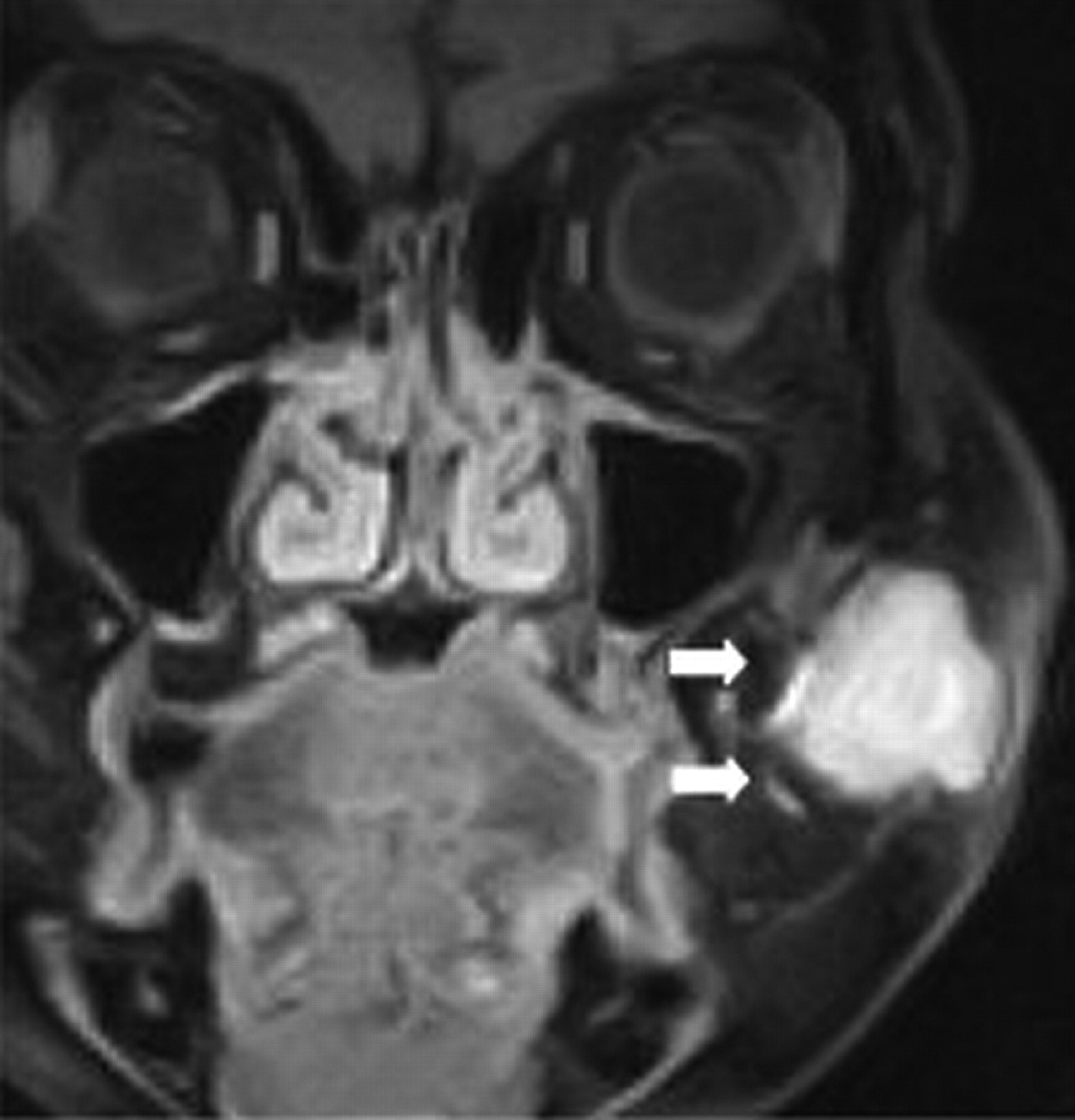
A 64-year-old woman was referred to our tumor ablation service with a complaint of progressive left facial swelling. The patient is from Puerto Rico and underwent an excisional biopsy of a left buccal space mass via a transoral approach approximately 1 year before presentation. A solid, encapsulated soft-tissue tumor was found intraoperatively. There was disruption of the capsule during removal, preventing complete excision. Pathology demonstrated a World Health Organization low-grade solitary fibrous tumor (SFT) of the buccal space with 2–3 mitotic cells per 10 high-power fields, which was CD34+ on immunohistochemical staining. Physical examination at current presentation revealed a firm, mobile, and nontender mass in the left malar soft tissues resulting in facial asymmetry. There was no numbness or facial paralysis. The overlying skin appeared normal. Oropharyngeal examination revealed no evidence of ulceration. Facial CT scan showed an enhancing mass within the left buccal space, which appeared isoattenuated to muscle. There was no calcification or necrosis, nor was there bony destruction of the mandible or maxilla. MR imaging at 0.3T revealed a well-circumscribed solid mass measuring 2.6 × 3.2 × 1.7 cm that was separate from the masseter muscle and that displaced the parotid duct and facial vein medially. The mass was isointense compared with muscle on T1-weighted precontrast images and was heterogeneous in appearance on T2-weighted sequences, containing areas of hyperintensity and hypointensity. There was homogeneous strong enhancement, most consistent with a presumed residual or recurrent SFT (Fig 1). The patient declined surgical reexcision because of the risk of facial nerve injury and for cosmetic reasons and, therefore, elected to have this lesion treated with percutaneous thermal ablation.

Fig 1.
  • Download figure
  • Open in new tab
  • Download powerpoint
Fig 1.

Coronal T1-weighted MR image after intravenous gadolinium-DTPA shows a homogeneously enhancing mass in the left buccal space (arrows).

Written, informed consent was obtained from the patient before the start of the procedure. Coagulation parameters were normal. Percutaneous cryoablation was performed in the supine position via a left transmalar approach with local anesthesia and moderate sedation using intravenous midazolam hydrochloride and fentanyl. CT fluoroscopy was used for imaging guidance. Cryoablation was performed with an argon-based cryoablation system (Endocare, Mountain View, Calif) with 1.7-mm–diameter percutaneous applicators. The applicators have a 3-cm active tip and create a freeze zone of approximately 3 cm (length) by 2 cm (width). Two cryoapplicators were placed simultaneously into the mass in a parallel fashion with the tips approximately 1 cm apart (Fig 2A). Temperatures were measured during the procedure with an attached thermocouple (range, −94°C to −136°C) at the tip. Active thawing of the freeze zone was achieved using helium. The patient underwent 2 consecutive 10-minute freeze-thaw cycles. Interval unenhanced CT evaluation showed a small crescent of soft tissue not optimally treated with the freeze zone. The cryoapplicators were then repositioned into this superolateral aspect of the mass, and 2 additional 8-minute freeze-thaw cycles were performed. The freeze zone measured 3.3 × 4.5 × 3.0 cm on immediate posttreatment CT imaging and encompassed the mass along with a peripheral 5-mm margin around the mass (Fig 2B). The patient was observed for a 2-hour period after the procedure and was discharged home in stable condition.

Fig 2.
  • Download figure
  • Open in new tab
  • Download powerpoint
Fig 2.

A, Procedural photograph during the CT-guided cryoablation showing 2 cryoapplicators positioned within the left cheek mass. B, Axial CT image after the last freeze shows the extent of the ice ball (arrows) beyond the medial margin of the mass. Note central needle tract from cryoapplicator.

At 2-day follow-up visit, there was some swelling of the malar soft tissues. A small eschar was noted on the skin surface along with sloughing of the buccal mucosal surface related to thermal injury. However, this completely resolved at 6-month follow-up. The patient experienced no residual pain, and there was no evidence of sensory or motor nerve deficit or duct injury. Routine follow-up MR imaging at 6 months revealed an organized fluid collection within the buccal soft tissues measuring approximately 2 cm compatible with posttreatment changes, with only a small area of peripheral enhancement. A sonography-guided fine needle aspiration was performed to evaluate for recurrence. Cytology revealed necrotic cells without evidence of tumor. Follow-up MR imaging at 14 months showed near-complete collapse of the treatment cavity with minimal posterior residual peripheral enhancement (Fig 3). The patient's facial swelling had resolved, and her external appearance returned to normal. Despite the low-grade pathology of the tumor, continued imaging surveillance will be performed, given the recurrent nature of the original neoplasm.

Fig 3.
  • Download figure
  • Open in new tab
  • Download powerpoint
Fig 3.

Fourteen-month follow-up coronal T1-weighted MR image after intravenous gadolinium-diethylene-triaminepentaacetic acid shows a small area of residual enhancement (arrows) along with central hypointensity representing coagulative necrosis.

Discussion

SFTs are spindle cell tumors that are typically associated with serosal surfaces, especially the pleura, but have been found to arise in various extrapleural locations, most notably the oral cavity. They typically present as a slow-growing, painless mass. SFTs of the thorax usually have a favorable prognosis. However, they have been reported to be malignant in 13%–23% of cases.2,3 Factors associated with malignancy include high cellularity, more than 4 mitoses per 10 high-power fields, pleomorphism, hemorrhage, and necrosis.3 Extrapleural SFTs, however, are almost always benign and are cured with simple surgical excision. In this patient, who developed a recurrence despite low-grade pathology and who refused surgery, percutaneous thermal ablation was proposed as a treatment alternative.

Additional diagnostic considerations for a mass involving the buccal space include tumors of minor salivary gland origin, such as pleomorphic adenoma, adenoid cystic carcinoma, or mucoepidermoid carcinoma, along with hemangioma, lymphoma, squamous cell carcinoma, lipoma, soft tissue sarcoma, abscess, lymph node, and nerve sheath tumor. However, because other buccal space masses can present in the same fashion, and because the imaging appearance of many of these processes is nonspecific, the diagnosis is based on the microscopic appearance and characteristic immunohistochemical staining. CD34, a transmembrane glycoprotein, has been found to be a highly sensitive marker for SFTs. Radiographically, they are characteristically isoattenuated to muscle on noncontrast CT and isointense to muscle and brain on noncontrast T1- and T2-weighted MR imaging. They typically enhance avidly in a homogeneous fashion after contrast administration. Some variability in MR imaging characteristics has been described, however, with T2 heterogeneity being the most commonly described alternative imaging appearance,4–6 as was seen in our case subject.

Percutaneous cryoablative techniques have been described for the treatment of primary neoplastic and metastatic disease in various organ systems, most notably the liver and prostate. Recent literature has also shown benefit in palliating painful bony and extra-abdominal soft-tissue metastases using percutaneous image-guided cryoablation.7,8 Benefits of this minimally invasive treatment option include the absence of a surgical scar, reduced recovery time, and the ability to visualize the treated tumor both during and immediately after the procedure to determine treatment success. Probe diameters have also decreased in recent years, further making use of this technique to other sites of disease safer and easier with less postprocedural bleeding. Cryoablation offers a few specific advantages over heat-based therapies. These include less procedural pain due to the anesthetic effect of freezing; direct visualization of the ice ball with CT, MR imaging, or ultrasound so that the treatment margins can be monitored in real time, with less disruption of underlying supportive tissue making it safer for lesions next to skin, nerve, and hollow viscera; and the ability to treat lesions with multiple applicators so that large and irregularly shaped masses can be ablated at one sitting.

Within the head and neck region, thermal ablative techniques using radiofrequency energy have been described to treat adenoid cystic carcinoma and recurrent thyroid carcinoma.9,10 However, to our knowledge, this is the first reported use of percutaneous cryoablation to treat a mass in this region. Further investigation of the possible benefits of cryoablation over excision for benign masses would be beneficial.

References

  1. ↵
    Goodlad JR, Fletcher CD. Solitary fibrous tumor arising at unusual sites: analysis of a series. Histopathology 1991;19:512–22
  2. ↵
    Hanau CA, Miettinen M. Solitary fibrous tumor: histological and immunohistochemical spectrum of benign and malignant variants presenting at different sites. Hum Pathol 1995;26:440–49
    CrossRefPubMed
  3. ↵
    England DM, Hochholzer L, McCarthy MJ. Localized benign and malignant fibrous tumors of the pleura: a clinicopathologic review of 223 cases. Am J Surg Pathol 1989;13:640–58
    CrossRefPubMed
  4. ↵
    Ganly I, Patel SG, Stambuk HE, et al. Solitary fibrous tumors of the buccal space. Arch Otolaryngol Head Neck Surg 2006;132:517–25
    CrossRefPubMed
  5. Shin JH, Sung IY, Suh JH, et al. Solitary fibrous tumor in the buccal space: MR findings with pathologic correlation. AJNR Am J Neuroradiol 2001;22:1890–92
    Abstract/FREE Full Text
  6. ↵
    Dunfee BL, Sakai O, Spiegel JH, et al. Solitary fibrous tumor of the buccal space. AJNR Am J Neuroradiol 2005;26:2114–16
    Abstract/FREE Full Text
  7. ↵
    Callstrom MR, Atwell TD, Charboneau JW, et al. Painful metastases involving bone: percutaneous image-guided cryoablation–prospective trial interim analysis. Radiology 2006;241:572–80
    CrossRefPubMed
  8. ↵
    Beland MD, Dupuy DE, Mayo-Smith WW. Percutaneous cryoablation of symptomatic extraabdominal metastatic disease: preliminary results. AJR Am J Roentgenol 2005;184:926–30
    PubMed
  9. ↵
    Monchik JM, Donatini G, Iannuccilli J, et al. Radiofrequency ablation and percutaneous ethanol injection treatment for recurrent local and distant well-differentiated thyroid carcinoma. Ann Surg 2006;244:296–304
    CrossRefPubMed
  10. ↵
    Bui Q, Dupuy DE. Percutaneous CT-guided radiofrequency ablation of an adenoid cystic carcinoma of the head and neck. AJR Am J Roentgenol 2002;179:1333–35
    PubMed
  • Received February 11, 2007.
  • Accepted after revision April 20, 2007.
  • Copyright © American Society of Neuroradiology
View Abstract
PreviousNext
Back to top

In this issue

American Journal of Neuroradiology: 28 (9)
American Journal of Neuroradiology
Vol. 28, Issue 9
October 2007
  • Table of Contents
  • Index by author
Advertisement
Print
Download PDF
Email Article

Thank you for your interest in spreading the word on American Journal of Neuroradiology.

NOTE: We only request your email address so that the person you are recommending the page to knows that you wanted them to see it, and that it is not junk mail. We do not capture any email address.

Enter multiple addresses on separate lines or separate them with commas.
Solitary Fibrous Tumor of the Buccal Space: Treatment with Percutaneous Cryoablation
(Your Name) has sent you a message from American Journal of Neuroradiology
(Your Name) thought you would like to see the American Journal of Neuroradiology web site.
CAPTCHA
This question is for testing whether or not you are a human visitor and to prevent automated spam submissions.
Cite this article
T.C. Schirmang, L.M. Davis, P.T. Nigri, D.E. Dupuy
Solitary Fibrous Tumor of the Buccal Space: Treatment with Percutaneous Cryoablation
American Journal of Neuroradiology Oct 2007, 28 (9) 1728-1730; DOI: 10.3174/ajnr.A0683

Citation Manager Formats

  • BibTeX
  • Bookends
  • EasyBib
  • EndNote (tagged)
  • EndNote 8 (xml)
  • Medlars
  • Mendeley
  • Papers
  • RefWorks Tagged
  • Ref Manager
  • RIS
  • Zotero
0 Responses
Respond to this article
Share
Bookmark this article
Solitary Fibrous Tumor of the Buccal Space: Treatment with Percutaneous Cryoablation
T.C. Schirmang, L.M. Davis, P.T. Nigri, D.E. Dupuy
American Journal of Neuroradiology Oct 2007, 28 (9) 1728-1730; DOI: 10.3174/ajnr.A0683
del.icio.us logo Twitter logo Facebook logo Mendeley logo
  • Tweet Widget
  • Facebook Like
  • Google Plus One
Purchase

Jump to section

  • Article
    • Abstract
    • Discussion
    • References
  • Figures & Data
  • Info & Metrics
  • Responses
  • References
  • PDF

Related Articles

  • No related articles found.
  • PubMed
  • Google Scholar

Cited By...

  • CT-guided cryoablation for palliation of secondary trigeminal neuralgia from head and neck malignancy
  • Crossref (26)
  • Google Scholar

This article has been cited by the following articles in journals that are participating in Crossref Cited-by Linking.

  • Solitary Fibrous Tumor of the Oral Cavity: Clinicopathologic and Immunohistochemical Study of 21 Cases
    Esther M. O’Regan, Vijay Vanguri, Carl M. Allen, Lewis Roy Eversole, John M. Wright, Sook-Bin Woo
    Head and Neck Pathology 2009 3 2
  • Soft-Tissue Cryoablation in Diffuse Locations: Feasibility and Intermediate Term Outcomes
    Peter J. Littrup, Hyun J. Bang, Brandt P. Currier, Dylan J. Goodrich, Hussein D. Aoun, Lance K. Heilbrun, Barbara A. Adam
    Journal of Vascular and Interventional Radiology 2013 24 12
  • Updated anatomy of the buccal space and its implications for plastic, reconstructive and aesthetic procedures
    Thilo L. Schenck, Konstantin C. Koban, Alexander Schlattau, Konstantin Frank, Anthony P. Sclafani, Riccardo E. Giunta, Malcolm Z. Roth, Alexander Gaggl, Robert H. Gotkin, Sebastian Cotofana
    Journal of Plastic, Reconstructive & Aesthetic Surgery 2018 71 2
  • Interventional Neuroradiology of the Head and Neck
    D. Gandhi, J.J. Gemmete, S.A. Ansari, S.K. Gujar, S.K. Mukherji
    American Journal of Neuroradiology 2008 29 10
  • Percutaneous Image-Guided Cryoablation of Head and Neck Tumors for Local Control, Preservation of Functional Status, and Pain Relief
    Jeffrey P. Guenette, Kemal Tuncali, Nathan Himes, Paul B. Shyn, Thomas C. Lee
    American Journal of Roentgenology 2017 208 2
  • CT-guided cryoablation for palliation of secondary trigeminal neuralgia from head and neck malignancy
    Suhail A Dar, Zachary Love, John D Prologo, Daniel Pierce Hsu
    Journal of NeuroInterventional Surgery 2013 5 3
  • Buccal space tumours
    Zubair Hasan, Darius Tan, Malcolm Buchanan, Carsten Palme, Faruque Riffat
    Auris Nasus Larynx 2019 46 2
  • Exceptionally large solitary fibrous tumor arising from the cheek: an immunohistochemical and ultrastructural study with a review of the literature
    Takafumi Satomi, On Hasegawa, Harutsugu Abukawa, Michihide Kohno, Ai Enomoto, Daichi Chikazu, Jun Matsubayashi, Toshitaka Nagao
    Medical Molecular Morphology 2014 47 2
  • Solitary fibrous tumor of the buccal space resected in combination with coronoidectomy
    Hiroyuki Yamada, Yoshiki Hamada, Hisako Fujihara, Kaori Fukami, Kenji Mishima, Kazutoshi Nakaoka, Kenichi Kumagai, Eisaku Imamura
    Oral Surgery, Oral Medicine, Oral Pathology and Oral Radiology 2012 114 1
  • Novel Application of Percutaneous Cryotherapy for the Treatment of Recurrent Oral Bleeding From a Noninvoluting Congenital Hemangioma Involving the Right Buccal Space and Maxillary Tuberosity
    Sepand Salehian, Joseph J. Gemmete, Steven Kasten, Sean P. Edwards
    CardioVascular and Interventional Radiology 2011 34 S2

More in this TOC Section

  • WHO Classification Update: Nasal&Skull Base Tumors
  • Peritumoral Signal in Vestibular Schwannomas
  • Chondrosarcoma vs Synovial Chondromatosis: Imaging
Show more HEAD & NECK

Similar Articles

Advertisement

Indexed Content

  • Current Issue
  • Accepted Manuscripts
  • Article Preview
  • Past Issues
  • Editorials
  • Editors Choice
  • Fellow Journal Club
  • Letters to the Editor

Cases

  • Case Collection
  • Archive - Case of the Week
  • Archive - Case of the Month
  • Archive - Classic Case

Special Collections

  • Special Collections

Resources

  • News and Updates
  • Turn around Times
  • Submit a Manuscript
  • Author Policies
  • Manuscript Submission Guidelines
  • Evidence-Based Medicine Level Guide
  • Publishing Checklists
  • Graphical Abstract Preparation
  • Imaging Protocol Submission
  • Submit a Case
  • Become a Reviewer/Academy of Reviewers
  • Get Peer Review Credit from Publons

Multimedia

  • AJNR Podcast
  • AJNR SCANtastic
  • Video Articles

About Us

  • About AJNR
  • Editorial Board
  • Not an AJNR Subscriber? Join Now
  • Alerts
  • Feedback
  • Advertise with us
  • Librarian Resources
  • Permissions
  • Terms and Conditions

American Society of Neuroradiology

  • Not an ASNR Member? Join Now

© 2025 by the American Society of Neuroradiology All rights, including for text and data mining, AI training, and similar technologies, are reserved.
Print ISSN: 0195-6108 Online ISSN: 1936-959X

Powered by HighWire