Advertisement

Main menu

  • Home
  • Content
    • Current Issue
    • Accepted Manuscripts
    • Article Preview
    • Past Issue Archive
    • AJNR Case Collection
    • Case of the Week Archive
    • Classic Case Archive
    • Case of the Month Archive
  • Special Collections
    • Spinal CSF Leak Articles (Jan 2020-June 2024)
    • 2024 AJNR Journal Awards
    • Most Impactful AJNR Articles
  • Multimedia
    • AJNR Podcast
    • AJNR Scantastics
    • Video Articles
  • For Authors
    • Submit a Manuscript
    • Author Policies
    • Fast publishing of Accepted Manuscripts
    • Graphical Abstract Preparation
    • Manuscript Submission Guidelines
    • Imaging Protocol Submission
    • Submit a Case for the Case Collection
  • About Us
    • About AJNR
    • Editorial Board
  • More
    • Become a Reviewer/Academy of Reviewers
    • Subscribers
    • Permissions
    • Alerts
    • Feedback
    • Advertisers
    • ASNR Home
  • Other Publications
    • ajnr

User menu

  • Alerts
  • Log in

Search

  • Advanced search
American Journal of Neuroradiology

American Journal of Neuroradiology

ASHNR American Society of Functional Neuroradiology ASHNR American Society of Pediatric Neuroradiology ASSR
  • Alerts
  • Log in

Advanced Search

  • Home
  • Content
    • Current Issue
    • Accepted Manuscripts
    • Article Preview
    • Past Issue Archive
    • AJNR Case Collection
    • Case of the Week Archive
    • Classic Case Archive
    • Case of the Month Archive
  • Special Collections
    • Spinal CSF Leak Articles (Jan 2020-June 2024)
    • 2024 AJNR Journal Awards
    • Most Impactful AJNR Articles
  • Multimedia
    • AJNR Podcast
    • AJNR Scantastics
    • Video Articles
  • For Authors
    • Submit a Manuscript
    • Author Policies
    • Fast publishing of Accepted Manuscripts
    • Graphical Abstract Preparation
    • Manuscript Submission Guidelines
    • Imaging Protocol Submission
    • Submit a Case for the Case Collection
  • About Us
    • About AJNR
    • Editorial Board
  • More
    • Become a Reviewer/Academy of Reviewers
    • Subscribers
    • Permissions
    • Alerts
    • Feedback
    • Advertisers
    • ASNR Home
  • Follow AJNR on Twitter
  • Visit AJNR on Facebook
  • Follow AJNR on Instagram
  • Join AJNR on LinkedIn
  • RSS Feeds

Welcome to the new AJNR, Updated Hall of Fame, and more. Read the full announcements.


AJNR is seeking candidates for the position of Associate Section Editor, AJNR Case Collection. Read the full announcement.


OtherHEAD & NECK

Unsuspected Swallowing of a Partial Denture

A. Haidary, J.S. Leider and R. Silbergleit
American Journal of Neuroradiology October 2007, 28 (9) 1734-1735; DOI: https://doi.org/10.3174/ajnr.A0658
A. Haidary
  • Find this author on Google Scholar
  • Find this author on PubMed
  • Search for this author on this site
J.S. Leider
  • Find this author on Google Scholar
  • Find this author on PubMed
  • Search for this author on this site
R. Silbergleit
  • Find this author on Google Scholar
  • Find this author on PubMed
  • Search for this author on this site
  • Article
  • Figures & Data
  • Info & Metrics
  • Responses
  • References
  • PDF
Loading

Abstract

SUMMARY: We report a case of a swallowed partial denture in a 51-year-old man who presented with progressive dysphagia, odynophagia, and fevers. Imaging studies were initially interpreted as supraglottitis with laryngeal inflammation, which was confirmed by direct visualization with flexible endoscopy. Despite appropriate therapy, the patient's symptoms persisted and rigid laryngoscopy was performed, which revealed a partial denture in the hypopharynx and upper esophagus. The subtle imaging appearance of a swallowed denture is discussed.

Aspiration or ingestion of a foreign body has frequently been reported in the pediatric population; however, it is not uncommon in the adult population. In children, coins account for most esophageal foreign bodies, whereas in adults, bones and boluses of meat have commonly been reported as the cause. However, in the elderly population, dental prostheses account for most esophageal foreign bodies.1–3 The base used in the construction of dental prostheses is methylmethacrylate, which is radiolucent. This radiolucency complicates early diagnosis of an impacted or ingested dental prosthesis. We present the radiologic findings of unsuspected impaction of a partial denture in a 51-year-old man with perforation of the posterior cervical esophageal wall.

Case Report

A 51-year-old white man with a history of alcohol abuse and hepatitis C presented to the emergency department with complaints of dysphagia and odynophagia. He reported that his symptoms started suddenly 2 days before and had progressively worsened to the point that he was unable to swallow solid foods and had difficulty with liquids. Despite his symptoms, he continued to drink up to half a pint of vodka daily. He reported subjective fevers and chills and denied a history of hematemesis or hemoptysis. His past medical history was otherwise unremarkable.

On physical examination, the patient was noted to be afebrile and vital signs were stable. Physical examination of the head and neck demonstrated no evidence for pharyngeal erythema, edema, or exudates. The patient did have mild tenderness to palpation in the lower cervical and laryngeal regions bilaterally, left greater than right. Several 5-mm cervical lymph nodes were palpable on the left side. Initial laboratory studies demonstrated slightly elevated WBC of 13.9 with left shift (neutrophils, 10.9). He also had a blood alcohol level of 45 mg/dL (0.045%).

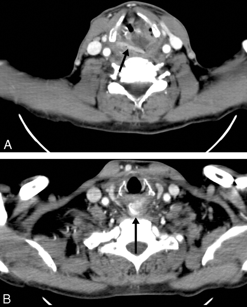
Given the above findings, there was clinical concern for an infectious cause and formation of a retropharyngeal abscess. Anteroposterior and lateral radiographs of the neck were obtained and demonstrated mild prevertebral soft tissue swelling in the lower cervical region. Contrast-enhanced CT scans in Figs 1A and B demonstrated distortion of the epiglottis and aryepiglottic folds. No radiopaque foreign bodies were prospectively identified, and there was no definite formation of an abscess. Given the concern for a neoplastic process, an ENT consultation was obtained. A laryngoscopy was performed, which demonstrated inflammatory change involving the epiglottis. The patient was admitted for supraglottitis and was started on intravenous ampicillin/sulbactam and dexamethasone.

Fig 1
  • Download figure
  • Open in new tab
  • Download powerpoint
Fig 1

A,B. Contrast-enhanced CT scan of the neck demonstrates extensive soft tissue edema. At the level of the vocal cords, there is a linear attenuation (arrow on A), which was not initially noted but, in retrospect, is felt to likely represent the base plate of the partial denture. In addition, a radiopaque attenuation (arrow on B) is seen in the esophagus at the level of the thoracic inlet, likely representing a tooth.

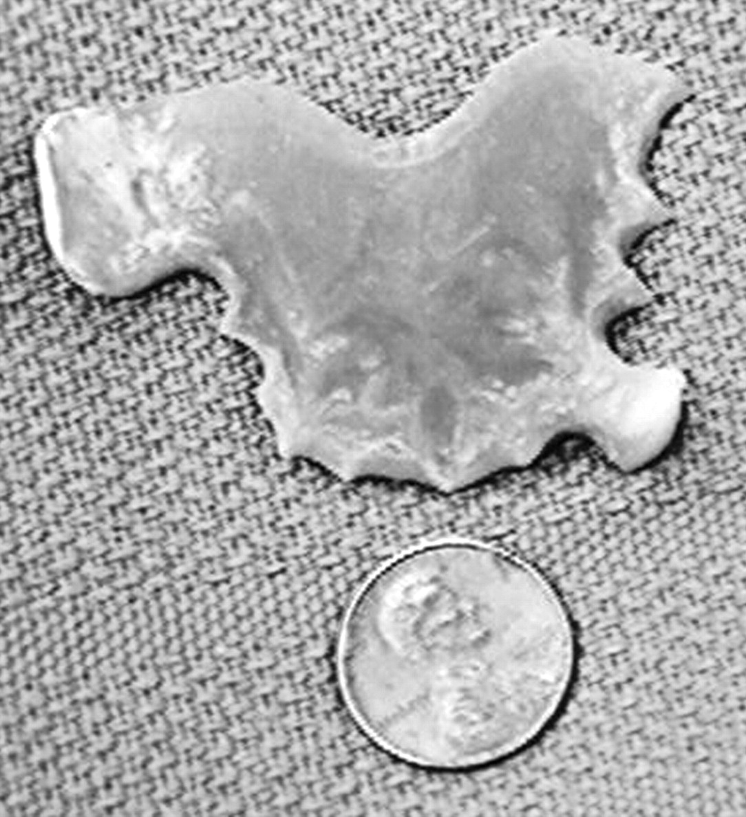
Despite adequate treatment, his symptoms persisted 5 days later and a flexible endoscopy was performed. On second examination, there was a concern for a masslike lesion and the patient underwent intraoperative rigid endoscopy, which revealed a tooth attached to a partial denture in the larynx (Fig 2). This obstruction was removed without difficulty.

Fig 2.
  • Download figure
  • Open in new tab
  • Download powerpoint
Fig 2.

Retrieved removable partial denture.

On retrospective review of CT scan, the linear attenuation seen in Fig 1A is felt to represent the base plate of the partial denture and the rounded radiodensity at the thoracic inlet, the single tooth. These findings could not be appreciated on the radiographs, likely because of superimposition with the thoracic spine.

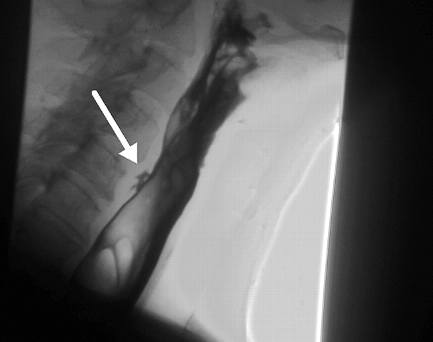
After the partial denture was retrieved, a water-soluble esophagram was performed, which showed contrast extravasation posteriorly (Fig 3). The remainder of the patient's hospital stay was uneventful, and repeat esophagram showed the leak to be contained and significantly decreased in size. On discharge, his symptoms had resolved and he was tolerating oral intake well.

Fig 3.
  • Download figure
  • Open in new tab
  • Download powerpoint
Fig 3.

Water-soluble esophagram (Omnipaque 240; GE Healthcare, Piscataway, NJ) demonstrates a small intramural tear (arrow) of the cervical esophagus at the C5 level with contrast extravasation.

Discussion

According to the National Health and Nutrition Examination Survey (NHANES III), 1 of every 5 persons between the ages of 18 and 74 years has full or partial dentures.4 The inadvertent swallowing of a dental prosthesis is not uncommon in the adult population. In a study by Abdullah et al2 of 200 patients with a known history of an impacted tracheal or esophageal foreign body, dental prostheses accounted for 11.5% of the cases.

Patients can present with vague symptoms of neck pain, dysphagia, odynophagia, and excessive salivation. The clinical history may be vague, and patients may not report a definite history of swallowing their dentures secondary to trauma, intoxication, or loss of consciousness. The diagnosis is further complicated by the fact that polymethylmethacrylate (PMMA), the resin base used for construction of partial dentures, is radiolucent. Even the porcelain and plastic artificial teeth are difficult to visualize on plain radiographs. Although attempts have been made to create a radiopaque resin base, they have not been able to match the physical and esthetic qualities of PMMA.5–8 Complete dentures as well as some temporary partial dentures do not have a metallic-based reinforcement, unlike implant-supported full dentures and permanent removable partial dentures, which have a metallic base for reinforcement and retention. Some temporary partial dentures (ie, stayplates or flippers) may have clasps, which are metal based for improved retention. If the patient has cosmetic concerns and does not want the clasps to be visible and retention is adequate, then a metallic component may not be present, as was the case in our patient.9

The delay in diagnosis can result in significant morbidity. If undetected, retained foreign bodies can lead to significant edema that causes obstruction or perforation, or both. Multiple case reports have documented the development of tracheoesophageal, aortoesophageal, and even esophago-broncho-aortic fistulas.10–13 In addition, there have been reports of aortic erosion as well as complications distally in the gastrointestinal tract, such as enterocolonic fistulas and colonic perforation.13–14 In a cross-sectional study of 103 patients with foreign-body ingestion, Khan et al15 reported that complications were more common in adults (37.1%) compared with children (8.8%) and that the most severe complications occurred with ingested dentures.

In the absence of radiologic findings, management should be based on history and clinical symptoms. In a patient with dysphagia and odynophagia or new-onset neck pain, a referral to an ENT physician should be considered.16 Direct visualization with flexible or rigid endoscopy should be performed. Knowledge of the subtle imaging findings of these partial dentures and interpretation of these studies with sufficient clinical history may lead to earlier diagnosis and treatment.

Footnotes

  • Paper presented previously at: Annual Meeting of the American Society of Head and Neck Radiology, September 28, 2006; Phoenix, Ariz.

References

  1. ↵
    Sittitrai P, Pattarasakulchai T, Tapatiwong H. Esophageal foreign bodies. J Med Assoc Thai 2000;83:1514–18
    PubMed
  2. ↵
    Abdullah BJ, Teong LK, Mahadevan J, et al. Dental prosthesis ingested and impacted in the esophagus and orolaryngopharynx. J Otolaryngol 1998;27:190–94
    PubMed
  3. ↵
    Herranz-Gonzalez J, Martinez-Vidal J, Garcia-Sarandeses A, et al. Esophageal foreign bodies in adults. Otolaryngol Head Neck Surg 1991 Nov;105:649–54
    PubMed
  4. ↵
    Redford M, Drury TF, Kingman A, et al. Denture use and the technical quality of dental prostheses among persons 18–74 years of age: United States, 1988–1991. J Dent Res 1996;75:714–25
    PubMed
  5. ↵
    McCabe JF, Wilson HJ. A radio-opaque denture material. J Dent 1976;4:211–17
    CrossRefPubMed
  6. Chandler HH, Bowen RL, Paffenbarger GC. Development of a radiopaque denture base material. J Biomed Mater Res 1971;5:253–65
    CrossRefPubMed
  7. Davy KW, Causton BE. Radio-opaque denture base: a new acrylic co-polymer. J Dent 1982;10:254–64
    CrossRefPubMed
  8. ↵
    Tsao DH, Guilford HJ, Kazanoglu A, et al. Clinical evaluation of a radiopaque denture base resin. J Prosthet Dent 1984;51:456–58
    CrossRefPubMed
  9. ↵
    Krol AJ, Jacobson TE, Finzen FC. Removable Partial Denture Design: Outline Syllabus, 5th ed. San Rafael, Calif: Indent; 1999:1–3, 111–15, 139–40
  10. ↵
    Singh B, Kantu M, Har-El G, et al. Complications associated with 327 foreign bodies of the pharynx, larynx, and esophagus. Ann Otol Rhinol Laryngol 1997;106:301–04
    Abstract/FREE Full Text
  11. Taha AS, Nakshabendi I, Russell RI. Vocal cord paralysis and oesophago-broncho-aortic fistula complicating foreign body-induced oesophageal perforation. Postgrad Med J 1992;68:277–78
    Abstract/FREE Full Text
  12. Rajesh PB, Goiti JJ. Late onset tracheo-oesophageal fistula following a swallowed dental plate. Eur J Cardiothorac Surg 1993;7:661–62
    Abstract/FREE Full Text
  13. ↵
    Athanassiadi K, Gerazounis M, Metaxas E, et al. Management of esophageal foreign bodies: a retrospective review of 400 cases. Eur J Cardiothorac Surg 2002;21:653–56
    Abstract/FREE Full Text
  14. ↵
    Velitchkov NG, Grigorov GI, Losanoff JE, et al. Ingested foreign bodies of the gastrointestinal tract: retrospective analysis of 542 cases. World J Surg 1996;20:1001–05
    CrossRefPubMed
  15. ↵
    Khan MA, Hameed A, Choudhry AJ. Management of foreign bodies in the esophagus. J Coll Physicians Surg Pak 2004;14:218–20
    PubMed
  16. ↵
    Firth AL, Moor J, Goodyear PW, et al. Dentures may be radiolucent. Emerg Med J 2003;20:562–63
    Abstract/FREE Full Text
  • Received February 4, 2007.
  • Accepted after revision April 3, 2007.
  • Copyright © American Society of Neuroradiology
View Abstract
PreviousNext
Back to top

In this issue

American Journal of Neuroradiology: 28 (9)
American Journal of Neuroradiology
Vol. 28, Issue 9
October 2007
  • Table of Contents
  • Index by author
Advertisement
Print
Download PDF
Email Article

Thank you for your interest in spreading the word on American Journal of Neuroradiology.

NOTE: We only request your email address so that the person you are recommending the page to knows that you wanted them to see it, and that it is not junk mail. We do not capture any email address.

Enter multiple addresses on separate lines or separate them with commas.
Unsuspected Swallowing of a Partial Denture
(Your Name) has sent you a message from American Journal of Neuroradiology
(Your Name) thought you would like to see the American Journal of Neuroradiology web site.
CAPTCHA
This question is for testing whether or not you are a human visitor and to prevent automated spam submissions.
Cite this article
A. Haidary, J.S. Leider, R. Silbergleit
Unsuspected Swallowing of a Partial Denture
American Journal of Neuroradiology Oct 2007, 28 (9) 1734-1735; DOI: 10.3174/ajnr.A0658

Citation Manager Formats

  • BibTeX
  • Bookends
  • EasyBib
  • EndNote (tagged)
  • EndNote 8 (xml)
  • Medlars
  • Mendeley
  • Papers
  • RefWorks Tagged
  • Ref Manager
  • RIS
  • Zotero
0 Responses
Respond to this article
Share
Bookmark this article
Unsuspected Swallowing of a Partial Denture
A. Haidary, J.S. Leider, R. Silbergleit
American Journal of Neuroradiology Oct 2007, 28 (9) 1734-1735; DOI: 10.3174/ajnr.A0658
del.icio.us logo Twitter logo Facebook logo Mendeley logo
  • Tweet Widget
  • Facebook Like
  • Google Plus One
Purchase

Jump to section

  • Article
    • Abstract
    • Case Report
    • Discussion
    • Footnotes
    • References
  • Figures & Data
  • Info & Metrics
  • Responses
  • References
  • PDF

Related Articles

  • No related articles found.
  • PubMed
  • Google Scholar

Cited By...

  • Neglected partial denture in the lower oesophagus presenting after 7 days
  • Dysphagia, dysphonia and sore throat following cerebral infarction: an unexpected cause
  • Crossref (19)
  • Google Scholar

This article has been cited by the following articles in journals that are participating in Crossref Cited-by Linking.

  • Manufacturing of an immediate removable partial denture with an intraoral scanner and CAD-CAM technology: a case report
    Francois Virard, Laurent Venet, Raphaël Richert, Daniel Pfeffer, Gilbert Viguié, Alexandre Bienfait, Jean-Christophe Farges, Maxime Ducret
    BMC Oral Health 2018 18 1
  • Unnoticed swallowing of a unilateral removable partial denture
    Mercedes Gallas, Montserrat Blanco, David Martinez‐Ares, Eduardo Rivo, Eva García‐Gontán, Miguel Cañizares
    Gerodontology 2012 29 2
  • Imaging findings of swallowed dentures: a case series
    Ashish Chawla, Jerome Bosco, Manickam Subramanian, Kabilan Chokkapan, Jagadish Shenoy, Tze Chwan Lim
    Emergency Radiology 2015 22 6
  • Delayed presentation of a small bowel perforation secondary to an ingested denture
    Pranav H Patel, Alistair Ap Slesser, Praveena Idaikkadar, Ioanna Kostourou, Reda W Awad
    JRSM Short Reports 2012 3 9
  • Neglected partial denture in the lower oesophagus presenting after 7 days
    Vinoth Boopathy, Satvinder Singh Bakshi, Padhmini Balasubramanian, Suganthy Dakshinamoorthy
    BMJ Case Reports 2015 2015
  • Sigmoid perforation caused by dentures—A rare case report
    Mariya Abdullah, Jason Cui, Rasika Hendaheva
    International Journal of Surgery Case Reports 2017 41
  • Delayed diagnosis of impacted partial denture in a patient with mental retardation
    MJ Lee
    Singapore Medical Journal 2013 54 9
  • Dysphagia, dysphonia and sore throat following cerebral infarction: an unexpected cause
    Peter Michael Edward Slade, Matthew Peter Larsen
    BMJ Case Reports 2015 2015
  • An unusual case of a misplaced denture
    Namita V. Nayyer, Michaelina Macluskey
    Oral Surgery 2014 7 S1
  • Shackelford's Surgery of the Alimentary Tract, 2 Volume Set
    Stephanie Scurci, Robert Kozol
    2019

More in this TOC Section

  • WHO Classification Update: Nasal&Skull Base Tumors
  • Peritumoral Signal in Vestibular Schwannomas
  • Chondrosarcoma vs Synovial Chondromatosis: Imaging
Show more HEAD & NECK

Similar Articles

Advertisement

Indexed Content

  • Current Issue
  • Accepted Manuscripts
  • Article Preview
  • Past Issues
  • Editorials
  • Editors Choice
  • Fellow Journal Club
  • Letters to the Editor

Cases

  • Case Collection
  • Archive - Case of the Week
  • Archive - Case of the Month
  • Archive - Classic Case

Special Collections

  • Special Collections

Resources

  • News and Updates
  • Turn around Times
  • Submit a Manuscript
  • Author Policies
  • Manuscript Submission Guidelines
  • Evidence-Based Medicine Level Guide
  • Publishing Checklists
  • Graphical Abstract Preparation
  • Imaging Protocol Submission
  • Submit a Case
  • Become a Reviewer/Academy of Reviewers
  • Get Peer Review Credit from Publons

Multimedia

  • AJNR Podcast
  • AJNR SCANtastic
  • Video Articles

About Us

  • About AJNR
  • Editorial Board
  • Not an AJNR Subscriber? Join Now
  • Alerts
  • Feedback
  • Advertise with us
  • Librarian Resources
  • Permissions
  • Terms and Conditions

American Society of Neuroradiology

  • Not an ASNR Member? Join Now

© 2025 by the American Society of Neuroradiology All rights, including for text and data mining, AI training, and similar technologies, are reserved.
Print ISSN: 0195-6108 Online ISSN: 1936-959X

Powered by HighWire