Advertisement

Main menu

  • Home
  • Content
    • Current Issue
    • Accepted Manuscripts
    • Article Preview
    • Past Issue Archive
    • AJNR Case Collection
    • Case of the Week Archive
    • Classic Case Archive
    • Case of the Month Archive
  • Special Collections
    • Spinal CSF Leak Articles (Jan 2020-June 2024)
    • 2024 AJNR Journal Awards
    • Most Impactful AJNR Articles
  • Multimedia
    • AJNR Podcast
    • AJNR Scantastics
    • Video Articles
  • For Authors
    • Submit a Manuscript
    • Author Policies
    • Fast publishing of Accepted Manuscripts
    • Graphical Abstract Preparation
    • Manuscript Submission Guidelines
    • Imaging Protocol Submission
    • Submit a Case for the Case Collection
  • About Us
    • About AJNR
    • Editorial Board
  • More
    • Become a Reviewer/Academy of Reviewers
    • Subscribers
    • Permissions
    • Alerts
    • Feedback
    • Advertisers
    • ASNR Home
  • Other Publications
    • ajnr

User menu

  • Alerts
  • Log in

Search

  • Advanced search
American Journal of Neuroradiology

American Journal of Neuroradiology

ASHNR American Society of Functional Neuroradiology ASHNR American Society of Pediatric Neuroradiology ASSR
  • Alerts
  • Log in

Advanced Search

  • Home
  • Content
    • Current Issue
    • Accepted Manuscripts
    • Article Preview
    • Past Issue Archive
    • AJNR Case Collection
    • Case of the Week Archive
    • Classic Case Archive
    • Case of the Month Archive
  • Special Collections
    • Spinal CSF Leak Articles (Jan 2020-June 2024)
    • 2024 AJNR Journal Awards
    • Most Impactful AJNR Articles
  • Multimedia
    • AJNR Podcast
    • AJNR Scantastics
    • Video Articles
  • For Authors
    • Submit a Manuscript
    • Author Policies
    • Fast publishing of Accepted Manuscripts
    • Graphical Abstract Preparation
    • Manuscript Submission Guidelines
    • Imaging Protocol Submission
    • Submit a Case for the Case Collection
  • About Us
    • About AJNR
    • Editorial Board
  • More
    • Become a Reviewer/Academy of Reviewers
    • Subscribers
    • Permissions
    • Alerts
    • Feedback
    • Advertisers
    • ASNR Home
  • Follow AJNR on Twitter
  • Visit AJNR on Facebook
  • Follow AJNR on Instagram
  • Join AJNR on LinkedIn
  • RSS Feeds

Welcome to the new AJNR, Updated Hall of Fame, and more. Read the full announcements.


AJNR is seeking candidates for the position of Associate Section Editor, AJNR Case Collection. Read the full announcement.


OtherINTERVENTIONAL

CT Angiographic Appearance of In-Stent Restenosis of Intracranial Arteries Treated with the Wingspan Stent

A.S. Turk, H.A. Rowley, D.B. Niemann, D. Fiorella, B. Aagaard-Kienitz, K. Pulfer and C.M. Strother
American Journal of Neuroradiology October 2007, 28 (9) 1752-1754; DOI: https://doi.org/10.3174/ajnr.A0713
A.S. Turk
  • Find this author on Google Scholar
  • Find this author on PubMed
  • Search for this author on this site
H.A. Rowley
  • Find this author on Google Scholar
  • Find this author on PubMed
  • Search for this author on this site
D.B. Niemann
  • Find this author on Google Scholar
  • Find this author on PubMed
  • Search for this author on this site
D. Fiorella
  • Find this author on Google Scholar
  • Find this author on PubMed
  • Search for this author on this site
B. Aagaard-Kienitz
  • Find this author on Google Scholar
  • Find this author on PubMed
  • Search for this author on this site
K. Pulfer
  • Find this author on Google Scholar
  • Find this author on PubMed
  • Search for this author on this site
C.M. Strother
  • Find this author on Google Scholar
  • Find this author on PubMed
  • Search for this author on this site
  • Article
  • Figures & Data
  • Info & Metrics
  • Responses
  • References
  • PDF
Loading

Abstract

SUMMARY: Four patients underwent angioplasty and stenting of medically refractory symptomatic intracranial atherosclerosis with the new Wingspan stent system. In all 4 patients, CT angiography (CTA) showed an abnormality within the stented segment that was suggestive of nonocclusive in-stent thrombus. However, subsequent conventional angiography findings were typical for in-stent restenosis. The CTA imaging features of in-stent restenosis are important to recognize, and the misinterpretation of in-stent restenosis as in-stent thrombus may result in inappropriate management.

Intracranial atherosclerosis accounts for up to 10% of the 700,000 new strokes that occur in the United States each year.1 Medical therapy has a poor prognosis, and endovascular therapy of this disease has previously been limited due to device technology.2–4 Recently, the Gateway balloon and Wingspan stent (Boston Scientific, Natick, Mass) have been released for treatment of medically refractory significant intracranial stenosis.5 Little is known about the noninvasive imaging appearance of this device in follow-up after treatment.

CT angiography (CTA) appears promising as a noninvasive tool for evaluating the status of intracranial stents, but limited literature on this topic exists.6 Extensive case series are beginning to appear in the coronary literature.7, 8 We report the CTA appearance of in-stent restenosis in 4 patients who underwent Wingspan placement.

Materials and Methods

All studies were reviewed under an institutional review board–approved protocol. CT studies were performed on either a 16- or 64-section GE LightSpeed CT scanner (GE Healthcare, Waukesha, Wis). Postprocessing was performed on an Advantage Windows Workstation using CT perfusion software version 3.0 (GE Healthcare).

CTA data were acquired with a helical acquisition by using a 0.5-second rotation speed and a collimation of 2.5 mm with 50% overlap. The data were prospectively reconstructed to 1.25 collimation with a 0.625 interval. For technique, we used a kilovolt of 120 and automilliampere, setting the highest automilliampere value at 440 mA by using 12 as a noise factor (220 mAs). A pitch of 1.35 and a table speed of 13.75 allow tracking the contrast bolus in the arterial phase. CTA was performed with a 100-mL bolus of 370-mg/dL nonionic contrast media at an injection rate of 3 mL/s, followed by a 50-mL saline flush. The timing bolus requires an additional 20 mL of contrast. Axial, sagittal, and coronal overlapping thick-slab 2D maximum-intensity-projection reformations were made by using 10-mm sections with a 2.5-mm overlap. Additional magnified curved multiplanar reformations (MPRs) were performed focusing on the stented vessel segment, using a 0.625-mm slab thickness.

Sample Case

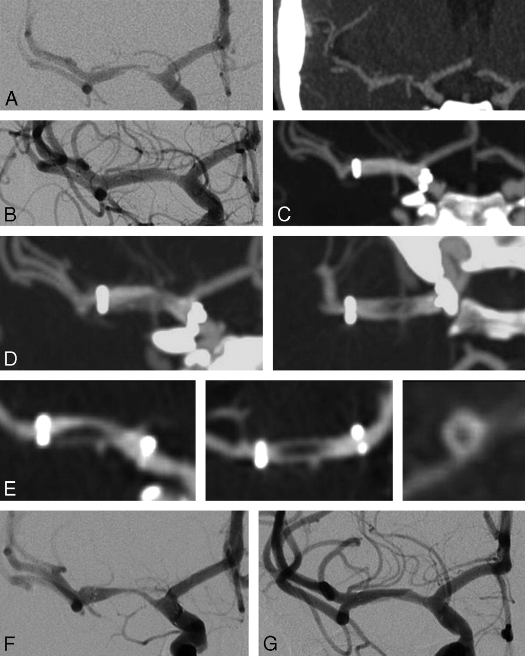
A 38-year-old woman with a history of diabetes, hypertension, and hypercholesterolemia presented with recurrent paresthesias, refractory to medical management. Angioplasty of a middle cerebral artery (MCA) stenosis was not durable; symptomatic restenosis occurred within weeks. She was then referred to our institution for intracranial stent placement. CTA showed a critical stenosis of the right M1 segment of the MCA, which was verified with digital subtraction angiography (DSA) (Fig 1A). She was continued on clopidogrel and aspirin and underwent angioplasty and stent placement (3 × 9 mm Gateway balloon and a 3.5 × 15 mm Wingspan stent) with good results (Fig 1B). CTA follow-up at 1 month showed the stented segment to be widely patent (Fig 1C). Three-month follow-up CTA performed because of recurrent symptoms showed a filling defect within the stent (Fig 1D). Curved thin-section MPRs focusing on the stented segment revealed the filling defect along the caudal aspect of the stent with contrast seen circumferentially around the filling defect, suggestive of a thrombus (Fig 1E).

Fig 1.
  • Download figure
  • Open in new tab
  • Download powerpoint
Fig 1.

A–G, A 38-year-old woman with right MCA stenosis refractory to medical therapy and prior angioplasty. Serial imaging details restenosis and eventual angioplasty.

Because of the potential risk of distal emboli associated with an attempt at repeat angioplasty, the patient's medication was changed to warfarin (Coumadin) and aspirin, and the patient was followed for an additional 4 months. CTA again showed an irregular nonocclusive filling defect within the stent. DSA demonstrated a significant smooth tapering in-stent restenosis (Fig 1F). Balloon angioplasty (3 × 9 mm Gateway balloon) resulted in resolution of the restenosis and symptoms (Fig 1G).

Other Cases

Three other patients with restenosis following successful angioplasty and stent placement with the Wingspan system (2 MCAs and 1 supraclinoid internal carotid artery) showed similar findings with irregular nonocclusive filling defects seen on CTA within the Wingspan stent; DSA in all of these patients confirmed the restenosis but showed no evidence of intraluminal thrombus.

Discussion

There is meager literature describing the CTA appearance of in-stent restenosis associated with the Wingspan stent. In our early experience with this device, we have encountered 4 patients who, at the time of follow-up with CTA, were found to have an intraluminal filling defect suggestive of thrombus. However, on further DSA evaluation, these filling defects were found to represent in-stent restenosis.

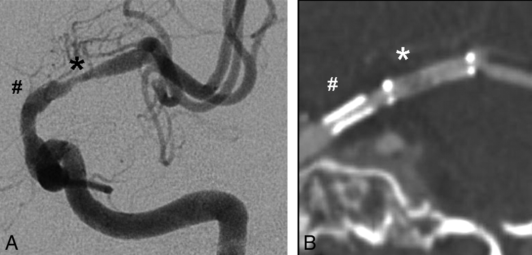
The ability of CTA to image the lumen within cerebrovascular stents is primarily limited by CT-related artifacts.6 Blooming effects result in stent struts that appear thicker and are exaggerated by any adjacent vascular calcification.9 The stent composition and design can also play a role in the blooming effect and overall degree of artifact.10 The Wingspan stent is composed of nitinol and has an open cell design, thin struts, and a relatively low metal surface area coverage, which results in less beam hardening and scatter artifacts compared with conventional coronary stents (Fig 2A, B). Optimal intraluminal contrast enhancement is also important for reducing intrastent artifact and accurately imaging the intrastent lumen. A final limitation of CTA lies with partial volume averaging due to the small size of the cerebral vessels. All CTA scans use 0.625-mm isotropic voxels with additional thin-section curved MPRs from either a 16- or 64-section CT scanner to minimize volume-averaging effects.

Fig 2.
  • Download figure
  • Open in new tab
  • Download powerpoint
Fig 2.

MCA in-stent restenosis with correlative CTA (A) and DSA (B). This patient previously required a coronary stent (number sign) for critical supraclinoid stenosis proximal to the Wingspan stent (asterisk) in the adjacent MCA. Note the extensive blooming effect related to the coronary stent in comparison with the adjacent Wingspan stent.

Because CTA is increasingly used as a technique for evaluation and follow-up of patients who have undergone intracranial stent placement, it is important that the possibility of false-positive studies indicating intraluminal thrombus be appreciated. Because of the management implications when a finding suggestive of an intraluminal thrombus is seen on a CTA in these patients, confirmation with DSA should be strongly considered. Further studies are required to determine both the cause and the frequency of this CTA finding.

References

  1. ↵
    American Heart Association. Heart Disease and Stroke Statistics: 2005 Update. Dallas: American Heart Association;2005
  2. ↵
    SSYLVIA Study Investigators. Stenting of symptomatic atherosclerotic lesions in the vertebral or intracranial arteries (SSYLVIA): study results. Stroke 2004;35:1388–92
    Abstract/FREE Full Text
  3. Kessler IM, Mounayer C, Piotin M, et al. The use of balloon-expandable stents in the management of intracranial arterial diseases: a 5-year single-center experience. AJNR Am J Neuroradiol 2005;26:2342–48
    Abstract/FREE Full Text
  4. ↵
    Wojak JC, Dunlap DC, Hargrave KR, et al. Intracranial angioplasty and stenting: long-term results from a single center. AJNR Am J Neuroradiol 2006;27:1882–92
    Abstract/FREE Full Text
  5. ↵
    Henkes H, Miloslavski E, Lowens S, et al. Treatment of intracranial atherosclerotic stenoses with balloon dilatation and self-expanding stent deployment (WingSpan). Neuroradiology 2005;47:222–28. Epub 2005 Mar 15
    CrossRefPubMed
  6. ↵
    Trossbach M, Hartmann M, Braun C, et al. Small vessel stents for intracranial angioplasty: in vitro evaluation of in-stent stenoses using CT angiography. Neuroradiology 2004;46:459–63
    PubMed
  7. ↵
    Roguin A, Resar JR. CT angiography is here: are we expected to see a change of angiography referral pattern? Int J Cardiovasc Intervent 2005;7:152–54
    PubMed
  8. ↵
    Pugliese F, Cademartiri F, van Mieghem C, et al. Multidetector CT for visualization of coronary stents. Radiographics 2006;26:887–904
    CrossRefPubMed
  9. ↵
    Nieman K, Cademartiri F, Raaijmakers R, et al. Noninvasive angiographic evaluation of coronary stents with multi-slice spiral computed tomography. Herz 2003;28:136–42
    CrossRefPubMed
  10. ↵
    Maintz D, Juergens KU, Wichter T, et al. Imaging of coronary artery stents using multislice computed tomography: in vitro evaluation. Eur Radiol 2003;13:830–35
    PubMed
  • Received May 1, 2007.
  • Accepted after revision May 3, 2007.
  • Copyright © American Society of Neuroradiology
View Abstract
PreviousNext
Back to top

In this issue

American Journal of Neuroradiology: 28 (9)
American Journal of Neuroradiology
Vol. 28, Issue 9
October 2007
  • Table of Contents
  • Index by author
Advertisement
Print
Download PDF
Email Article

Thank you for your interest in spreading the word on American Journal of Neuroradiology.

NOTE: We only request your email address so that the person you are recommending the page to knows that you wanted them to see it, and that it is not junk mail. We do not capture any email address.

Enter multiple addresses on separate lines or separate them with commas.
CT Angiographic Appearance of In-Stent Restenosis of Intracranial Arteries Treated with the Wingspan Stent
(Your Name) has sent you a message from American Journal of Neuroradiology
(Your Name) thought you would like to see the American Journal of Neuroradiology web site.
CAPTCHA
This question is for testing whether or not you are a human visitor and to prevent automated spam submissions.
Cite this article
A.S. Turk, H.A. Rowley, D.B. Niemann, D. Fiorella, B. Aagaard-Kienitz, K. Pulfer, C.M. Strother
CT Angiographic Appearance of In-Stent Restenosis of Intracranial Arteries Treated with the Wingspan Stent
American Journal of Neuroradiology Oct 2007, 28 (9) 1752-1754; DOI: 10.3174/ajnr.A0713

Citation Manager Formats

  • BibTeX
  • Bookends
  • EasyBib
  • EndNote (tagged)
  • EndNote 8 (xml)
  • Medlars
  • Mendeley
  • Papers
  • RefWorks Tagged
  • Ref Manager
  • RIS
  • Zotero
0 Responses
Respond to this article
Share
Bookmark this article
CT Angiographic Appearance of In-Stent Restenosis of Intracranial Arteries Treated with the Wingspan Stent
A.S. Turk, H.A. Rowley, D.B. Niemann, D. Fiorella, B. Aagaard-Kienitz, K. Pulfer, C.M. Strother
American Journal of Neuroradiology Oct 2007, 28 (9) 1752-1754; DOI: 10.3174/ajnr.A0713
del.icio.us logo Twitter logo Facebook logo Mendeley logo
  • Tweet Widget
  • Facebook Like
  • Google Plus One
Purchase

Jump to section

  • Article
    • Abstract
    • Materials and Methods
    • Discussion
    • References
  • Figures & Data
  • Info & Metrics
  • Responses
  • References
  • PDF

Related Articles

  • No related articles found.
  • PubMed
  • Google Scholar

Cited By...

  • Quantitative analysis of high-resolution, contrast-enhanced, cone-beam CT for the detection of intracranial in-stent hyperplasia
  • Surveillance imaging after intracranial stent implantation: non-invasive imaging compared with digital subtraction angiography
  • Successful detection of embologenic ulceration in a symptomatic non-hemodynamic intracranial stenosis using C-arm cone beam CT
  • Double-barrel entanglement of intracranial Enterprise stents resulting from undetected incomplete stent apposition
  • Contrast-Enhanced Angiographic Cone-Beam CT of Cerebrovascular Stents: Experimental Optimization and Clinical Application
  • The Utility of Quantitative Magnetic Resonance Angiography in the Assessment of Intracranial In-Stent Stenosis
  • Crossref (29)
  • Google Scholar

This article has been cited by the following articles in journals that are participating in Crossref Cited-by Linking.

  • Intracranial atherosclerotic disease: An update
    Adnan I. Qureshi, Edward Feldmann, Camilo R. Gomez, S. Claiborne Johnston, Scott E. Kasner, Donald C. Quick, Peter A. Rasmussen, M. Fareed K. Suri, Robert A. Taylor, Osama O. Zaidat
    Annals of Neurology 2009 66 6
  • Contrast-Enhanced Angiographic Cone-Beam CT of Cerebrovascular Stents: Experimental Optimization and Clinical Application
    N.V. Patel, M.J. Gounis, A.K. Wakhloo, N. Noordhoek, J. Blijd, D. Babic, D. Takhtani, S.-K. Lee, A. Norbash
    American Journal of Neuroradiology 2011 32 1
  • Stent Application for the Treatment of Cerebral Aneurysms
    Byung Moon Kim, Dong Joon Kim, Dong Ik Kim
    Neurointervention 2011 6 2
  • The Utility of Quantitative Magnetic Resonance Angiography in the Assessment of Intracranial In-Stent Stenosis
    Shyam Prabhakaran, Lakshmi Warrior, Kalani R. Wells, Miral D. Jhaveri, Michael Chen, Demetrius K. Lopes
    Stroke 2009 40 3
  • Optimized intravenous Flat Detector CT for non-invasive visualization of intracranial stents: first results
    Tobias Struffert, Stephan Kloska, Tobias Engelhorn, Yu Deuerling-Zheng, Sabine Ott, Marc Doelken, Marc Saake, Martin Köhrmann, Arnd Doerfler
    European Radiology 2011 21 2
  • Quantitative analysis of high-resolution, contrast-enhanced, cone-beam CT for the detection of intracranial in-stent hyperplasia
    Thomas F Flood, Imramsjah M J van der Bom, Lara Strittmatter, Ajit S Puri, Gregory M Hendricks, Ajay K Wakhloo, Matthew J Gounis
    Journal of NeuroInterventional Surgery 2015 7 2
  • Spatial Resolution Limits of Multislice Computed Tomography (MS-CT), C-arm-CT, and Flat Panel-CT (FP-CT) Compared to MicroCT for Visualization of a Small Metallic Stent
    Mircea Ionescu, Ralph W. Metcalfe, Dianna Cody, Miguel Valdivia y Alvarado, John Hipp, Goetz Benndorf
    Academic Radiology 2011 18 7
  • Sub-Nyquist acquisition and constrained reconstruction in time resolved angiography
    Charles A. Mistretta
    Medical Physics 2011 38 6Part1
  • Retrograde Stent Placement for Coil Embolization of a Wide-Necked Posterior Inferior Cerebellar Artery Aneurysm
    Hong Gee Roh, Young Il Chun, Jin Woo Choi, Joon Cho, Won-Jin Moon, Sten Solander
    Korean Journal of Radiology 2012 13 4
  • Hemodynamic Changes Following Wingspan Stent Placement-A Quantitative Magnetic Resonance Angiography Study
    Shyam Prabhakaran, Kalani R. Wells, Miral D. Jhaveri, Demetrius K. Lopes
    Journal of Neuroimaging 2011 21 2

More in this TOC Section

  • SAVE vs. Solumbra Techniques for Thrombectomy
  • CT Perfusion&Reperfusion in Acute Ischemic Stroke
  • Delayed Reperfusion Post-Thrombectomy&Thrombolysis
Show more Interventional

Similar Articles

Advertisement

Indexed Content

  • Current Issue
  • Accepted Manuscripts
  • Article Preview
  • Past Issues
  • Editorials
  • Editors Choice
  • Fellow Journal Club
  • Letters to the Editor

Cases

  • Case Collection
  • Archive - Case of the Week
  • Archive - Case of the Month
  • Archive - Classic Case

Special Collections

  • Special Collections

Resources

  • News and Updates
  • Turn around Times
  • Submit a Manuscript
  • Author Policies
  • Manuscript Submission Guidelines
  • Evidence-Based Medicine Level Guide
  • Publishing Checklists
  • Graphical Abstract Preparation
  • Imaging Protocol Submission
  • Submit a Case
  • Become a Reviewer/Academy of Reviewers
  • Get Peer Review Credit from Publons

Multimedia

  • AJNR Podcast
  • AJNR SCANtastic
  • Video Articles

About Us

  • About AJNR
  • Editorial Board
  • Not an AJNR Subscriber? Join Now
  • Alerts
  • Feedback
  • Advertise with us
  • Librarian Resources
  • Permissions
  • Terms and Conditions

American Society of Neuroradiology

  • Not an ASNR Member? Join Now

© 2025 by the American Society of Neuroradiology All rights, including for text and data mining, AI training, and similar technologies, are reserved.
Print ISSN: 0195-6108 Online ISSN: 1936-959X

Powered by HighWire