
Article Figures & Data
Figures
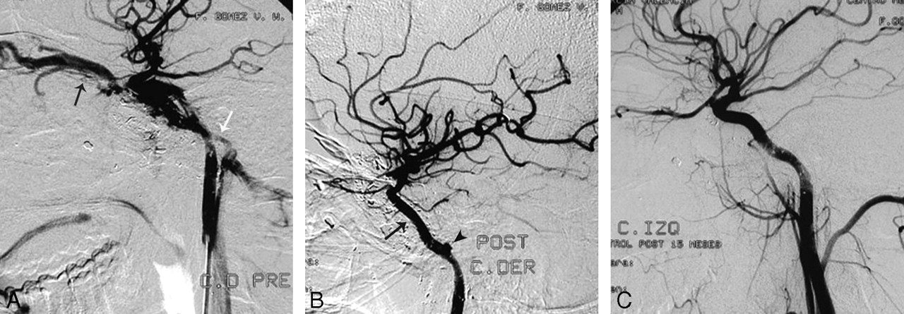
- Fig 1.
A, Patient 2. Selective left ICA angiogram demonstrates a CCF after a gunshot wound, with filling of the superior ophthalmic vein (black arrow) and inferior petrosal sinus (white arrow). B, Immediate control post-covered stent deployment (black arrow) shows complete occlusion of the fistula. A small pseudoaneurysm is noticed in the petrous carotid artery (black arrowhead), which was managed conservatively. C, Follow-up after 15 months shows a normal artery without recanalization of the fistula. There has been spontaneous resolution of the small pseudoaneurysm, and there is no intimal hyperplasia.
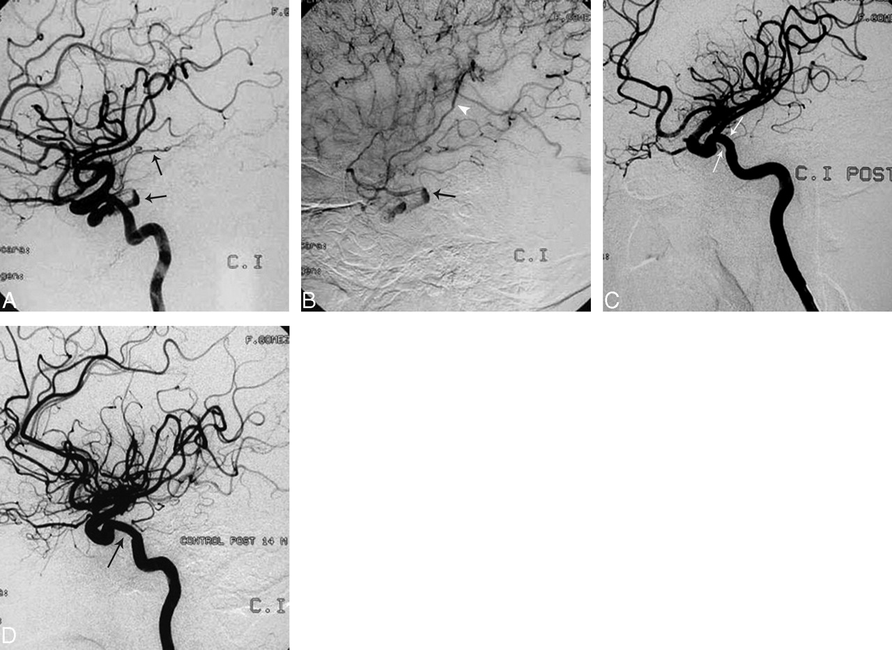
- Fig 2.
A and B, Patient 4. Angiogram with arterial (A) and venous (B) phase images showing a CCF with retrograde cortical venous drainage (black arrows and white arrowhead). Notice the separation of the cortical veins due to the intracerebral hematoma identified in a previous MR image (not shown). In this case, a covered stent was used because the fistula was too small to accept a balloon. C, Control poststent graft deployment (white arrows). There is complete occlusion of the fistula. D, Fifteen-month control angiogram demonstrates persistent occlusion of the fistula with minimal intrastent intimal hyperplasia (black arrow), which remained stable in the 42-month angiographic follow-up (not shown).
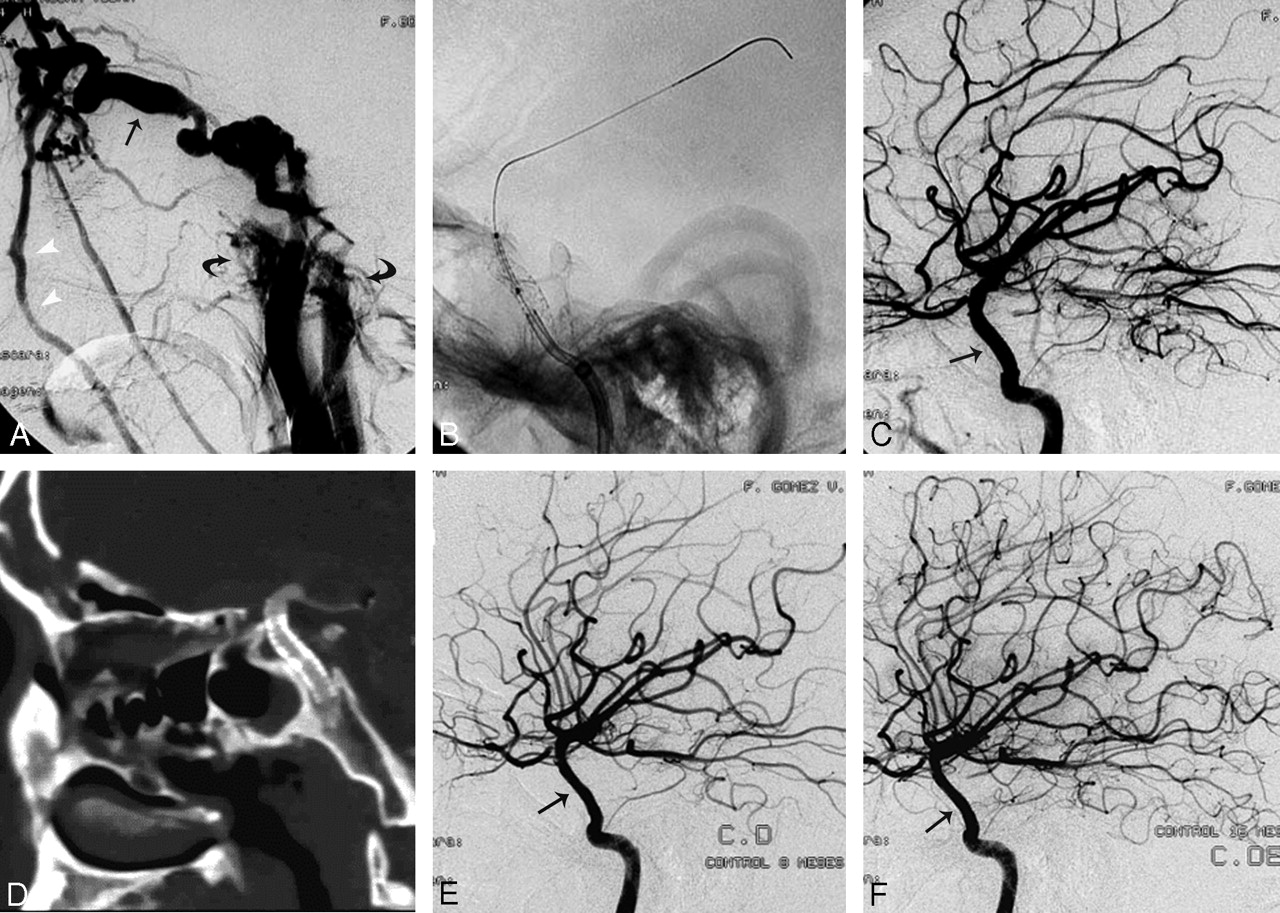
- Fig 3.
A, Patient 5. Selective right ICA angiogram demonstrates a high-flow CCF with arterial phase enhancement of a markedly enlarged superior ophthalmic vein (black arrow), facial veins (white arrowheads), and both inferior petrosal sinuses (curved arrows). This is a case of near-complete cavernous ICA transection with nonvisualization of the right anterior and middle cerebral arteries due to complete deviation of the flow into the fistula. B, There is an exchange wire stabilized in one of the distal middle cerebral artery branches. A bare stent has just been deployed in the cavernous carotid artery to reconstruct the vessel wall and provide stability to the covered stent, which is being positioned inside the bare stent at the exact location of the fistula. C, Control postdeployment shows occlusion of the fistula with re-establishment of the intracranial flow through the right ICA. There is straightening (arrow) of the cavernous ICA with no hemodynamic consequence in the control angiogram. D, Three-month follow-up with CT angiography. Here the sagittal reformat shows the straightening of the cavernous carotid artery with the stent in place and preserved patency. E and F, Angiographic follow-up at 8 and 15 months demonstrates a normal intracranial ICA without recurrence of the fistula or significant intrastent intimal hyperplasia (black arrows).


Tables
Patient demographics, fistula characteristics, angiographic follow-up, and results
Patient No./ Age (y)/Sex Presentation Mechanism of Trauma Venous Drainage Reason Not to Use Balloon Covered Stent (mm) Additional Material Angiographic Follow-Up (months) Result 1/23/M Chemosis, proptosis PHBC Ophthalmic, petrous No balloon available 4 × 12 - 1 Occluded fistula, asymptomatic occlusion ICA 2/21/M Chemosis, proptosis GSW Ophthalmic, petrous Bony fragments 4 × 12, 4 × 16 - 23.5 Occluded fistula, preserved ICA 3/21/M Chemosis, proptosis BHBC Ophthalmic, petrous No balloon available 3.5 × 12 Coil, 8 × 20, Histoacryl 37.2 Occluded fistula, preserved ICA 4/39/M Right hemiplegia MVC Cortical vein Small cavernous sinus 4 × 12 42 Occluded fistula, preserved ICA 5/21/M Chemosis, proptosis, ophthalmoplegia, blindness BHBC Ophthalmic, petrous ICA transection 4 × 12 Bare stent, 5 × 24 14.9 Occluded fistula, preserved ICA 6/54/M ASDH, chemosis, proptosis PHBC Ophthalmic Stent, first choice 4 × 9 - 7.2 Occluded fistula, preserved ICA 7/50/F Chemosis, proptosis, CNP MCC Ophthalmic, petrous Stent, first choice 4 × 12 - 3 Occluded fistula, preserved ICA Note:—CNP indicates cranial nerve palsy; ASDH, acute subdural hematoma; GSW, gunshot wound; ICA, internal carotid artery; MCC, motorcycle crash; MVC, motor vehicle crash; BHBC, bicycle hit by car; PHBC, pedestrian hit by car.
In this issue


Jump to section
Related Articles
Cited By...
- Treatment of direct carotid-cavernous fistulas with a double lumen balloon
- Multiple Unilateral Traumatic Carotid-Cavernous Sphenoid Sinus Fistulas with Associated Massive Epistaxis: A Consequence of Parkour
- 4D flow preliminary investigation of a direct carotid cavernous fistula due to a ruptured intracavernous aneurysm
- Use of Onyx for Transarterial Balloon-Assisted Embolization of Traumatic Carotid Cavernous Fistulas: A Report of 23 Cases
- Treatment of a traumatic carotid-cavernous fistula by the sole use of a flow diverting stent