
Article Figures & Data
Figures
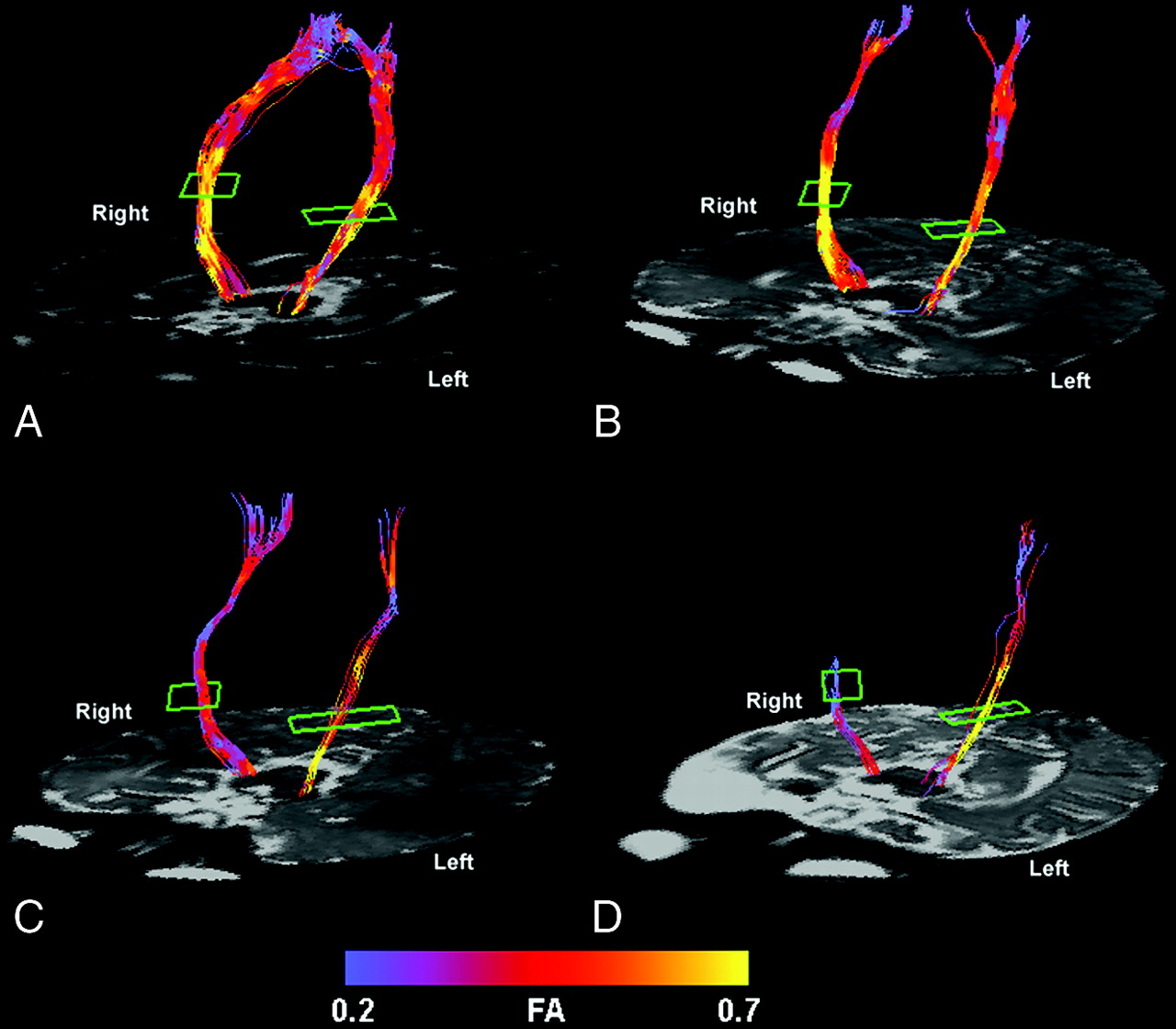
- Fig 1.
Color-coded representation of fractional anisotropy in the pyramidal tracts of a control subject (A), a patient with mild left hemiparesis (B), a patient with moderate left hemiparesis (C), and a patient with severe left hemiparesis (D). Lower fractional anisotropy values are seen in the affected pyramidal tract compared with the unaffected tract of the patients with moderate and severe hemiparesis. The patient with mild hemiparesis and the control subject show no appreciable asymmetry. Although the entire tract is shown from the level of the cerebral peduncle to the centrum semiovale, the analysis was limited to that portion of the pyramidal tract between the posterior limb of the internal capsule (outlined in green) and the cerebral peduncle (see Methods).
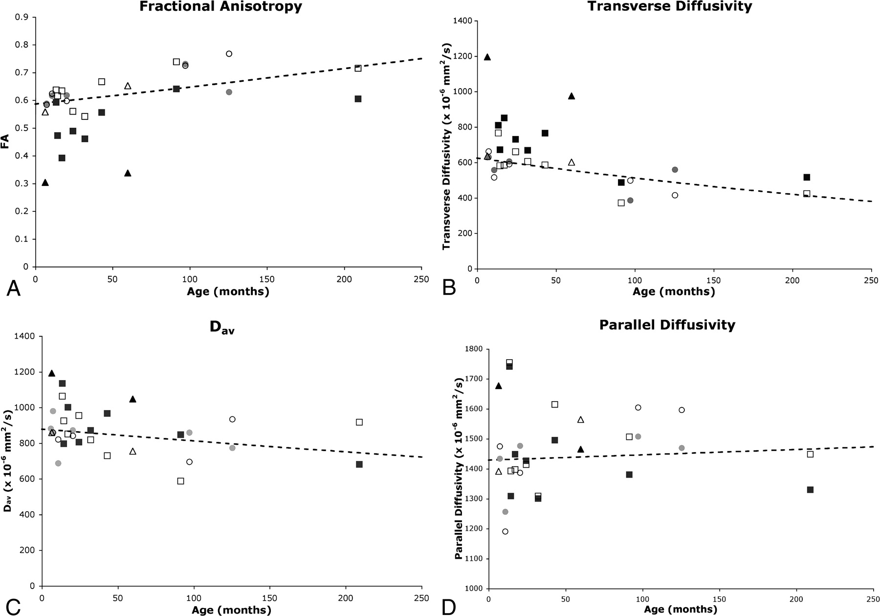
- Fig 2.
Comparison of diffusion parameters in affected and unaffected tracts of the patients with normative curves. Fractional anisotropy (A), transverse diffusivity (B), Dav (C), and parallel diffusivity (D) are plotted against age for the patients with mild hemiparesis (circles), those with moderate hemiparesis (squares), and those with severe hemiparesis (triangles). Both affected (solid symbol) and unaffected (open symbol) pyramidal tract diffusion values are plotted for each patient. Values are compared with the normative curves (dashed curve) of the natural logarithm of the diffusion metric versus age (see Methods).


Tables
Case Gender GA at Birth (wks) Age at MR Imaging (mos) Severity of Hemiparesis Hemiparetic side Conventional MR Imaging Results 1 Male Term 7.32 Mild Right Normal 2 Female Term 10.84 Mild Left Normal 3 Male 27 20.46 Mild Right Normal 4 Female Term 97.23 Mild Right Focal volume loss in left posterior frontal PVMW and caudate body with mild ex vacuo dilation of lateral ventricle consistent with venous infarct 5 Female Term 125.6 Mild Left Focal T2 hyperintensity right posterior frontal PVWM with mild ex vacuo dilation of lateral ventricle consistent with venous infarct 6 Female 36 13.63 Moderate Right Small focal dilation of right frontal horn in region of caudate head 7 Male Term 14.58 Moderate Left Volume loss in right posterior frontal PVWM with evidence of prior hemorrhage and small right thalamus, consistent with venous infarct 8 Female 37 17.25 Moderate Left Right parietotemporal encephalomalacia consistent with MCA angular branch infarct; small right thalamus; small right CP 9 Female Term 24.55 Moderate Left Right frontoparietotemporal polymicrogyria; small right thalamus; small right CP 10 Female 37 32.17 Moderate Left Punctate T2 hyperintensity in right parietal PVWM; thin posterior body & splenium of corpus callosum 11 Male Term 43.16 Moderate Right Left frontoparietal polymicrogyria; small left thalamus; small left CP 12 Male Term 91.55 Moderate Right Encephalomalacia left paramedian frontal & parietal lobes with ex vacuo dilation of lateral ventricle consistent with ACA infarct; thin body & splenium of corpus callosum; small left CP 13 Male Term 209.27 Moderate Left Right frontoparietal polymicrogyria; small right CP 14 Male Term 6.49 Severe Left Right frontoparietotemporal cystic encephalomalacia with ex vacuo dilation of lateral ventricle consistent with MCA infarct; small right deep gray nuclei; small right CP 15 Male 37 59.98 Severe Left Right frontoparietotemporal cystic encephalomalacia with ex vacuo dilation of lateral ventricle consistent with MCA infarct; small right deep gray nuclei; small right CP Note:—GA indicates gestational age; PVWM, periventricular white matter; CP, cerebral peduncle; MCA, middle cerebral artery; ACA, anterior cerebral artery.
Subject Category Fractional Anisotropy Transverse Diffusivity Parallel Diffusivity Dav Control 0.3% (3.8, −3.2) −1.3% (4.9, −7.5) −0.5% (2.6, −3.7) −1.9% (6.5, −10.3) Mild hemiparesis −2.8% (3.6, −9.3) 3.6% (15.1, −7.9) −0.9% (4.8, −6.7) 1.6% (17.1, −14.0) Moderate hemiparesis −17.7% (−12.6, −22.8) 21.5% (30.6, 12.4) −3.3% (1.2, −7.9) 6.6% (18.8, −5.7) Severe hemiparesis −46.9% (−36.7, −57.1) 75.2% (93.4, 57.1) 7.2% (16.3, −2.0) 38.8% (63.4, 14.2) P value P < .0001†‡ P < .0001†‡ P = .23† P < .03†‡ * 95% confidence intervals are listed in parentheses.
† P values are listed for one-way ANOVA analyses.
‡ P values <.05.
In this issue


Jump to section
Related Articles
Cited By...
- Whole-Brain DTI Assessment of White Matter Damage in Children with Bilateral Cerebral Palsy: Evidence of Involvement beyond the Primary Target of the Anoxic Insult
- Advanced Fiber Tracking in Early Acquired Brain Injury Causing Cerebral Palsy
- Does Diffusion Tensor Imaging-Based Tractography at 3 Months of Age Contribute to the Prediction of Motor Outcome After Perinatal Arterial Ischemic Stroke?
- Diffusion MRI in corticofugal fibers correlates with hand function in unilateral cerebral palsy
- Diffusion tensor imaging of the pyramidal tracts in infants with motor dysfunction