
Article Figures & Data
Figures
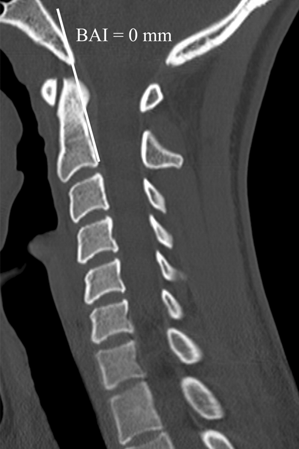
- Fig 1.
BAI. Midsagittal MDCT image of the craniocervical junction demonstrates the posterior axial line drawn along the posterior cortex of the body of the axis and extended cranially. The BAI is the distance between the basion and this line.
- Fig 2.
Midsagittal MDCT image of the craniocervical junction demonstrates the BDI as the distance from the most inferior portion of the basion to the closest point of the superior aspect of the dens.
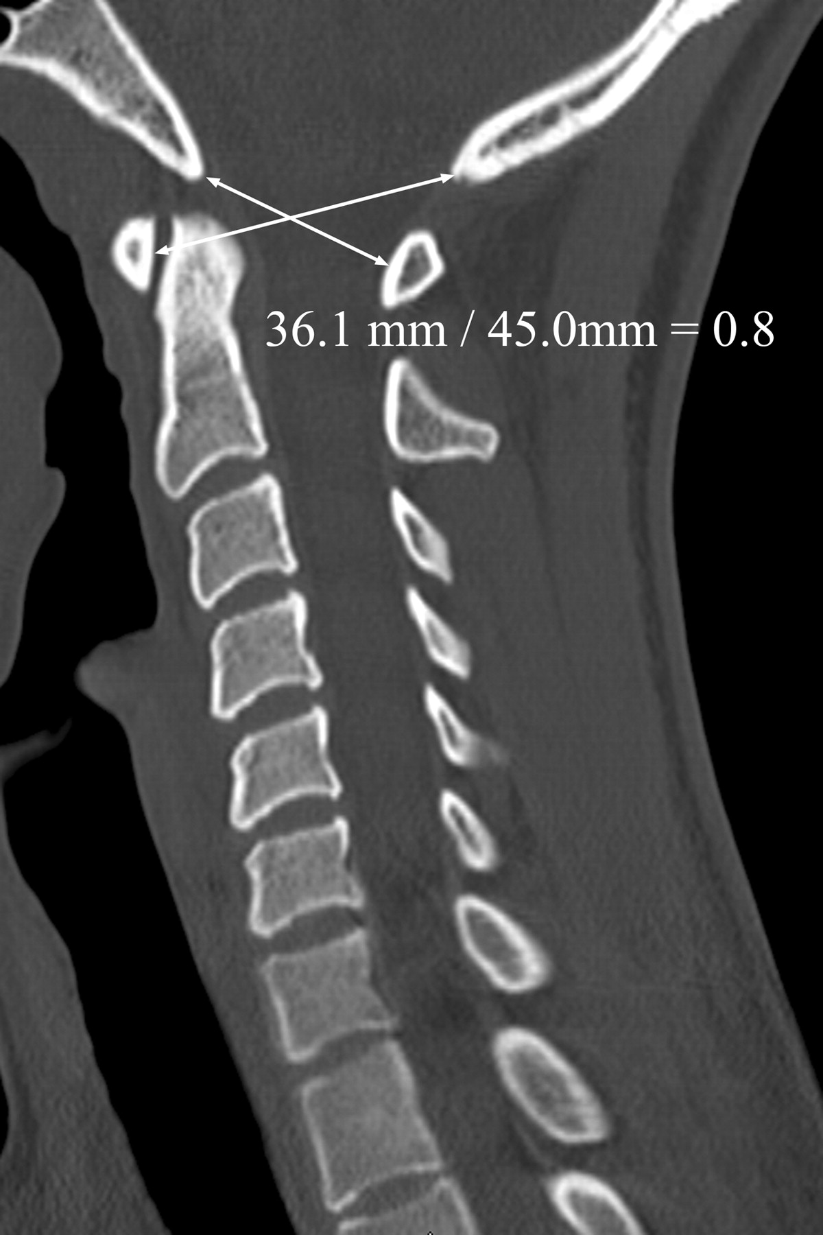
- Fig 3.
Midsagittal MDCT image of the craniocervical junction demonstrates the Powers ratio, which is calculated by dividing the distance between the tip of the basion to the spinolaminar line by the distance from the tip of the opisthion to the midpoint of the posterior aspect of the anterior arch of C1.
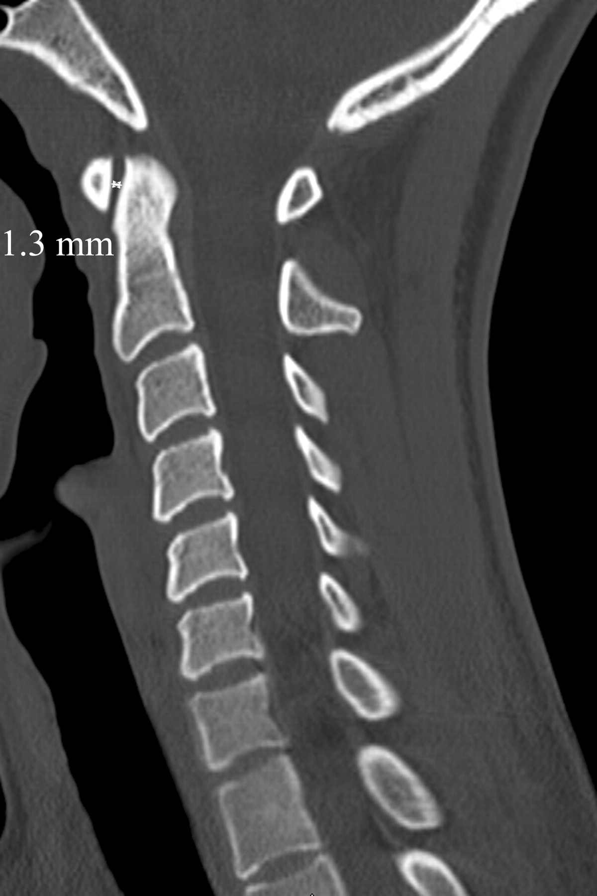
- Fig 4.
Midsagittal MDCT image of the craniocervical junction demonstrates the ADI, which is calculated by drawing a line from the posterior aspect of the anterior arch of C1 to the most anterior aspect of the dens at the midpoint of the thickness of the arch in craniocaudal dimension.
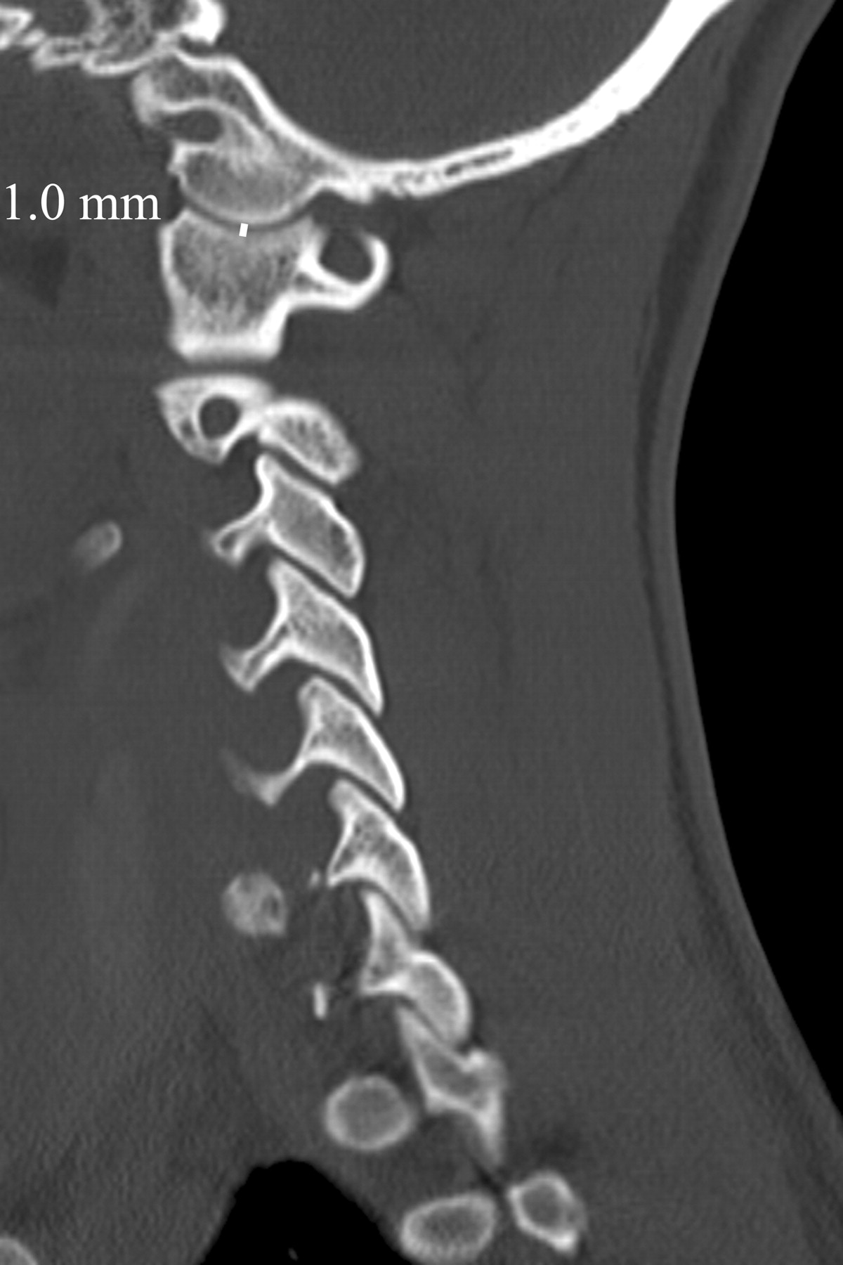
- Fig 5.
Sagittal MDCT image of the craniocervical junction demonstrates the AOI, which is calculated by drawing a line perpendicular to the articular surfaces of the occipital condyle and the lateral mass of C1. This line is drawn at the center of the articulation by correlating the sagittal and coronal images.
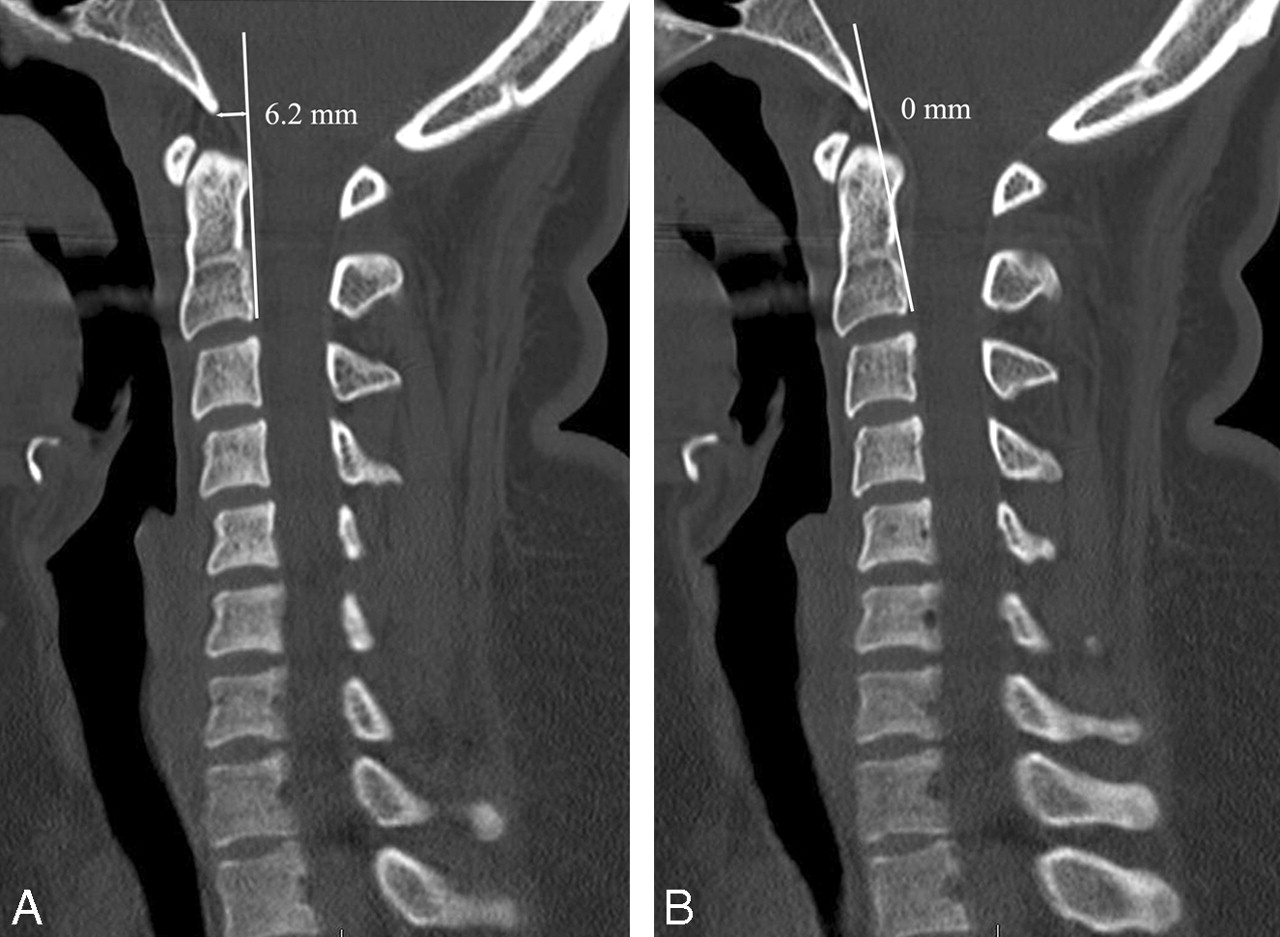
- Fig 6.
Interexaminer variability of the BAI. Midsagittal MDCT image of the craniocervical junction demonstrates the posterior axial line (PAL) drawn along the posterior cortex of the body of the axis and extended cranially. A, Examiner A draws the PAL on the basis of a single interpretation of the posterior cortex of the body of the axis. B, Examiner B draws the PAL using a different interpretation of the posterior cortex of the body of the axis.


Tables
- Table 1:
Normal anatomic relationships of the craniocervical junction on MDCT in 200 patients and comparison with accepted values on plain radiographs*
Method Mean SD Range MDCT Normal Value† Plain Radiograph Normal Value2,5,6 BAI 3.4 4.64 −8.7–26.0 Not reliable <12.0 BDI 5.7 1.39 1.4–9.1 <8.5 <12.0 Powers ratio 0.8 0.08 0.6–1.2 <0.9 <1.0 ADI 1.3 0.37 0.5–2.4 <2.0 in both sexes <3.0 men <2.5 women AOI 1.0 0.23 0.5–1.8 <1.4 No data in adults Note:—MDCT indicates multidetector row CT; BAI; basion-axial interval; BDI, basion-dens interval; ADI, atlantodental interval; AOI, atlanto-occipital interval.
* Results are given in millimeters with the exception of the Powers ratio.
† Normal value is the maximum value for 97.5% of the population.
Method Shrout-Fleiss Reliability BAI 0.54 BDI 0.84 Powers ratio 0.87 ADI 0.65 AOI 0.77 Note:—BAI indicates basion-axial interval; BDI, basion-dens interval; ADI, atlantodental interval; AOI, atlanto-occipital interval.
* Value of 1 represents maximum reliability.


