
Article Figures & Data
Figures
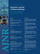
- Fig 1.
Ventricular enlargement in MR image of a patient with NPH with a positive Evans Index.
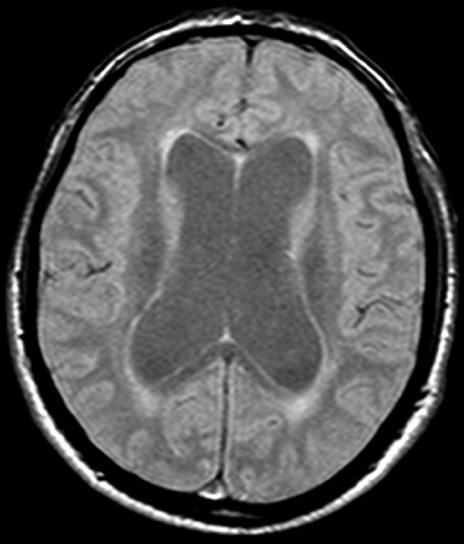
- Fig 2.
PCCMR imaging CSF dynamic study in a 74-year-old patient with NPH. A, Midline sagittal T1-weighted MR imaging is used to graphically describe the phase-contrast cine series. The section is placed at the level of the inferior colliculus, perpendicular to a line drawn through the distal aqueduct. B, Axial section in which region of interest is drawn as close as possible to the aqueduct border. C, Respective absolute values of CSF during 16 cardiac phases are reported on the graph. The flow plot demonstrates sinusoidal pattern of flow where negative values represent aqueductal systolic CSF volume (microliter) outflow and positive values represent diastolic CSF volume inflow.
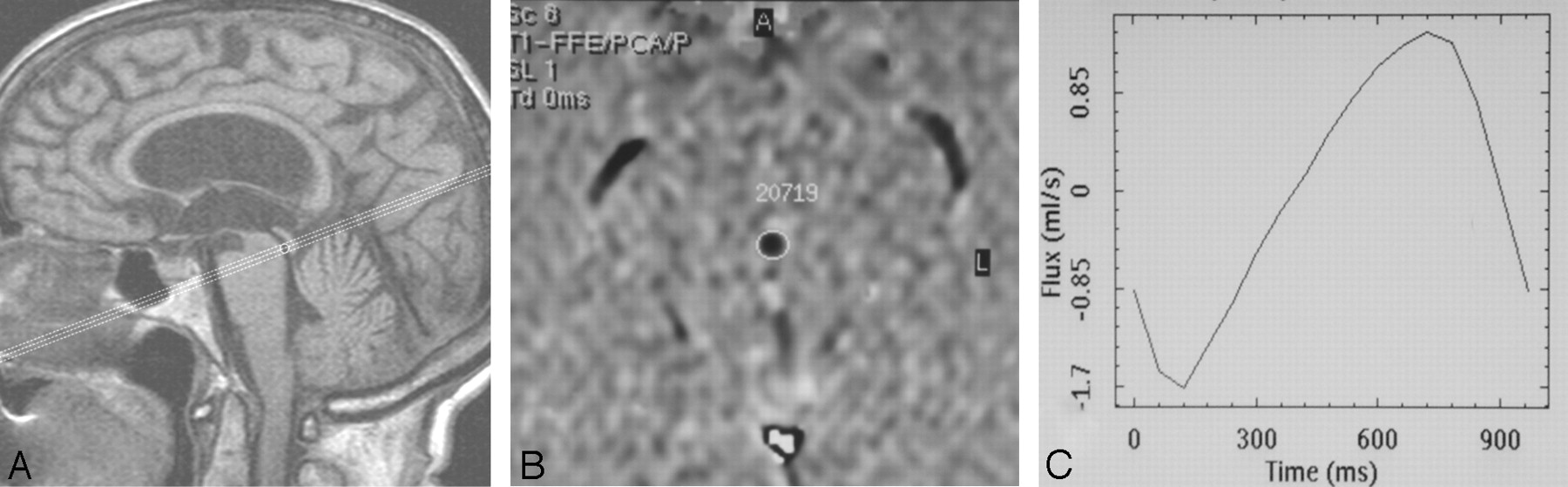
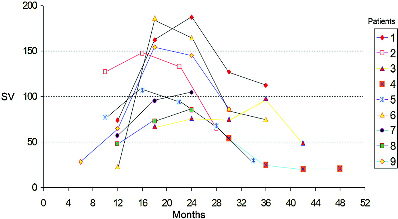
- Fig 3.
Changes in SV values at 6, 12, 18, and 24 months for each of the 9 patients.
- Fig 4.
Changes in SV values (Table 1) standardized for the estimated onset of NPH, as per the reported first symptoms of NPH.
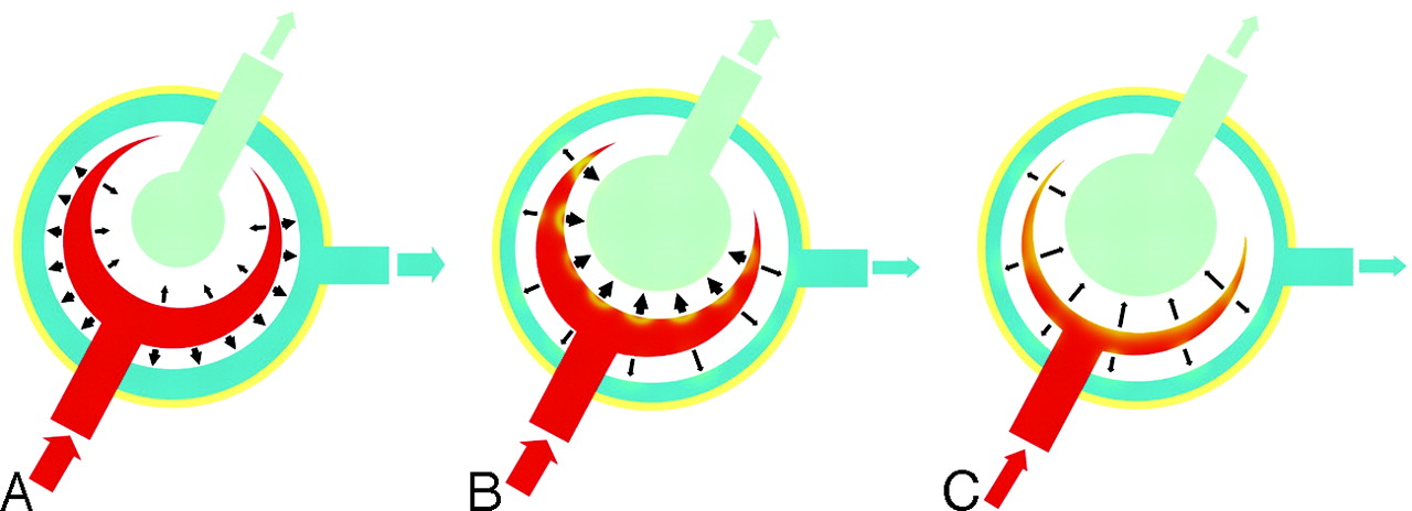
- Fig 5.
Proposed model demonstrating SV changes in NPH. A, In healthy subjects, expansion of the cerebral hemispheres occurs both outward and inward. The outward expansion produces venous blood outflow as a result of compression on the cortical veins. Inward expansion produces flow of CSF into the aqueduct as a result of compression of the lateral and third ventricles. B, In communicating hydrocephalus, the brain has already expanded outward during diastole, compressing the cortical veins. However, during systole, with arterial blood entering, the systolic expansion is directed inwards, resulting in a much greater SV in the aqueduct. C, Progressive ischemia and a reduction of arterial inflow results in a decreased “ventricular CSF pump.”


Tables
- Table 1:
Symptoms present at first evaluation and duration of presence of each of these symptoms since their onset
Patient No. Duration (mos.) of Symptoms at First Evaluation Gait Urinary Cognitive 1 11 0 7 2 6 4 6 3 18 8 6 4 30 10 18 5 10 0 6 6 12 7 0 7 6 2 12 8 12 6 12 9 5 1 0 Patient No. GS (mos.) UIS (mos.) MMSE (mos.) 0 6 12 18 24 0 6 12 18 24 0 6 12 18 24 1 1 1 2 2 2 0 2 2 1 2 27 25 19 20 17 2 1 1 1 2 NE 1 1 1 2 NE 16 16 15 16 NE 3 2 2 2 2 2 2 2 2 2 3 18 17 16 13 13 4 2 2 2 2 NE 2 2 3 3 NE 17 18 15 13 NE 5 1 1 1 2 2 0 0 1 2 1 26 24 23 23 22 6 1 1 2 2 3 2 3 2 2 3 29 27 25 22 18 7 1 1 2 NE NE 1 1 2 NE NE 21 21 18 NE NE 8 1 1 1 NE NE 1 2 2 NE NE 19 19 17 NE NE 9 1 1 1 1 2 0 0 1 1 2 27 27 25 22 18 Note:—GS indicates Gait Scale; UIS, Urinary Incontinence Scale; MMSE, Mini-Mental State Examination; NE, not evaluated.
Patient No. CSF Aqueductal SV (mos.) 0 6 12 18 24 1 74 162 187 127 112 2 127 147 133 65 NE 3 67 76 75 98 49 4 54 25 20 21 NE 5 77 107 94 68 30 6 23 186 165 86 75 7 57 95 104 NE NE 8 48 73 85 NE NE 9 28 65 154 145 86 Note:—SV indicates stroke volume; NE, not evaluated.
In this issue


Jump to section
Related Articles
Cited By...
- Aqueductal CSF Stroke Volume Is Increased in Patients with Idiopathic Normal Pressure Hydrocephalus and Decreases after Shunt Surgery
- Does Phase-Contrast Imaging through the Cerebral Aqueduct Predict the Outcome of Lumbar CSF Drainage or Shunt Surgery in Patients with Suspected Adult Hydrocephalus?
- High-Convexity Tightness Predicts the Shunt Response in Idiopathic Normal Pressure Hydrocephalus
- Intracranial Pressure versus Phase-Contrast MR Imaging for Normal Pressure Hydrocephalus
- Aqueductal Stroke Volume: Comparisons with Intracranial Pressure Scores in Idiopathic Normal Pressure Hydrocephalus
- CSF Flow in the Brain in the Context of Normal Pressure Hydrocephalus
- Current and Emerging MR Imaging Techniques for the Diagnosis and Management of CSF Flow Disorders: A Review of Phase-Contrast and Time-Spatial Labeling Inversion Pulse
- Natural course of idiopathic normal pressure hydrocephalus
- Differential Diagnosis of Normal Pressure Hydrocephalus by MRI Mean Diffusivity Histogram Analysis
- Assessment of Craniospinal Pressure-Volume Indices