
Article Figures & Data
Figures
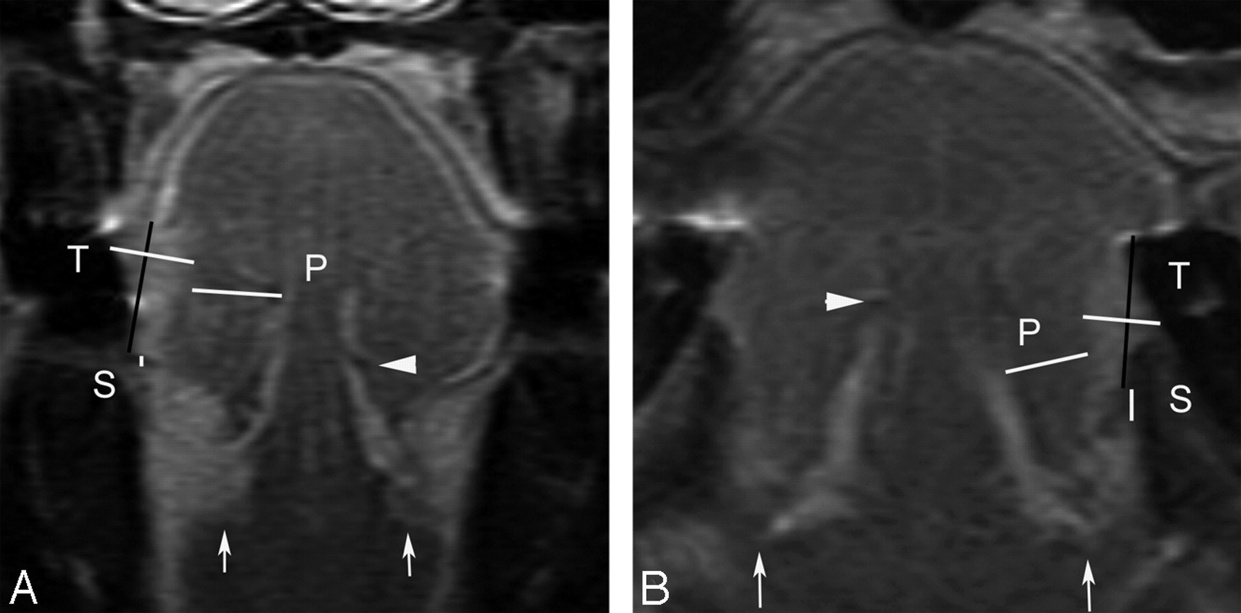
- Fig 1.
Coronal contrast-enhanced T1-weighted images show measured tumor thickness (T), sublingual distance (S), and paralingual distance (P). White arrows show sublingual glands, and a white arrowhead shows the contralateral deep lingual artery. A, MR image of a 51-year-old woman with T2N0 disease shows a vertical black line connecting 2 tumor-mucosa junctions as a reference line. A horizontal white line drawn perpendicular to the reference line represents radiologically determined tumor thickness (T) of 8.7 mm. White line (S) between the tumor and the sublingual space demonstrates the sublingual distance of 0.6 mm. The line (P) between the tumor and the paralingual space demonstrates the paralingual distance of 8.9 mm. B, A 73-year-old man with T1N0 disease (T = 6.2 mm, S = 2.8 mm, P = 6.6 mm). Both patients had no evidence of lymph node metastases for their follow-up duration of >1 year.
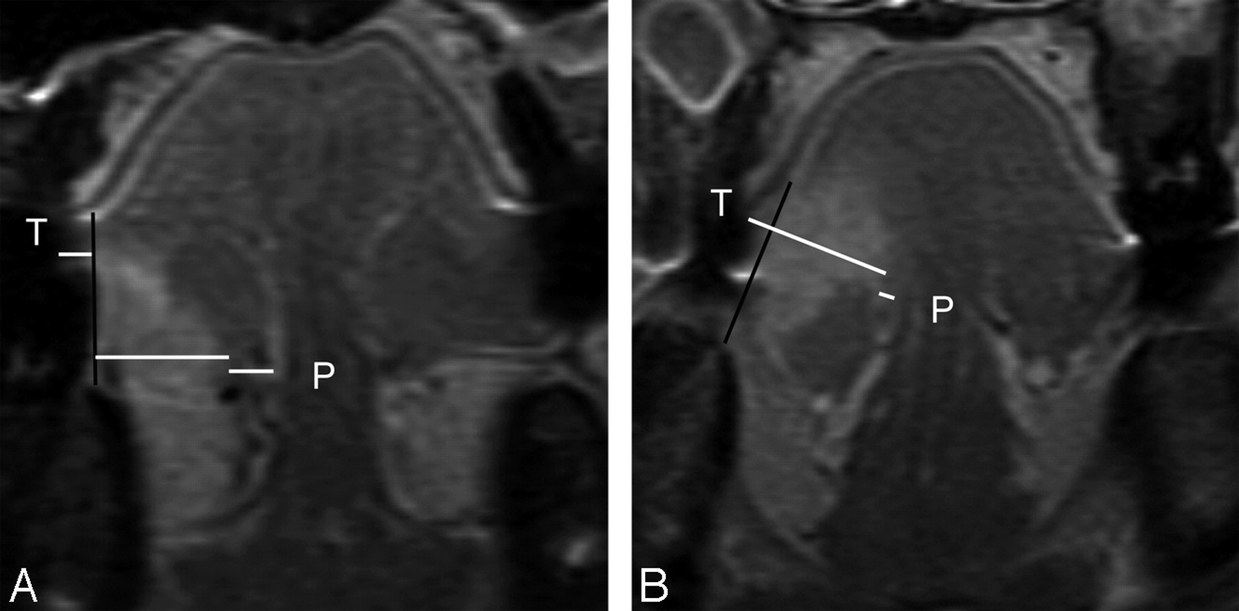
- Fig 2.
A, MR image of a 54-year-old man with T2N0 disease shows a vertical black line connecting 2 tumor-mucosa junctions as a reference line. Horizontal white lines are drawn perpendicular to the reference line. Tumor thickness (T) is the sum of both of these horizontal lines and is determined as 10.1 mm (sublingual distance = 0 mm, paralingual distance [P] = 3.8 mm). The elective dissected neck specimen revealed no pathologically positive lymph node. B, MR image of a 41-year-old woman with T3N0 disease demonstrates T of 15.5 mm, sublingual distance of 0 mm, and P of 0.8 mm. Elective dissected neck specimen revealed 1 metastatic node in level III.
- Fig 3.
A, MR image of a 22-year-old man with T2N0 disease and tumor thickness (T) of 13.8 mm, sublingual distance of 0 mm, and paralingual distance (P) of 2.7 mm. Elective dissected neck specimen revealed 1 pathologically positive node in level I. B, MR image of a 33-year-old woman with T2N0 disease demonstrates T of 8.4 mm, S of 4.4 mm, and P of 5.3 mm. Late lymph node metastasis developed 2 months after glossectomy, and 2 pathologically positive nodes (level II and III) were verified with neck dissection.
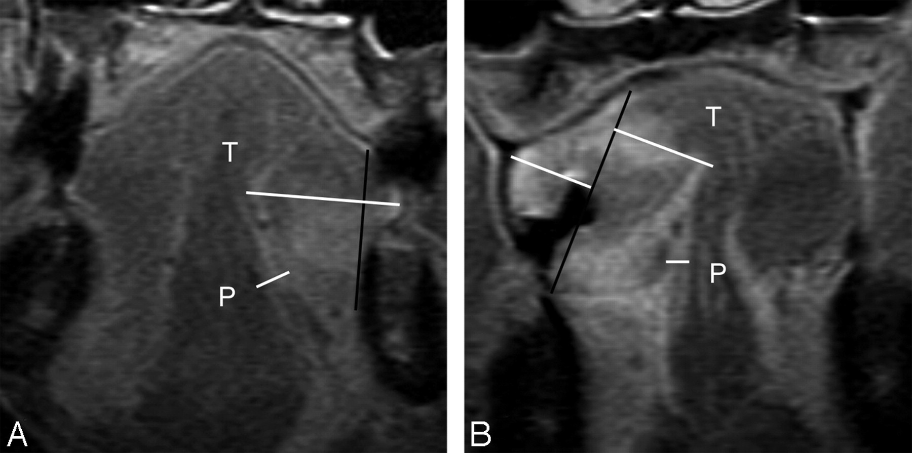
- Fig 4.
A, MR image of a 74-year-old man with T4N2b disease, which invaded the mandible, demonstrates a tumor thickness (T) of 19.0 mm, sublingual distance of 0 mm, and paralingual distance (P) of −5.8 mm. Therapeutic neck dissection revealed 9 metastatic nodes in levels I-V. B, MR image of a 61-year-old man with T4N1 disease demonstrates tumor thickness (T) of 27.2 mm, sublingual distance of 0 mm, and paralingual distance (P) of −3.1 mm. The T is the sum of both of these horizontal white lines perpendicular to the reference line. Therapeutic neck dissection revealed 1 metastatic node in level I.


Tables
- Table 1:
Patient characteristics and associations between lymph node metastases and variables
Patient Characteristics Lymph Node Metastases Absent (n = 28) Present (n = 15) P value Sex, N (%) Male 29 (67) 20 (69) 9 (31) .67 Female 14 (33) 8 (57) 6 (43) Age (mean ± SD) 58 ± 15 57 ± 14 59 ± 16 .48 Performance status <2 35 (81) 23 (68) 12 (32) >.99 ≥2 8 (19) 5 (63) 3 (38) T classification, N (%) T1–2 34 (79) 26 (76) 8 (24) <.01 T3–4 9 (21) 2 (22) 7 (78) N classification, N (%) N0 32 (74) 25 (78) 7 (22) <.01 N1–3 11 (26) 3 (27) 8 (72) Differentiation, N(%) Well-moderate 38 (88) 25 (66) 13 (34) >.99 Poor 5 (12) 3 (60) 2 (40) Tumor thickness (mm) (mean ± SD) 11.7 ± 7.3 8.5 ± 4.5 17.8 ± 7.8 <.0001 Sublingual distance (mm) (mean ± SD) 4.0 ± 4.4 5.5 ± 4.4 1.1 ± 2.4 <.005 Paralingual distance (mm) (mean ± SD) 4.7 ± 5.1 7.2 ± 3.3 0 ± 4.5 <.0001 Parameter β Coefficient SE Odds Ratio (95% CI) P Sex, male −0.51 0.67 0.60 (0.16–2.24) .60 Age (years/10) 0.07 0.22 1.07 (0.69–1.65) .77 Performance status, ≥2 0.14 0.81 1.15 (0.23–5.65 .86 T classification, T3–4 2.43 0.90 11.38 (1.96–66.13) <.01 N classification, N1–3 2.25 0.80 9.52 (1.98–45.76) <.005 Differentiation, poor 0.25 0.98 1.28 (0.19–8.67) .80 Tumor thickness (mm) 0.31 0.10 1.36 (1.12–1.65) <.005 Sublingual distance (mm) −0.34 0.12 0.71 (0.56–0.91) <.01 Paralingual distance (mm) −0.64 0.22 0.53 (0.34–0.80) <.005 Note:—SE indicates standard error.
Parameter β Coefficient SE Odds Ratio (95% CI) P Model 1 T classification, T3–4 1.89 1.40 6.60 (0.43–101.90) .18 N classification, N1–3 −2.23 1.75 0.11 (0.01–3.30) .20 Tumor thickness 0.36 0.16 1.43 (1.05–1.96) <.05 Sublingual distance −0.13 0.13 0.88 (0.68–1.14) .32 Model 2 T classification, T3–4 0.25 1.69 1.28 (0.05–35.15) .88 N classification, N1–3 −1.37 1.39 0.25 (0.02–3.89) .32 Sublingual distance 0.06 0.18 1.07 (0.75–1.50) .72 Paralingual distance −0.84 0.37 0.43 (0.21–0.89) <.05 - Table 4:
Correlation between measured MR imaging distance and cervical lymph node metastasis
MR Imaging Distance (mm) No. of Patients (%) by Presence of Metastases Absent (n = 28) Present (n = 15) Tumor thickness <8.3 15 (100) 0 (0) 8.3–22.5 13 (52) 12 (48) >22.5 0 (0) 3 (100) Sublingual distance 0 8 (42) 11 (58) 0–8.5 10 (71) 4 (29) >8.5 10 (100) 0 (0) Paralingual distance <0 0 (0) 6 (100) 0–5.3 8 (47) 9 (53) >5.3 20 (100) 0 (0)


