
Article Figures & Data
Figures
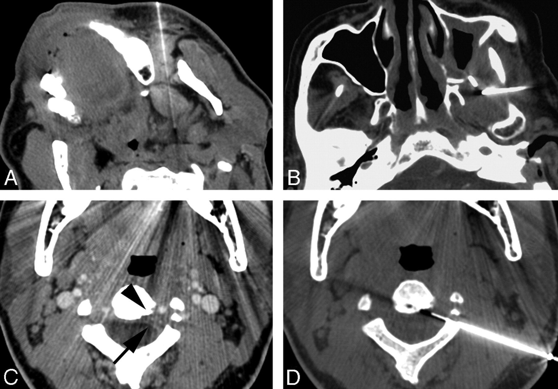
- Fig 1.
A, Paramaxillary approach to the left parapharyngeal space mass, proven to be an oncocytoma. Slight turning of the head to the opposite side simplifies the approach to this parapharyngeal space lesion. B, Subzygomatic approach to the masticator space mass via the intercondylar notch. The core specimens in this patient with previously treated squamous cell carcinoma revealed scar tissue with no evidence of malignant cells. C, CT image in a patient with a mass at the C2 level reveals a subtle left-sided epidural soft-tissue (arrow) and cortical irregularity of the vertebral body (arrowhead). This image was acquired with contrast to map the location of the adjacent vertebral artery. D, A posterolateral approach to the epidural mass was planned. A 22-gauge Franseen needle is advanced through a guiding needle, and aspiration biopsy is performed. Aspiration biopsy was consistent with a diagnosis of chordoma.
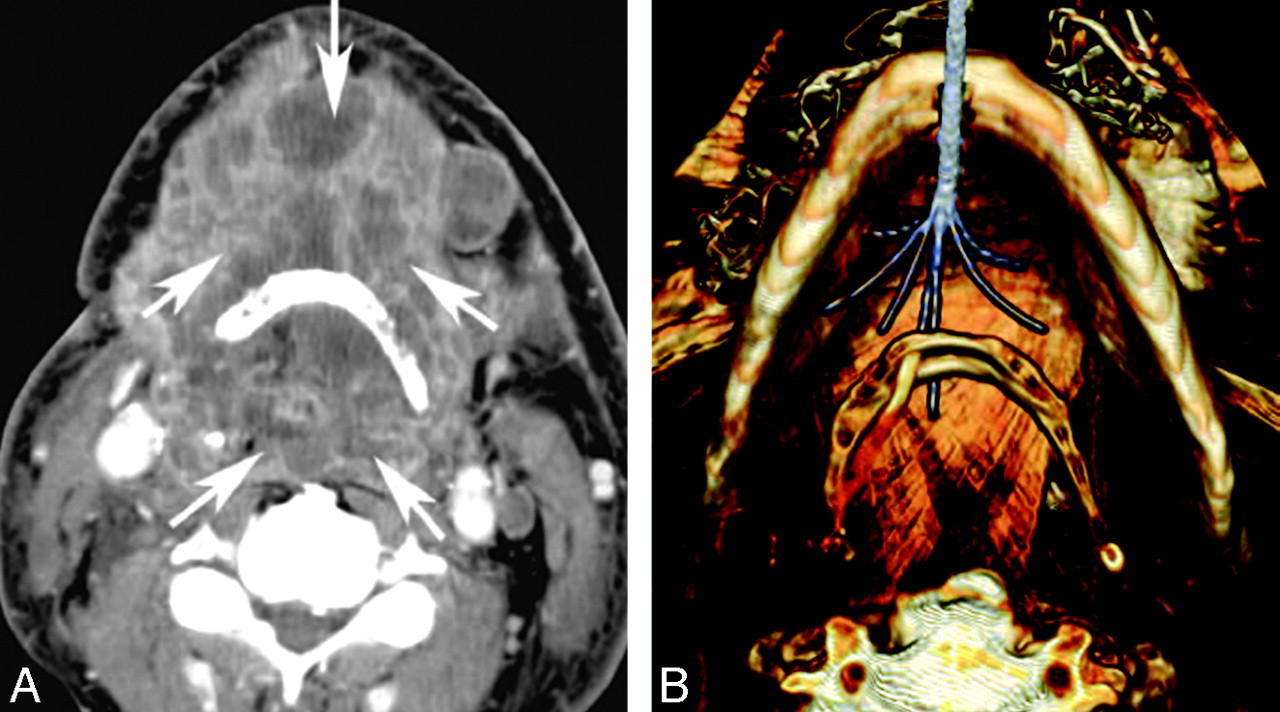
- Fig 2.
A 59-year-old man with severe dyspnea and dysphagia secondary to a large squamous cell carcinoma treated with radio-frequency ablation. A, Axial contrast-enhanced CT scan demonstrates a large necrotic tumor (arrows) in the floor of the mouth and hypopharynx. B, 3D volume-rendered reconstruction demonstrates the radio-frequency probe (Starburst XLi; RITA Medical Systems, Mountainview, Calif) and electrode deployment within the tumor by means of a submental approach. Note that the tumor anterior and posterior to the hyoid bone could be ablated simultaneously. Reproduced with permission from the Journal of Vascular and Interventional Radiology.6 Copyright 2008, Elsevier.
- Fig 3.
A, Clinical photograph of the posterior aspect of the neck in an adolescent with a known venous malformation. The arrows in the picture point to the scar from a previous attempt at resection. Note the soft-tissue bulge of the residual venous malformation. B, Percutaneous injection of contrast into the venous malformation performed via an 18-gauge Teflon cannula shows the clustered and dilated venous spaces. The malformation was treated by using 3% STS that was injected after aspiration of the contrast. C, Clinical photograph on a follow-up visit shows complete disappearance of the soft-tissue bulge.
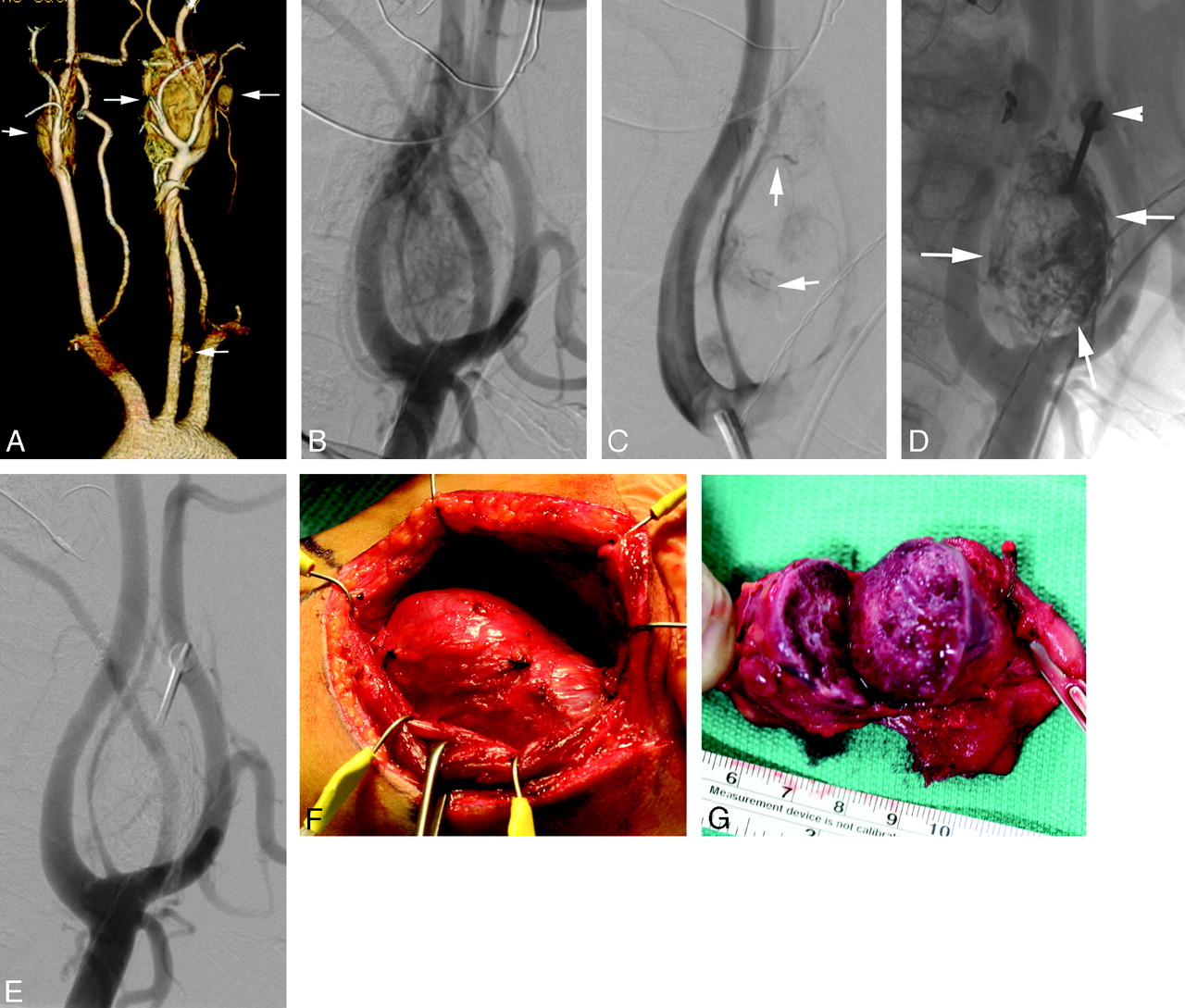
- Fig 4.
A 33-year-old man with a family history of multiple paragangliomas and a palpable left neck mass. Preoperative embolization was planned before resection of the left carotid body tumor. A, Volume-rendered reconstruction of a CTA of the neck shows bilateral carotid body tumors as well as 2 other glomus tumors along the left carotid sheath (arrows). B, Lateral view of the left common carotid angiogram reveals a large carotid body tumor supplied by multiple external carotid branches and splaying the ICA and ECA. C, After transarterial embolization of the occipital and ascending pharyngeal contributions to the tumor, residual tumor blush is noted. The vessels that remain are too small to be catheterized with a microcatheter (arrows). D, Fluoroscopic puncture of the tumor performed with an 18-gauge needle (arrowhead) and tumor embolized percutaneously by using a 30% mixture of n-BCA glue and ethiodol oil (arrows). E, Postembolization angiogram reveals near-complete devascularization of the carotid body tumor. F, Intraoperative photograph shows the exposed carotid body tumor. Intraoperative bleeding was <130 mL for the entire procedure. G, The cut surface of the excised specimen shows grossly a thrombosed tumor.
- Fig 5.
A 12-year-old girl with a large periorbital AVM. A, Anteroposterior view of the right ICA shows the arterial supply to the AVM (arrowheads) via small branches of the ophthalmic artery (arrow). B, External carotid angiogram in the lateral projections demonstrates a large AVM fed by multiple branches of the internal maxillary artery and the anterior branch of the superficial temporal artery. A large venous varix is seen early in the arterial phase draining into the supraorbital and facial veins. C, Percutaneous access to the venous varix is shown with placement of coils and injection of a 1:1 mixture of n-BCA glue and ethiodol oil. Small remaining feeders were then embolized transarterially with Onyx (not shown). D, Postembolizarion right ICA angiogram shows complete cessation of supply from the ophthalmic artery. E, Right external carotid angiogram also demonstrates near-complete thrombosis of the AVM and the venous varix. F, Intraoperative photograph showing the thrombosed AVM mass. Total blood loss at surgery was <200 mL. Intraoperative photograph courtesy of A. Kahana, MD, Department of Ophthalmology, University of Michigan, Ann Arbor, Mich.
- Fig 6.
An elderly man with a recurrent head and neck cancer presenting with pulsatile bleeding through the oral cavity. A, CT angiogram of the neck shows an ulcerated left oropharyngeal mass (arrowheads) that encases the left ECA (arrow). B, Common carotid angiogram reveals a long-segment tumor encasement of the left ECA. C, The ECA is embolized with fibered and detachable platinum coils. The patient did not have additional episodes of bleeding after the embolization.
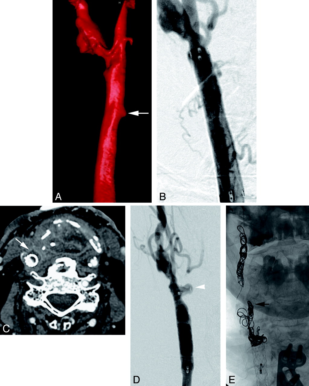
- Fig 7.
A 65-year-old patient with a previously treated supraglottic laryngeal carcinoma presenting with profuse hemoptysis. A, A volume-rendered 3D digital subtraction angiogram reveals a small pseudoaneurysm (arrow) arising from the medial aspect of the right common carotid artery. Also note the atherosclerotic changes at the carotid bifurcation. B, This image was obtained following deployment of an 8 × 40 mm stent-graft in the common carotid artery and excluding the pseduoaneurysm. C, The patient presented 11 weeks later with a recurrent hemorrhage from his tracheostomy site. Neck CT shows a rim-enhancing fluid collection (arrow) adjacent to the stent-graft suggestive of infection. D, Common carotid angiogram at this time shows a recurrent carotid blowout (arrowhead) at the distal end of the stent-graft. E, Following a successful temporary balloon occlusion test, the ICA and ECA (black arrow) were occluded with coils, and the common carotid artery was sacrificed proximal to the pseudoaneurysm. The patient has not had another bleed in the following 5 months.


In this issue


Jump to section
- Article
- Abstract
- Image-Guided Biopsies
- Radio-frequency Ablation and Cryoablation for Tumors
- Percutaneous Sclerotherapy
- Preoperative Tumor Embolization
- Embolization of Cervicofacial High-Flow Malformations
- Management of Bleeding from the Head and Neck
- Intra-Arterial Chemotherapy for Head and Neck Carcinoma
- References
- Figures & Data
- Info & Metrics
- Responses
- References