Advertisement

Main menu

  • Home
  • Content
    • Current Issue
    • Accepted Manuscripts
    • Article Preview
    • Past Issue Archive
    • Video Articles
    • AJNR Case Collection
    • Case of the Week Archive
    • Case of the Month Archive
    • Classic Case Archive
  • Special Collections
    • AJNR Awards
    • Low-Field MRI
    • Alzheimer Disease
    • ASNR Foundation Special Collection
    • Photon-Counting CT
    • View All
  • Multimedia
    • AJNR Podcasts
    • AJNR SCANtastic
    • Trainee Corner
    • MRI Safety Corner
    • Imaging Protocols
  • For Authors
    • Submit a Manuscript
    • Submit a Video Article
    • Submit an eLetter to the Editor/Response
    • Manuscript Submission Guidelines
    • Statistical Tips
    • Fast Publishing of Accepted Manuscripts
    • Graphical Abstract Preparation
    • Imaging Protocol Submission
    • Author Policies
  • About Us
    • About AJNR
    • Editorial Board
    • Editorial Board Alumni
  • More
    • Become a Reviewer/Academy of Reviewers
    • Subscribers
    • Permissions
    • Alerts
    • Feedback
    • Advertisers
    • ASNR Home

User menu

  • Alerts
  • Log in

Search

  • Advanced search
American Journal of Neuroradiology

American Journal of Neuroradiology

ASHNR American Society of Functional Neuroradiology ASHNR American Society of Pediatric Neuroradiology ASSR
  • Alerts
  • Log in

Advanced Search

  • Home
  • Content
    • Current Issue
    • Accepted Manuscripts
    • Article Preview
    • Past Issue Archive
    • Video Articles
    • AJNR Case Collection
    • Case of the Week Archive
    • Case of the Month Archive
    • Classic Case Archive
  • Special Collections
    • AJNR Awards
    • Low-Field MRI
    • Alzheimer Disease
    • ASNR Foundation Special Collection
    • Photon-Counting CT
    • View All
  • Multimedia
    • AJNR Podcasts
    • AJNR SCANtastic
    • Trainee Corner
    • MRI Safety Corner
    • Imaging Protocols
  • For Authors
    • Submit a Manuscript
    • Submit a Video Article
    • Submit an eLetter to the Editor/Response
    • Manuscript Submission Guidelines
    • Statistical Tips
    • Fast Publishing of Accepted Manuscripts
    • Graphical Abstract Preparation
    • Imaging Protocol Submission
    • Author Policies
  • About Us
    • About AJNR
    • Editorial Board
    • Editorial Board Alumni
  • More
    • Become a Reviewer/Academy of Reviewers
    • Subscribers
    • Permissions
    • Alerts
    • Feedback
    • Advertisers
    • ASNR Home
  • Follow AJNR on Twitter
  • Visit AJNR on Facebook
  • Follow AJNR on Instagram
  • Join AJNR on LinkedIn
  • RSS Feeds

AJNR Awards, New Junior Editors, and more. Read the latest AJNR updates

OtherSpine Imaging and Spine Image-Guided Interventions

Intraoperative 3T MR Imaging for Spinal Cord Tumor Resection: Feasibility, Timing, and Image Quality Using a “Twin” MR–Operating Room Suite

T.P. Duprez, A. Jankovski, C. Grandin, L. Hermoye, G. Cosnard and C. Raftopoulos
American Journal of Neuroradiology November 2008, 29 (10) 1991-1994; DOI: https://doi.org/10.3174/ajnr.A1134
T.P. Duprez
  • Find this author on Google Scholar
  • Find this author on PubMed
  • Search for this author on this site
A. Jankovski
  • Find this author on Google Scholar
  • Find this author on PubMed
  • Search for this author on this site
C. Grandin
  • Find this author on Google Scholar
  • Find this author on PubMed
  • Search for this author on this site
L. Hermoye
  • Find this author on Google Scholar
  • Find this author on PubMed
  • Search for this author on this site
G. Cosnard
  • Find this author on Google Scholar
  • Find this author on PubMed
  • Search for this author on this site
C. Raftopoulos
  • Find this author on Google Scholar
  • Find this author on PubMed
  • Search for this author on this site
  • Article
  • Figures & Data
  • Info & Metrics
  • Responses
  • References
  • PDF
Loading

Article Figures & Data

Figures

  • Fig 1.
    • Download figure
    • Open in new tab
    • Download powerpoint
    Fig 1.

    Case 1. A, Preoperative midsagittal FSE T2-weighted image at 1.5T (TR, 4000 ms; TE, 75.8 ms; echo train length (ETL) 20; 2 NEX; acquisition time (AT), 3′36“ for 11 4-mm-thick sections): swelling and abnormal hyperintensity of the diseased segment of the cord were obvious. B, Intraoperative midsagittal FSE T2-weighted image at 3T after closure of the surgical wound. The 2 elements had been put on the midline of the patient's back in the cranial-caudal direction without capability of SENSE acquisition because of insufficient overlapping between coils. Inflatable balloon of the laryngeal tube was visible (arrow). IQ was rated excellent by using following PSPs: TR, 2209 ms; TE, 120 ms; ETL 20, 4 NEX; no SENSE acceleration factor; AT: 4′56” for 11 4-mm-thick sections.

  • Fig 2.
    • Download figure
    • Open in new tab
    • Download powerpoint
    Fig 2.

    Case 2. Preoperative images. Midsagittal contrast-enhanced FSE T1- (A) and T2-weighted (B) views showed the usual features of spinal cord ependymoma at the T1-T2 level exhibiting pathognomonic hemosiderin-containing epidural “caps” (ball arrowheads in B). PSPs for T1-weighted sequence were as follows: TR, 530 ms; TE, 7.8 ms; ETL, 4; 4 NEX; SENSE acceleration factor, 1.4; AT, 4′04“ for 11 4-mm-thick sections. PSPs for T2-weighted sequence were as follows: TR, 2209 ms; TE, 120 ms; ETL, 20 ms; 4 NEX; SENSE acceleration factor, 1.4; AT: 3′28” for 11 4-mm-thick sections. On the T2-weighted image (B), SNR for cord and CSF were 119 and 301, respectively. Cord/CSF CNR was 182. C, Intraoperative midsagittal contrast-enhanced FSE T1-weighted view (similar section location and PSP as A) showed trapped air bubble at upper pole of resection site (arrow) and unmodified appearance of inferior hemosiderin-containing epidural cap (ball arrowhead). D, Intraoperative midsagittal FSE T2-weighted view (similar section location and PSP as B) again showed air bubble mimicking epidural (arrow) and unchanged true inferior epidural cap (ball arrowhead). SNRs for cord and CSF were 58 and 194, respectively. Cord/CSF CNR was 135.
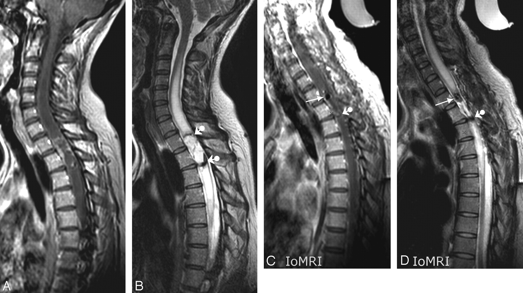
  • Fig 3.
    • Download figure
    • Open in new tab
    • Download powerpoint
    Fig 3.

    Case 3. Preoperative images. Midsagittal postcontrast FSE T1- (A) and T2-weighted (B) views showing usual features of a spinal cord glioma. C–E. Intraoperative images. Midsagittal precontrast FSE T1-weighted (C), postcontrast FSE T1-weighted (D), and FSE T2-weighted views: observe similar phenomenon of air bubble trapping within surgical field (arrow in C) as in case 2 and contrast-enhancement of the margins of the resection cavity (thin arrows in D) at this early postcontrast phase of the intraoperative MR procedure. Observe true intraoperative conditions with a Gelfoam roll plugged on open dura (ball arrowhead in C). Similar PSPs as for the previous patient were used. F–H, Intraoperative images. Serial transverse FSE T1-weighted sections at the level of C7, before contrast agent perfusion (F), at 8 minutes (G), and at 30 minutes (H) after perfusion: striking increase in thickness of enhanced margins of the resection cavity were obvious comparing F to G and H. PSPs were as follows: TR, 530 ms; TE, 7.8 ms; ETL, 4; 4 NEX; SENSE acceleration factor, 1.6; AT, 3′15“ for 16 4-mm-thick sections.

PreviousNext
Back to top

In this issue

American Journal of Neuroradiology: 29 (10)
American Journal of Neuroradiology
Vol. 29, Issue 10
November 2008
  • Table of Contents
  • Index by author
Advertisement
Print
Download PDF
Email Article

Thank you for your interest in spreading the word on American Journal of Neuroradiology.

NOTE: We only request your email address so that the person you are recommending the page to knows that you wanted them to see it, and that it is not junk mail. We do not capture any email address.

Enter multiple addresses on separate lines or separate them with commas.
Intraoperative 3T MR Imaging for Spinal Cord Tumor Resection: Feasibility, Timing, and Image Quality Using a “Twin” MR–Operating Room Suite
(Your Name) has sent you a message from American Journal of Neuroradiology
(Your Name) thought you would like to see the American Journal of Neuroradiology web site.
CAPTCHA
This question is for testing whether or not you are a human visitor and to prevent automated spam submissions.
Cite this article
T.P. Duprez, A. Jankovski, C. Grandin, L. Hermoye, G. Cosnard, C. Raftopoulos
Intraoperative 3T MR Imaging for Spinal Cord Tumor Resection: Feasibility, Timing, and Image Quality Using a “Twin” MR–Operating Room Suite
American Journal of Neuroradiology Nov 2008, 29 (10) 1991-1994; DOI: 10.3174/ajnr.A1134

Citation Manager Formats

  • BibTeX
  • Bookends
  • EasyBib
  • EndNote (tagged)
  • EndNote 8 (xml)
  • Medlars
  • Mendeley
  • Papers
  • RefWorks Tagged
  • Ref Manager
  • RIS
  • Zotero
0 Responses
Respond to this article
Share
Bookmark this article
Intraoperative 3T MR Imaging for Spinal Cord Tumor Resection: Feasibility, Timing, and Image Quality Using a “Twin” MR–Operating Room Suite
T.P. Duprez, A. Jankovski, C. Grandin, L. Hermoye, G. Cosnard, C. Raftopoulos
American Journal of Neuroradiology Nov 2008, 29 (10) 1991-1994; DOI: 10.3174/ajnr.A1134
del.icio.us logo Twitter logo Facebook logo Mendeley logo
  • Tweet Widget
  • Facebook Like
  • Google Plus One
Purchase

Jump to section

  • Article
    • Abstract
    • Technique
    • Discussion
    • References
  • Figures & Data
  • Info & Metrics
  • Responses
  • References
  • PDF

Related Articles

  • No related articles found.
  • PubMed
  • Google Scholar

Cited By...

  • No citing articles found.
  • Crossref
  • Google Scholar

This article has not yet been cited by articles in journals that are participating in Crossref Cited-by Linking.

More in this TOC Section

  • Management Outcomes For VO Spine Biopsy
  • Characteristics of SIH Type I Culprit Lesions
  • Advanced Imaging of Type 2 Spinal CSF Leaks
Show more Spine Imaging and Spine Image-Guided Interventions

Similar Articles

Advertisement

Indexed Content

  • Current Issue
  • Accepted Manuscripts
  • Article Preview
  • Past Issues
  • Editorials
  • Editor's Choice
  • Fellows' Journal Club
  • Letters to the Editor
  • Video Articles

Cases

  • Case Collection
  • Archive - Case of the Week
  • Archive - Case of the Month
  • Archive - Classic Case

More from AJNR

  • Trainee Corner
  • Imaging Protocols
  • MRI Safety Corner
  • Book Reviews

Multimedia

  • AJNR Podcasts
  • AJNR Scantastics

Resources

  • Turnaround Time
  • Submit a Manuscript
  • Submit a Video Article
  • Submit an eLetter to the Editor/Response
  • Manuscript Submission Guidelines
  • Statistical Tips
  • Fast Publishing of Accepted Manuscripts
  • Graphical Abstract Preparation
  • Imaging Protocol Submission
  • Evidence-Based Medicine Level Guide
  • Publishing Checklists
  • Author Policies
  • Become a Reviewer/Academy of Reviewers
  • News and Updates

About Us

  • About AJNR
  • Editorial Board
  • Editorial Board Alumni
  • Alerts
  • Permissions
  • Not an AJNR Subscriber? Join Now
  • Advertise with Us
  • Librarian Resources
  • Feedback
  • Terms and Conditions
  • AJNR Editorial Board Alumni

American Society of Neuroradiology

  • Not an ASNR Member? Join Now

© 2025 by the American Society of Neuroradiology All rights, including for text and data mining, AI training, and similar technologies, are reserved.
Print ISSN: 0195-6108 Online ISSN: 1936-959X

Powered by HighWire