Advertisement

Main menu

  • Home
  • Content
    • Current Issue
    • Accepted Manuscripts
    • Article Preview
    • Past Issue Archive
    • AJNR Case Collection
    • Case of the Week Archive
    • Classic Case Archive
    • Case of the Month Archive
  • Special Collections
    • Spinal CSF Leak Articles (Jan 2020-June 2024)
    • 2024 AJNR Journal Awards
    • Most Impactful AJNR Articles
  • Multimedia
    • AJNR Podcast
    • AJNR Scantastics
    • Video Articles
  • For Authors
    • Submit a Manuscript
    • Author Policies
    • Fast publishing of Accepted Manuscripts
    • Graphical Abstract Preparation
    • Manuscript Submission Guidelines
    • Imaging Protocol Submission
    • Submit a Case for the Case Collection
  • About Us
    • About AJNR
    • Editorial Board
  • More
    • Become a Reviewer/Academy of Reviewers
    • Subscribers
    • Permissions
    • Alerts
    • Feedback
    • Advertisers
    • ASNR Home
  • Other Publications
    • ajnr

User menu

  • Alerts
  • Log in

Search

  • Advanced search
American Journal of Neuroradiology

American Journal of Neuroradiology

ASHNR American Society of Functional Neuroradiology ASHNR American Society of Pediatric Neuroradiology ASSR
  • Alerts
  • Log in

Advanced Search

  • Home
  • Content
    • Current Issue
    • Accepted Manuscripts
    • Article Preview
    • Past Issue Archive
    • AJNR Case Collection
    • Case of the Week Archive
    • Classic Case Archive
    • Case of the Month Archive
  • Special Collections
    • Spinal CSF Leak Articles (Jan 2020-June 2024)
    • 2024 AJNR Journal Awards
    • Most Impactful AJNR Articles
  • Multimedia
    • AJNR Podcast
    • AJNR Scantastics
    • Video Articles
  • For Authors
    • Submit a Manuscript
    • Author Policies
    • Fast publishing of Accepted Manuscripts
    • Graphical Abstract Preparation
    • Manuscript Submission Guidelines
    • Imaging Protocol Submission
    • Submit a Case for the Case Collection
  • About Us
    • About AJNR
    • Editorial Board
  • More
    • Become a Reviewer/Academy of Reviewers
    • Subscribers
    • Permissions
    • Alerts
    • Feedback
    • Advertisers
    • ASNR Home
  • Follow AJNR on Twitter
  • Visit AJNR on Facebook
  • Follow AJNR on Instagram
  • Join AJNR on LinkedIn
  • RSS Feeds

Welcome to the new AJNR, Updated Hall of Fame, and more. Read the full announcements.


AJNR is seeking candidates for the position of Associate Section Editor, AJNR Case Collection. Read the full announcement.


Research ArticleINTERVENTIONAL

But Did You Use HydroCoil? Perianeurysmal Edema and Hydrocephalus with Bare Platinum Coils

J.B. White, H.J. Cloft and D.F. Kallmes
American Journal of Neuroradiology February 2008, 29 (2) 299-300; DOI: https://doi.org/10.3174/ajnr.A0877
J.B. White
  • Find this author on Google Scholar
  • Find this author on PubMed
  • Search for this author on this site
H.J. Cloft
  • Find this author on Google Scholar
  • Find this author on PubMed
  • Search for this author on this site
D.F. Kallmes
  • Find this author on Google Scholar
  • Find this author on PubMed
  • Search for this author on this site
  • Article
  • Figures & Data
  • Info & Metrics
  • Responses
  • References
  • PDF
Loading

Abstract

SUMMARY: Bioactive coils were developed to improve aneurysm packing and accelerate neointimal healing. Recent reports, however, have implicated these coils in the development of aseptic meningitis, perianeurysmal edema, and hydrocephalus. This report demonstrates that the same clinical phenomena can occur with the use of bare platinum coils.

High recurrence rates following coil embolization with bare platinum led to the development of second-generation “bioactive” coils. Coincident with increased use of these newer coils has been concern over untoward effects on the surrounding CSF and brain parenchyma. Recent reports have implicated newer coils in the development of aseptic meningitis, perianeurysmal edema, and hydrocephalus in the postprocedural setting.1–3

This report presents 2 patients whose aneurysms were treated with bare platinum coils who subsequently developed significant brain stem edema and hydrocephalus.

Case Reports

Case 1

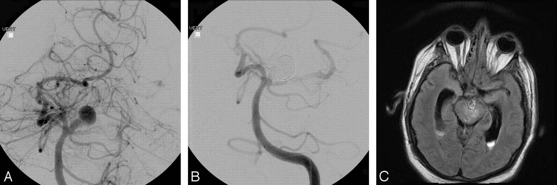
A 73-year-old man presented with an incidentally discovered right vertebral artery aneurysm that measured 17 × 17 × 15 mm (Fig 1A) as detected on CT angiography. The pretreatment CT scan revealed no evidence for pre-existing cerebral or brain stem edema. He was premedicated with aspirin and clopidogrel. Under general anesthesia, the patient underwent coiling of the aneurysm with a total of 17 bare platinum coils from 3 different manufacturers (Orbit Trufill, Cordis, Miami Lakes, Fla; HeliPaq, Micrus Endovascular, San Jose, Calif; and MicroPlex, MicroVention, Aliso Viejo, Calif; Fig 1B). The packing attenuation following coiling was calculated at 13%. During the ensuing 24 hours, the patient developed visual scintillations in the right eye, which were consistent with his well-documented migraine auras. A follow-up angiogram 1 day following the coil procedure showed complete aneurysm occlusion and no evidence for parent artery compromise or distal emboli. On the third hospital day, the patient was dismissed home and was maintained on aspirin and clopidogrel.

Fig 1.
  • Download figure
  • Open in new tab
  • Download powerpoint
Fig 1.

A, Pre-embolization angiogram showing a large right vertebrobasilar region aneurysm. B, Postembolization angiogram showing the coiled aneurysm with minimal residual neck. C, Axial flare MR image showing edema within the brain stem adjacent to the coiled aneurysm.

Approximately 1 week after the coiling, the patient presented to his local hospital with symptoms of headache, dysequilibrium, abducens palsy, and internuclear ophthalmoplegia. An MR imaging showed the coiled aneurysm in the setting of marked brain stem edema (Fig 1C). At that time, angiographic findings were unchanged as compared with the patient’s angiogram obtained at the time of his coiling. Corticosteroids were initiated, but during the course of the following 5 days, the patient developed hydrocephalus, necessitating placement of an external ventricular drain, as well as a posterior fossa hemorrhage. His neurologic status continued to decline with the family withdrawing support on postcoiling day 18.

Case 2

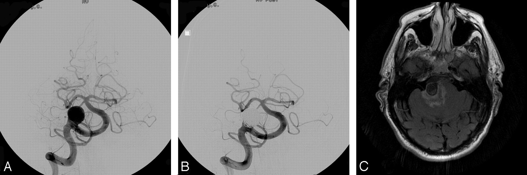
The patient was a 55-year-old man who presented clinically as a Hunt and Hess grade 1. CT imaging revealed a Fisher grade 3 subarachnoid hemorrhage with no evidence for cerebral or brain stem edema. Angiography revealed an aneurysm at the basilar apex measuring 13 × 10 × 9 mm (Fig 2A). Under general anesthesia, he underwent successful coiling of the aneurysm with a total of 12 bare platinum coils (MicroPlex, MicroVention; Fig 2B). The calculated packing attenuation of the aneurysm following coiling was 24%.

Fig 2.
  • Download figure
  • Open in new tab
  • Download powerpoint
Fig 2.

A, Pre-embolization angiogram showing a large basilar apex region aneurysm. B, Postembolization angiogram showing the coiled aneurysm with minimal residual neck. A large posterior communicating artery on the left produces washout of the left posterior cerebral artery. C, Axial flare MR image showing edema within the brain stem adjacent to the coiled aneurysm.

On the second day of his hospitalization, an external ventricular drain was placed for hydrocephalus, which was subsequently replaced with a ventriculoperitoneal shunt. For 1 month following the patient’s coiling procedure, he experienced significant hypersomnolence, which fluctuated with near-complete resolution before discharge. An MR imaging was performed 6 days following the coil procedure, which was remarkable only for profound edema within the brain stem adjacent to the coiled aneurysm (Fig 2C). Vasospasm surveillance by using transcranial Doppler sonography and angiography revealed no evidence for altered flow velocities. At 5 weeks from his initial hemorrhage, the patient was discharged to an inpatient rehabilitation unit, where he thrived until discharge to his home.

Discussion

This report shows that patients with aneurysms coiled with bare platinum alone are capable of developing the same clinical sequelae that have been previously identified with the use of bioactive coils.1–4 Both of our patients developed significant perianeurysmal edema, which was manifest in their brain stems. Both of our patients also developed hydrocephalus, one of whom did so in the setting of an unruptured aneurysm. Both patients developed a fluctuating level of consciousness that ultimately resolved in one and led to hemorrhage and death in another.

In previous cases in which patients were coiled with bioactive materials, the symptoms and signs at presentation were commonly headache, malaise, and cranial neuropathies.1–4 Other findings were fever, nuchal rigidity, visual loss, and photophobia. In nearly every patient, corticosteroids were implemented with near-complete resolution of the patients’ symptoms. Despite the use of steroids, the condition of one of our patients (case 1) continued to decline, with a subsequent hemorrhage and ultimately death. The etiology of hemorrhage in this patient is unclear, though one may conclude that the aneurysm ruptured in the setting of vast inflammation coupled with fluctuating transmural pressures associated with the development and treatment of his hydrocephalus. In the other patient (case 2), steroids were not implemented; rather, stimulants were used, leading to a gradual improvement.

The development of hydrocephalus has also been well documented in cases of unruptured aneurysms following treatment with bioactive coils.1–4 Some have proposed that the development of an aseptic meningitis and its inflammatory sequelae leads to a communicating form of hydrocephalus.3 This would seem a plausible explanation for the hydrocephalus that developed in our first patient. The development of hydrocephalus in the same set of treated patients with ruptured aneurysms is more difficult to interpret because a substantial percentage of those patients will also develop hydrocephalus in the absence of endovascular therapy. In the recently completed HELPS (HydroCoil Endovascular Aneurysm Occlusion and Packing Study), early hydrocephalus (<3 months post coiling) occurred in a small number of randomized unruptured aneurysms in both arms of the trial with no significant difference identified between the HydroCoil and control arms (p = 0.6) (unpublished data by P. White presented on behalf of HELPS trialists, World Federation of Interventional and Therapeutic Neuroradiology meeting, September 2007, Beijing, China).

A review of the recent reports highlighting the complications of hydrocephalus, aseptic meningitis, and perianeurysmal edema from coiling 11 aneurysms reveals that a substantial number of the aneurysms treated were large (6, 8, 8, 8, 10, 11, 15, 15, 16, 20, and 20 mm).1–5 It may be that the natural inflammatory response that occurs with the healing of aneurysms is proportional to the size of the aneurysm treated—both of the patients in our report harbored large aneurysms.

We conclude that adverse inflammatory reactions from coiling of aneurysms are not limited to bioactive coils. Bare platinum coils can also lead to perianeurysmal edema and hydrocephalus.

Acknowledgments

The authors acknowledge Nils Mueller-Kronast, MD, Director of Neurointerventional Radiology, Parkview Health, Ft. Wayne, Ind for providing images for this case report.

References

  1. ↵
    Deshaies EM, Adamo MA, Boulos AS. A prospective single-center analysis of the safety and efficacy of the HydroCoil embolization system for the treatment of intracranial aneurysms. J Neurosurg 2007;106:226–33
    CrossRefPubMed
  2. Im SH, Han MH, Kwon BJ, et al. Aseptic meningitis after embolization of cerebral aneurysms using hydrogel-coated coils: report of three cases. AJNR Am J Neuroradiol 2007;28:511–12
    Abstract/FREE Full Text
  3. ↵
    Stracke CP, Krings T, Moller-Hartmann W, et al. Severe inflammatory reaction of the optic system after endovascular treatment of a supraophthalmic aneurysm with bioactive coils. AJNR Am J Neuroradiol 2007;28:1401–02
    Abstract/FREE Full Text
  4. ↵
    Meyers PM, Lavine SD, Fitzsimmons BF, et al. Chemical meningitis after cerebral aneurysm treatment using two second-generation aneurysm coils: report of two cases. Neurosurgery 2004;55:1222
    CrossRefPubMed
  5. ↵
    Horie N, Kitagawa N, Morikawa M, et al. Progressive perianeurysmal edema induced after endovascular coil embolization: report of three cases and review of the literature. J Neurosurg 2007;106:916–20
    CrossRefPubMed
  • Received September 25, 2007.
  • Accepted after revision October 11, 2007.
  • Copyright © American Society of Neuroradiology
View Abstract
PreviousNext
Back to top

In this issue

American Journal of Neuroradiology: 29 (2)
American Journal of Neuroradiology
Vol. 29, Issue 2
February 2008
  • Table of Contents
  • Index by author
Advertisement
Print
Download PDF
Email Article

Thank you for your interest in spreading the word on American Journal of Neuroradiology.

NOTE: We only request your email address so that the person you are recommending the page to knows that you wanted them to see it, and that it is not junk mail. We do not capture any email address.

Enter multiple addresses on separate lines or separate them with commas.
But Did You Use HydroCoil? Perianeurysmal Edema and Hydrocephalus with Bare Platinum Coils
(Your Name) has sent you a message from American Journal of Neuroradiology
(Your Name) thought you would like to see the American Journal of Neuroradiology web site.
CAPTCHA
This question is for testing whether or not you are a human visitor and to prevent automated spam submissions.
Cite this article
J.B. White, H.J. Cloft, D.F. Kallmes
But Did You Use HydroCoil? Perianeurysmal Edema and Hydrocephalus with Bare Platinum Coils
American Journal of Neuroradiology Feb 2008, 29 (2) 299-300; DOI: 10.3174/ajnr.A0877

Citation Manager Formats

  • BibTeX
  • Bookends
  • EasyBib
  • EndNote (tagged)
  • EndNote 8 (xml)
  • Medlars
  • Mendeley
  • Papers
  • RefWorks Tagged
  • Ref Manager
  • RIS
  • Zotero
0 Responses
Respond to this article
Share
Bookmark this article
But Did You Use HydroCoil? Perianeurysmal Edema and Hydrocephalus with Bare Platinum Coils
J.B. White, H.J. Cloft, D.F. Kallmes
American Journal of Neuroradiology Feb 2008, 29 (2) 299-300; DOI: 10.3174/ajnr.A0877
del.icio.us logo Twitter logo Facebook logo Mendeley logo
  • Tweet Widget
  • Facebook Like
  • Google Plus One
Purchase

Jump to section

  • Article
    • Abstract
    • Case Reports
    • Discussion
    • Acknowledgments
    • References
  • Figures & Data
  • Info & Metrics
  • Responses
  • References
  • PDF

Related Articles

  • No related articles found.
  • PubMed
  • Google Scholar

Cited By...

  • Perianeurysmal vasogenic oedema (PAVO) following aneurysm embolisation: a unique case of asymptomatic long-term progression and review of the literature
  • A multicenter registry of hydrocephalus following coil embolization of unruptured aneurysms: which patients are at risk and why it occurs
  • Delayed Hydrocephalus after Embolization of Unruptured Aneurysms Using Bare Platinum Coils: Report of 2 Cases
  • Symptomatic Perianeurysmal Edema Following Bare Platinum Embolization of a Small Unruptured Cerebral Aneurysm
  • Crossref (29)
  • Google Scholar

This article has been cited by the following articles in journals that are participating in Crossref Cited-by Linking.

  • Materials and techniques for coiling of cerebral aneurysms: how much scientific evidence do we have?
    W. Kurre, J. Berkefeld
    Neuroradiology 2008 50 11
  • Aneurysmal wall enhancement and perianeurysmal edema after endovascular treatment of unruptured cerebral aneurysms
    I-Chang Su, Robert A. Willinsky, Noel F. Fanning, Ronit Agid
    Neuroradiology 2014 56 6
  • Cytokine and Growth Factor Concentration in Cerebrospinal Fluid from Patients with Hydrocephalus Following Endovascular Embolization of Unruptured Aneurysms in Comparison with Other Types of Hydrocephalus
    Monika Killer, Adam Arthur, Abdul Rahman Al-Schameri, John Barr, Donald Elbert, Gunther Ladurner, Julie Shum, Gregory Cruise
    Neurochemical Research 2010 35 10
  • Symptomatic Perianeurysmal Edema Following Bare Platinum Embolization of a Small Unruptured Cerebral Aneurysm
    I. Craven, U.J. Patel, A. Gibson, S.C. Coley
    American Journal of Neuroradiology 2009 30 10
  • Bare platinum coils vs. HydroCoil in the treatment of unruptured intracranial aneurysms—A single center randomized controlled study
    Wojciech Poncyljusz, Artur Zarzycki, Łukasz Zwarzany, Thomas H. Burke
    European Journal of Radiology 2015 84 2
  • A multicenter registry of hydrocephalus following coil embolization of unruptured aneurysms: which patients are at risk and why it occurs
    Raymond D Turner, LeoDante B da Costa, Karel G terBrugge
    Journal of NeuroInterventional Surgery 2013 5 3
  • Post-embolization perianeurysmal edema revealed by temporal lobe epilepsy in a case of unruptured internal carotid artery aneurysm treated with bare platinum coils
    L. Vu Dang, M. Aggour, A. Thiriaux, K. Kadziolka, L. Pierot
    Journal of Neuroradiology 2009 36 5
  • Unusual Delayed Hydrocephalus After Bare Platinum Coil Embolization of an Unruptured Aneurysm -Case Report-
    Kouichi MISAKI, Naoyuki UCHIYAMA, Masanao MOHRI, Yuichi HIROTA, Yutaka HAYASHI, Jun-ichiro HAMADA
    Neurologia medico-chirurgica 2010 50 7
  • A patient develops transient unique cerebral and cerebellar lesions after unruptured aneurysm coiling
    Kentaro Deguchi, Yuko Kawahara, Shoko Deguchi, Nobutoshi Morimoto, Tomoko Kurata, Yoshio Ikeda, Tomotsugu Ichikawa, Koji Tokunaga, Nobuyuki Kawai, Kenji Sugiu, Koji Abe
    BMC Neurology 2015 15 1
  • Delayed Hydrocephalus after Embolization of Unruptured Aneurysms Using Bare Platinum Coils: Report of 2 Cases
    M. Ozaki, Y. Murayama, M. Ebara, H. Takao, T. Abe
    American Journal of Neuroradiology 2011 32 10

More in this TOC Section

  • SAVE vs. Solumbra Techniques for Thrombectomy
  • Delayed Reperfusion Post-Thrombectomy&Thrombolysis
  • Contrast-Induced Encephalopathy after NeuroIR
Show more Interventional

Similar Articles

Advertisement

Indexed Content

  • Current Issue
  • Accepted Manuscripts
  • Article Preview
  • Past Issues
  • Editorials
  • Editors Choice
  • Fellow Journal Club
  • Letters to the Editor

Cases

  • Case Collection
  • Archive - Case of the Week
  • Archive - Case of the Month
  • Archive - Classic Case

Special Collections

  • Special Collections

Resources

  • News and Updates
  • Turn around Times
  • Submit a Manuscript
  • Author Policies
  • Manuscript Submission Guidelines
  • Evidence-Based Medicine Level Guide
  • Publishing Checklists
  • Graphical Abstract Preparation
  • Imaging Protocol Submission
  • Submit a Case
  • Become a Reviewer/Academy of Reviewers
  • Get Peer Review Credit from Publons

Multimedia

  • AJNR Podcast
  • AJNR SCANtastic
  • Video Articles

About Us

  • About AJNR
  • Editorial Board
  • Not an AJNR Subscriber? Join Now
  • Alerts
  • Feedback
  • Advertise with us
  • Librarian Resources
  • Permissions
  • Terms and Conditions

American Society of Neuroradiology

  • Not an ASNR Member? Join Now

© 2025 by the American Society of Neuroradiology All rights, including for text and data mining, AI training, and similar technologies, are reserved.
Print ISSN: 0195-6108 Online ISSN: 1936-959X

Powered by HighWire