
Article Figures & Data
Figures
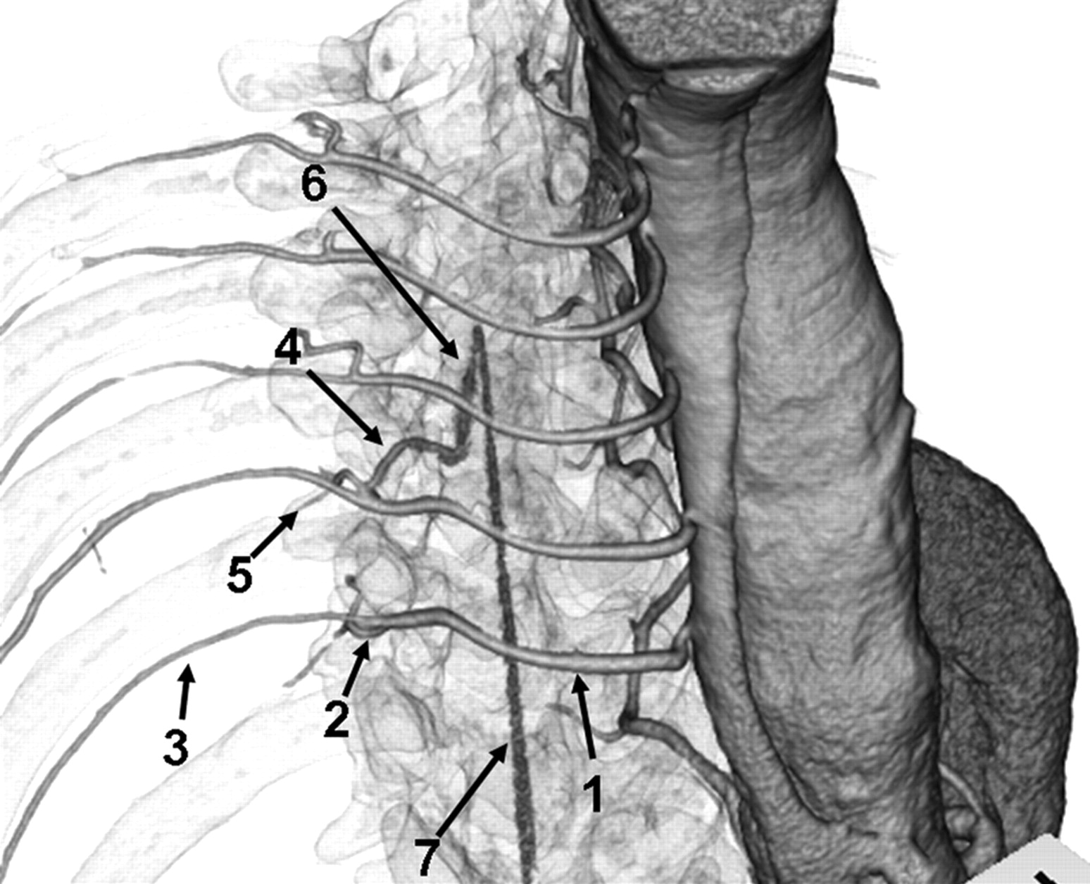
- Fig 1.
Anatomic course of the AKA. Right anterosuperior view of a 3D volume-rendered CT image of IACTA with semitransparent skeletal system. Intercostal and lumbar arteries (1) originate from the aorta, and divide into posterior (2) and anterior (3) branches. Anterior branches run through the intercostal groove. Posterior branches subdivide into the radiculomedullary artery (4) and muscular branch (5). Radiculomedullary artery courses to the spine and enters the vertebral foramen. The AKA (6) is the largest anterior radiculomedullary artery and joins the anterior spinal artery (7) in a characteristic hairpin curve.
- Fig 2.
IACTA image obtained from a 68-year-old man with TAA. A, CPR of the first phase shows a hairpin curved vessel continuously to the aorta through the intercostal artery (arrow). B, On a CPR image obtained from the second phase in the same profile as that of the first phase, enhancement of this vessel is decreased. This vessel is identified as the AKA.
- Fig 3.
IACTA image obtained from a 77-year-old man with TAA. Oblique coronal MPR of the first (A) and second (B) phases show a hairpin curved vessel (arrows). The vessel is more enhanced in the second than in the first phase and is not connected to arteries. This vessel is identified as the radiculomedullary vein.
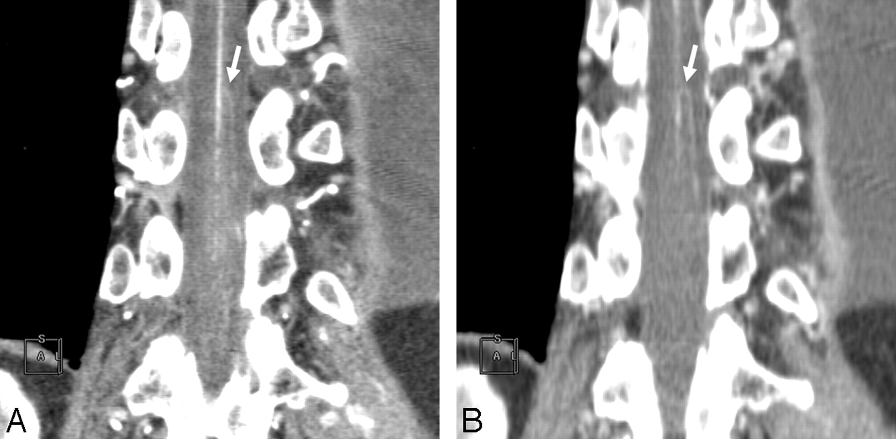
- Fig 4.
A, Oblique sagittal partial MIP image of IACTA obtained from a 73-year-old man with TAAA. Contrast material is not mixed throughout the aorta, and dorsal portion of the aorta and the intercostal or lumbar arteries (arrow) are enhanced over 1000 HU. B, Oblique sagittal partial MIP image of IVCTA obtained from a 58-year-old man with TAAA. The aorta is homogeneously enhanced, but intercostal or lumbar arteries (arrow) are poorly visualized in comparison with IACTA.
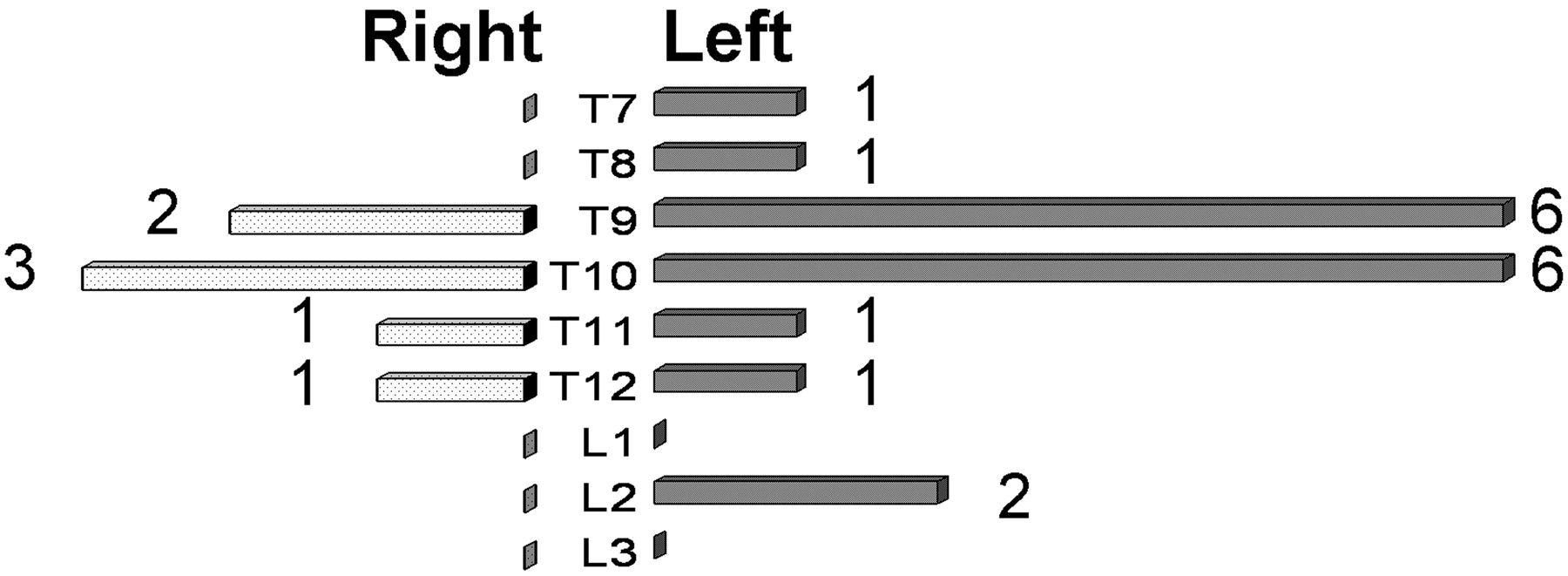
- Fig 5.
Laterality and levels of the AKA in all of the patients.
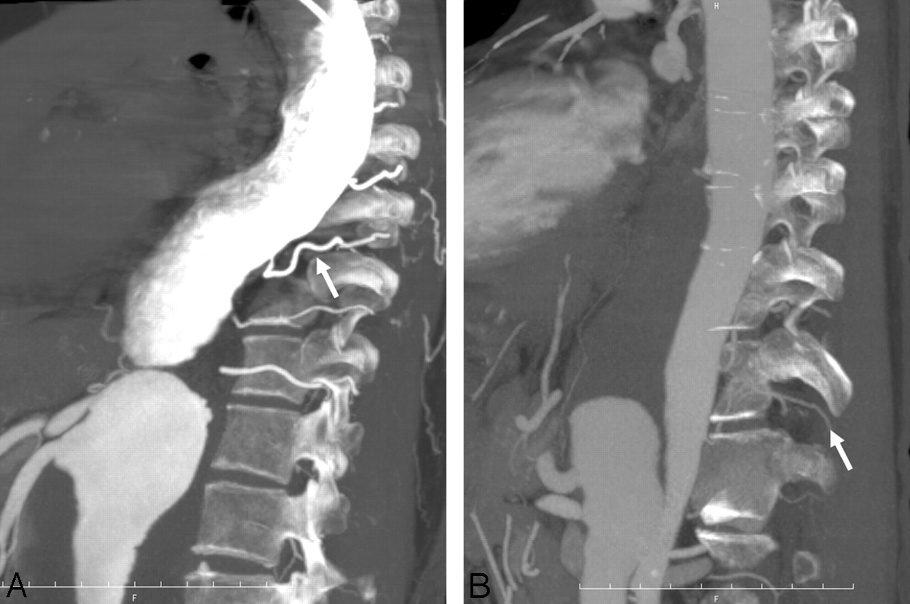
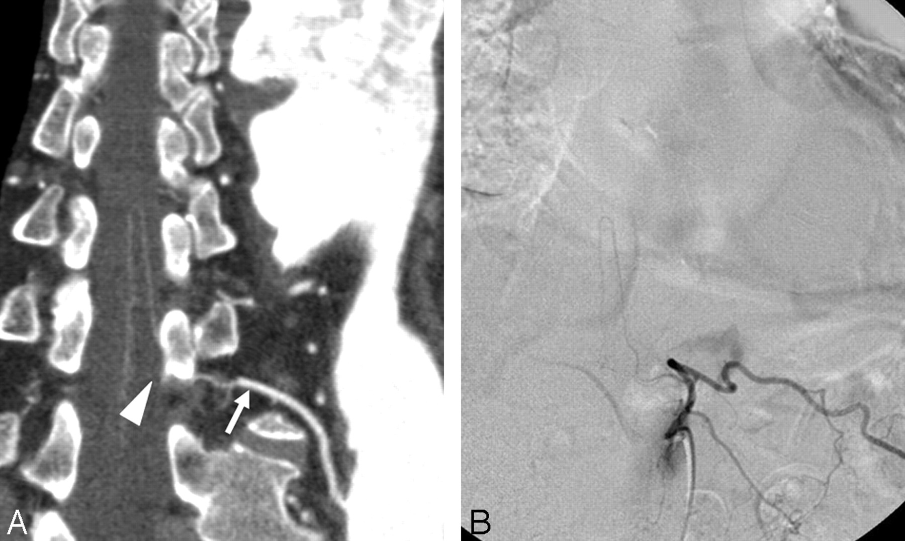
- Fig 6.
A, CPR image of the AKA obtained from IACTA. A hairpin curved vessel ascends to the midsagittal surface of the spinal cord, but continuity with the left 11th intercostal artery (arrow) is disturbed by a vertebral pedicle (arrowhead). B, Selective angiography of the left 11th intercostal artery demonstrates the AKA originating from this location.


Tables
Variable IVCTA IACTA P No. of patients 15 17 Age (range), years 70.6 (54–87) 65.9 (22–83) .168 Male:female 9:6 13:4 .450 TAA:TAAA 3:12 9:8 .125 Dissecting:true 7:8 4:13 .266 AKA detection, n/N (%) 9/15 (60.0) 16/17 (94.1) .033 Confirmation of continuity, n/N (%) 5/9 (55.6) 14/16 (87.5) .142 CT value of aorta, mean ± SD, HU 352.2 ± 46.0 965.4 ± 634.1 .002 Note:—IVCTA indicates intravenous CT angiography; IACTA, intra-arterial CT angiography; TAA, thoracic aortic aneurysm; TAAA, thoracoabdominal aortic aneurysm; HU, Hounsfield unit; AKA, artery of Adamkiewicz.
Method (Reference) Detection Rate, % (n/N) Confirmation Rate, % (n/N) Detectors Row Phase Scan Duration Injection Rate Seconds mL/s IVCTA (19) 90 (63/70) 32 (20/63) 4 1 40 3.5 IVCTA (20) 90 (9/10) 89 (8/9) 8 1 40 4.0–4.5 IVCTA (18) 83 (25/30) 72 (18/25) 16 1 NA 3.5 IACTA (24) 100 (39/39) 90 (35/39) 4 or 6 1 NA 4.0–7.0 IVCTA (present study) 60 (9/15) 56 (5/9) 16 2 25 5.0 IACTA (present study) 94 (16/17) 88 (14/16) 16 2 25 5.0 Note:—IVCTA indicates intravenous CT angiography; IACTA, intra-arterial CT angiography; NA, not assessed.
In this issue


Jump to section
Related Articles
Cited By...
- Advantages of 70-kV CT Angiography for the Visualization of the Adamkiewicz Artery: Comparison with 120-kV Imaging
- Bone-Subtracted Spinal CT Angiography Using Nonrigid Registration for Better Visualization of Arterial Feeders in Spinal Arteriovenous Fistulas
- Optimal Preoperative Imaging of Spinal Cord Blood Supply
- Reply: