Advertisement

Main menu

  • Home
  • Content
    • Current Issue
    • Accepted Manuscripts
    • Article Preview
    • Past Issue Archive
    • AJNR Case Collection
    • Case of the Week Archive
    • Classic Case Archive
    • Case of the Month Archive
  • Special Collections
    • Spinal CSF Leak Articles (Jan 2020-June 2024)
    • 2024 AJNR Journal Awards
    • Most Impactful AJNR Articles
  • Multimedia
    • AJNR Podcast
    • AJNR Scantastics
    • Video Articles
  • For Authors
    • Submit a Manuscript
    • Author Policies
    • Fast publishing of Accepted Manuscripts
    • Graphical Abstract Preparation
    • Manuscript Submission Guidelines
    • Imaging Protocol Submission
    • Submit a Case for the Case Collection
  • About Us
    • About AJNR
    • Editorial Board
  • More
    • Become a Reviewer/Academy of Reviewers
    • Subscribers
    • Permissions
    • Alerts
    • Feedback
    • Advertisers
    • ASNR Home
  • Other Publications
    • ajnr

User menu

  • Alerts
  • Log in

Search

  • Advanced search
American Journal of Neuroradiology

American Journal of Neuroradiology

ASHNR American Society of Functional Neuroradiology ASHNR American Society of Pediatric Neuroradiology ASSR
  • Alerts
  • Log in

Advanced Search

  • Home
  • Content
    • Current Issue
    • Accepted Manuscripts
    • Article Preview
    • Past Issue Archive
    • AJNR Case Collection
    • Case of the Week Archive
    • Classic Case Archive
    • Case of the Month Archive
  • Special Collections
    • Spinal CSF Leak Articles (Jan 2020-June 2024)
    • 2024 AJNR Journal Awards
    • Most Impactful AJNR Articles
  • Multimedia
    • AJNR Podcast
    • AJNR Scantastics
    • Video Articles
  • For Authors
    • Submit a Manuscript
    • Author Policies
    • Fast publishing of Accepted Manuscripts
    • Graphical Abstract Preparation
    • Manuscript Submission Guidelines
    • Imaging Protocol Submission
    • Submit a Case for the Case Collection
  • About Us
    • About AJNR
    • Editorial Board
  • More
    • Become a Reviewer/Academy of Reviewers
    • Subscribers
    • Permissions
    • Alerts
    • Feedback
    • Advertisers
    • ASNR Home
  • Follow AJNR on Twitter
  • Visit AJNR on Facebook
  • Follow AJNR on Instagram
  • Join AJNR on LinkedIn
  • RSS Feeds

Welcome to the new AJNR, Updated Hall of Fame, and more. Read the full announcements.


AJNR is seeking candidates for the position of Associate Section Editor, AJNR Case Collection. Read the full announcement.


OtherHEAD & NECK

Perineural Invasion through the Maxillary Division of the Right Trigeminal Nerve in a Rare Case of Nasolabial Malignant Peripheral Nerve Sheath Tumor

C. Sempere-Ortega and J. Martinez-San-Millan
American Journal of Neuroradiology February 2008, 29 (2) 396-397; DOI: https://doi.org/10.3174/ajnr.A0866
C. Sempere-Ortega
  • Find this author on Google Scholar
  • Find this author on PubMed
  • Search for this author on this site
J. Martinez-San-Millan
  • Find this author on Google Scholar
  • Find this author on PubMed
  • Search for this author on this site
  • Article
  • Figures & Data
  • Info & Metrics
  • Responses
  • References
  • PDF
Loading

Abstract

SUMMARY: We report the CT and MR imaging findings in a patient with malignant peripheral nerve sheath tumor in the right nasolabial area, which exhibited typical imaging features of cystic adenoid carcinoma.

We present a rare case of a patient without neurofibromatosis type 1 (NF-1) and with a malignant peripheral nerve sheath tumor (MPNST) arising in the terminal branches of the maxillary nerve in the right nasolabial area with retrograde perineural invasion of the entire nerve all the way to the pterygopalatine fossa. The location, behavior, and imaging features of our patient prompted the radiologic diagnosis of cystic adenoid carcinoma; however, pathologic analysis of the lesion confirmed the diagnosis of MPNST. To our knowledge, there is no previous imaging report of a lesion of this nature with this location and behavior.

Case Report

We present the case of a 62-year-old man with a previous history of facial trauma who complained of nose pain and nocturnal breathing problems. Findings of the clinical examination were completely normal. Several months later, the patient came back to our center because pain and breathing problems had increased and he could not continue his normal activities. In the new clinical examination, a soft-tissue mass was observed swelling on the right nasolabial fold and on the vestibule of the mouth, with no associated mucosal lesion; therefore, a submucosal lesion was suggested. CT and MR imaging revealed the presence of a homogeneous enhancing solid mass in the right nasolabial area (Fig 1A, -B), apparently located in the labial submucosa (Fig 1A), which invaded the right maxillary nerve all the way to the pterygopalatine fossa (Fig 1A, -B). The maxillary nerve was enlarged (Fig 2), with a homogeneous tubular-shaped increase of its diameter (Figs 3A and 1A, -B), resulting in a remodelling of the infraorbital canal (Fig 3B). A cystic adenoid carcinoma was suggested on the basis of the imaging and clinical findings. The patient underwent surgery, and the pathologic examination, surprisingly, was consistent with MPNST. Neither family history nor stigmata of NF-1 was found in the patient.

Fig 1.
  • Download figure
  • Open in new tab
  • Download powerpoint
Fig 1.

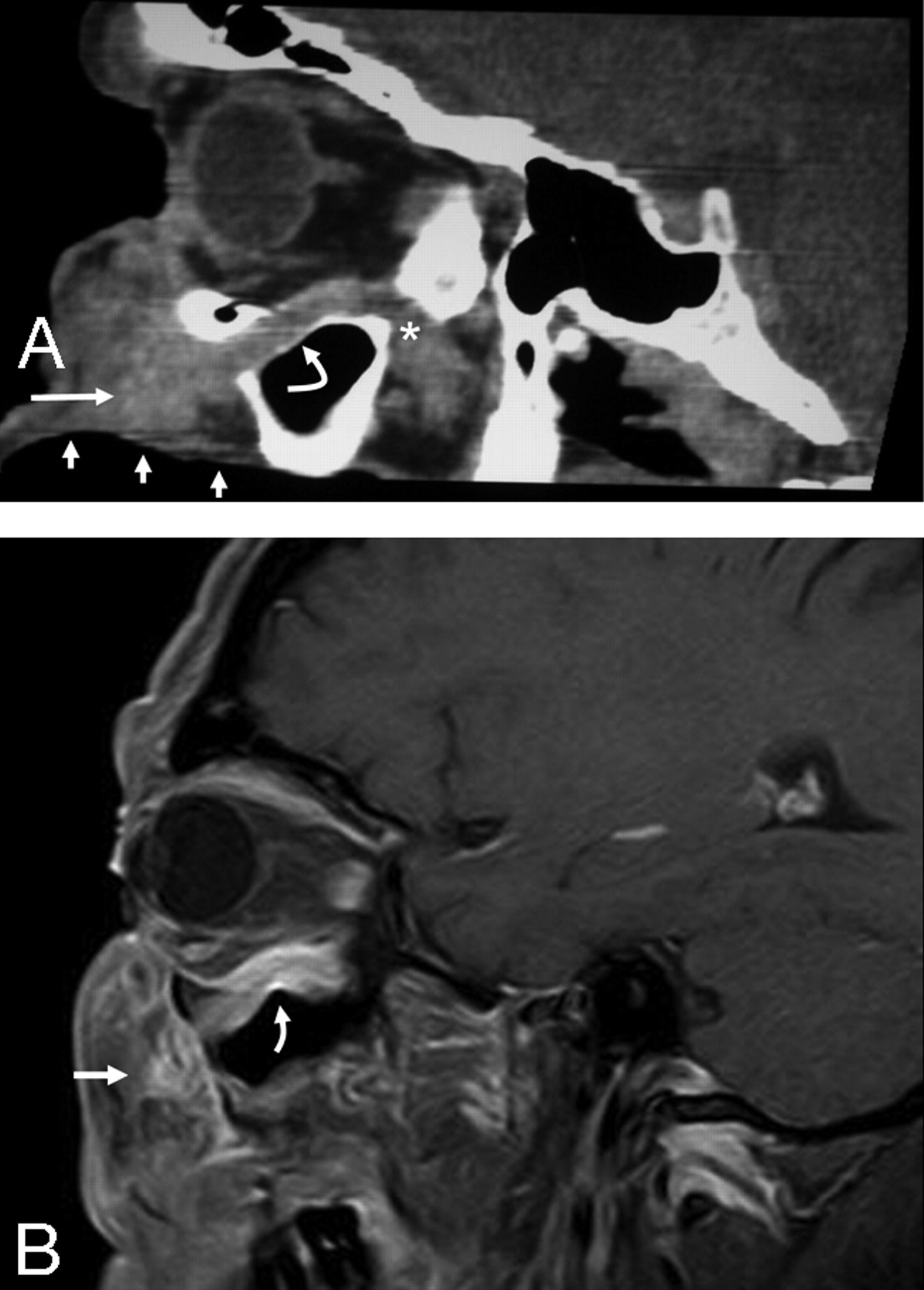
A, Curved saggital reconstruction image of a contrast-enhanced CT scan shows a submucose mass in the nasolabial area (straight arrow) with enlargement of the Vb nerve (curved arrow) all the way to the pterygopalatine fossa. Short arrows indicate buccal mucosa, and the asterisk indicates the pterygopalatine fossa. B, Contrast-enhanced oblique saggital fat-suppressed T1-weighted MR image shows an enhancing mass (straight arrow) in the nasolabial area with perineural spread through maxillary nerve (curved arrow).

Fig 2.
  • Download figure
  • Open in new tab
  • Download powerpoint
Fig 2.

Coronal reconstruction contrast-enhanced CT scan demonstrates enlargement of the right maxillary nerve (arrow).

Fig 3.
  • Download figure
  • Open in new tab
  • Download powerpoint
Fig 3.

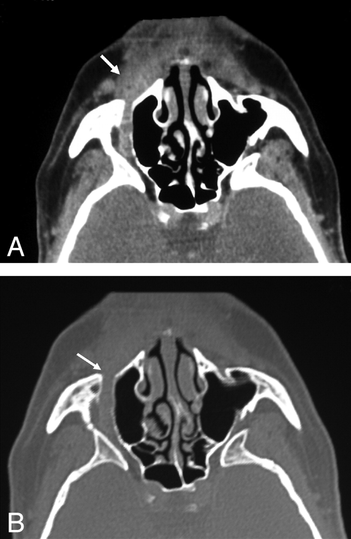
A, Curved axial reconstruction of a contrast-enhanced CT scan shows the original mass in the right nasolabial (arrow) area followed by a tubular-shaped enlargement of the maxillary nerve. B, Same image as in A with bone windowing. Note the infraorbital canal enlargement (arrow).

Discussion

MPNSTs are rare lesions derived from the mesenchymal cells of the neural crest. The term “MPNST” is now preferred instead of terms like “neurofibrosarcoma,” “neurogenic sarcoma,” or “malignant schwannoma” because the originating cell is not clearly understood.1 MPNSTs represent approximately 6% of all malignant soft-tissue tumors.2,3 These lesions have a tendency to affect the trunk and extremities, and only 10% of all MPNSTs are located in the head and neck region.2 The most common nerve affected is the sciatic nerve, followed by the brachial plexus.2 Between 38%–50%3,4 of these malignant tumors are associated with NF-1, and this entity is also related to multiplicity.3,4 The risk for developing an MPNST is 4600 times greater in patients with NF-1 than in the general population.4

The typical histologic features of these tumors are a monomorphic population of spindle cells arranged in fascicles, which usually demonstrate focal S-100 protein positivity, though 30% may be negative.3 They may show reactivity for Leu-7 or myelin basic protein, but they are negative for cytokeratins. Occasionally, they are positive for the melanocytic marker HMB-45.3

These lesions grow inside the nerve sheath, adopting a spindle or globular appearance,4 and though most of them present recognizable nerve roots entering and exiting the lesions, it is common to find intraneural invasion through the nerve bundles.2 The optimal treatment is combined surgical excision with radiation therapy.3 Radiation therapy is indicated because of the high recurrence rate of the tumor.3 Overall survival and disease-free survival are influenced by the patient's age (there is a better prognosis in patients >30 years old), tumor location, tumor size (patients with tumors <10 cm in diameter fared better than those with large tumors), and the quality of margins.5

Perineural spread involves the dissemination of a tumor through the planes of the neural sheath or along lymphatics of the epineurium and perineurium toward noncontiguous regions.6 It also carries a higher recurrence rate and a subsequent worse prognosis.6,7

The facial nerve and the second (V2) and third (V3) divisions of the trigeminal nerve are the most commonly affected nerves, with perineural invasion in the head and neck region, and perineural invasion usually occurs in a centripetal fashion, though centrifugal involvement has also been described.7 In head and neck tumors, perineural infiltration is a typical manifestation of adenoid cystic carcinoma,8 though it is most commonly found in squamous cell carcinoma because of its higher prevalence.9 Non-Hodgkin lymphomas and other sarcomas may also show this perineural invasion behavior.9 Both MR and CT imaging are capable of detecting perineural invasion, though medical literature has classically considered MR imaging as the technique of choice.7,9 The imaging findings of perineural spread depend on the head and neck area affected but usually include foraminal enlargement or destruction, obliteration of fat planes, nerve enlargement, nerve enhancement, replacement of the trigeminal subarachnoid cistern with soft tissue, convexity of the lateral dural margin of the cavernous sinus, etc.6

In our patient, the tumor arose from the terminal sensitive branches of the right infraorbital nerve in the nasolabial area and centripetally invaded this nerve all the way to the pterygoid fossa. The nerve infiltration resulted in a nerve enlargement and a homogeneous tubular-shaped increase of the nerve diameter, but not in the typical spindle shape of MPNST. Multidetector CT allowed curved reconstructions that were very illustrative of the particularly perineural invasion exhibited by the tumor. However, MR imaging is necessary in this type of lesion because CT is a limited tool for demonstrating intracranial extension to the cavernous sinus, Meckel cave, and the cisternal and pontine portions of the cranial nerve V.

The differential diagnoses in the presence of a swelling mass in the nasolabial fold involve nasolabial cyst, odontogenic inflammatory process, odontogenic tumor, squamous cell carcinoma, soft-tissue sarcoma, bone lesion, lymphoproliferative disorder, and salivary gland tumor.10 On the basis of the location, solid nature, and enhancing pattern of the lesion after intravenous contrast administration and the perineural invasion exhibited, the differential diagnosis was narrowed to solid malignant tumors. Given the absence of an associated mucosal lesion, cystic adenoid carcinoma was suggested as the first diagnostic possibility.

Conclusion

To our knowledge, there has been no previous case reported in the literature of an MPNST in this location showing this particular behavior and imaging features because it is usually seen in cystic adenoid and squamous cell carcinoma, but not in MPNSTs.

References

  1. ↵
    Hrehorovich PA, Franke HR, Maximin S, et al. Malignant peripheral nerve sheath tumor. Radiographics 2003;23:790–94
    CrossRefPubMed
  2. ↵
    Kransdorf MJ. Malignant soft-tissue tumors in a large referral population: distribution of diagnoses by age, sex, and location. AJRAmerican J Roentgenol 1995;164:129–34
  3. ↵
    Martinez-Devesa P, Mitchell TE, Scott I, et al. Malignant peripheral nerve sheath tumors of the head and neck: two cases and a review of the literature. Ear Nose Throat J 2006;85:392–96
    PubMed
  4. ↵
    Ducatman BS, Scheihauer BW, Piepgras DG, et al. Malignant peripheral nerve sheath tumors: a clinicopathologic study of 120 cases. Cancer 1986;57:2006–21
    CrossRefPubMed
  5. ↵
    Ojiri H. Perineural spread in head and neck malignancies. Radiat Med 2006;24:18
  6. ↵
    Caldemeyer KS, Mathews VP, Righi PD, et al. Imaging features and clinical significance of perineural spread or extension of head and neck tumors. Radiographics 1998;18:97
    PubMed
  7. ↵
    Mendenhall WM, Amdur RJ, Williams LS, et al. Carcinoma of the skin of the head and neck with perineural invasion. Head Neck 2002;24:78–83
    CrossRefPubMed
  8. ↵
    Parker GD, Harnsberger HR. Clinical-radiologic issues in perineural tumor spread of malignant diseases of the extracranial head and neck. Radiographics 1991;11:383
    CrossRefPubMed
  9. ↵
    Wanebo JE, Malik JM, VandenBerg SR, et al. Malignant peripheral nerve sheath tumors: a clinicopathologic study of 28 cases. Cancer 1993;71:1247–53
    CrossRefPubMed
  10. ↵
    Rinaggio J, Aguirre A, Zeid M, et al. Swelling of the nasolabial area. Oral Surg Oral Med Oral Pathol Oral Radiol Endod 2000;89:669–73
    CrossRefPubMed
  • Received September 7, 2007.
  • Accepted after revision September 23, 2007.
  • Copyright © American Society of Neuroradiology
View Abstract
PreviousNext
Back to top

In this issue

American Journal of Neuroradiology: 29 (2)
American Journal of Neuroradiology
Vol. 29, Issue 2
February 2008
  • Table of Contents
  • Index by author
Advertisement
Print
Download PDF
Email Article

Thank you for your interest in spreading the word on American Journal of Neuroradiology.

NOTE: We only request your email address so that the person you are recommending the page to knows that you wanted them to see it, and that it is not junk mail. We do not capture any email address.

Enter multiple addresses on separate lines or separate them with commas.
Perineural Invasion through the Maxillary Division of the Right Trigeminal Nerve in a Rare Case of Nasolabial Malignant Peripheral Nerve Sheath Tumor
(Your Name) has sent you a message from American Journal of Neuroradiology
(Your Name) thought you would like to see the American Journal of Neuroradiology web site.
CAPTCHA
This question is for testing whether or not you are a human visitor and to prevent automated spam submissions.
Cite this article
C. Sempere-Ortega, J. Martinez-San-Millan
Perineural Invasion through the Maxillary Division of the Right Trigeminal Nerve in a Rare Case of Nasolabial Malignant Peripheral Nerve Sheath Tumor
American Journal of Neuroradiology Feb 2008, 29 (2) 396-397; DOI: 10.3174/ajnr.A0866

Citation Manager Formats

  • BibTeX
  • Bookends
  • EasyBib
  • EndNote (tagged)
  • EndNote 8 (xml)
  • Medlars
  • Mendeley
  • Papers
  • RefWorks Tagged
  • Ref Manager
  • RIS
  • Zotero
0 Responses
Respond to this article
Share
Bookmark this article
Perineural Invasion through the Maxillary Division of the Right Trigeminal Nerve in a Rare Case of Nasolabial Malignant Peripheral Nerve Sheath Tumor
C. Sempere-Ortega, J. Martinez-San-Millan
American Journal of Neuroradiology Feb 2008, 29 (2) 396-397; DOI: 10.3174/ajnr.A0866
del.icio.us logo Twitter logo Facebook logo Mendeley logo
  • Tweet Widget
  • Facebook Like
  • Google Plus One
Purchase

Jump to section

  • Article
    • Abstract
    • Case Report
    • Discussion
    • Conclusion
    • References
  • Figures & Data
  • Info & Metrics
  • Responses
  • References
  • PDF

Related Articles

  • No related articles found.
  • PubMed
  • Google Scholar

Cited By...

  • No citing articles found.
  • Crossref (5)
  • Google Scholar

This article has been cited by the following articles in journals that are participating in Crossref Cited-by Linking.

  • Malignant peripheral nerve sheath tumors of the trigeminal nerve: a systematic review of 36 cases
    Richard F. Schmidt, Frederick Yick, Zain Boghani, Jean Anderson Eloy, James K. Liu
    Neurosurgical Focus 2013 34 3
  • Malignant peripheral nerve sheath tumour of the oral and maxillofacial region—A systematic review
    Lauren Frenzel Schuch, Laura Borges Kirschnick, José Alcides Almeida de Arruda, Isadora Peres Klein, Felipe Martins Silveira, Ana Carolina Uchoa Vasconcelos, Alan Roger Santos‐Silva, Márcio Ajudarte Lopes, Vinicius Coelho Carrard, Pablo Agustin Vargas, Marco Antonio Trevizani Martins, Vivian Petersen Wagner, Manoela Domingues Martins
    Oral Diseases 2022 28 8
  • Spontaneous malignant transformation of trigeminal schwannoma: consideration of responsible gene alterations for tumorigenesis—a case report
    Natsuki Ogasawara, Shinji Yamashita, Koji Yamasaki, Tomoki Kawano, Tomohiro Kawano, Junichiro Muta, Fumitaka Matsumoto, Takashi Watanabe, Hajime Ohta, Kiyotaka Yokogami, Tsuyoshi Fukushima, Yuichiro Sato, Hideo Takeshima
    Brain Tumor Pathology 2023 40 4
  • Diagnostic usefulness of panoramic imaging of the pterygopalatine fossa: case of a schwannoma causing pterygopalatine fossa expansion
    Shinya Kotaki, Shoko Gamoh, Hiroaki Yoshida, Chihoko Ikeda, Kazuya Tominaga, Masahiro Wato, Yutaka Ueno, Hironori Akiyama, Kimishige Shimizutani
    Oral Radiology 2019 35 3
  • Trigeminal malignant peripheral nerve-sheath tumor: Systematic review, and case report treated by proton-beam therapy
    Mathieu Lozouet, Lesueur Paul, Mazen Kallel, Gregoire Braux, Arthur Leclerc, Evelyne Emery
    Neurochirurgie 2025 71 2

More in this TOC Section

  • WHO Classification Update: Nasal&Skull Base Tumors
  • Peritumoral Signal in Vestibular Schwannomas
  • Chondrosarcoma vs Synovial Chondromatosis: Imaging
Show more HEAD & NECK

Similar Articles

Advertisement

Indexed Content

  • Current Issue
  • Accepted Manuscripts
  • Article Preview
  • Past Issues
  • Editorials
  • Editors Choice
  • Fellow Journal Club
  • Letters to the Editor

Cases

  • Case Collection
  • Archive - Case of the Week
  • Archive - Case of the Month
  • Archive - Classic Case

Special Collections

  • Special Collections

Resources

  • News and Updates
  • Turn around Times
  • Submit a Manuscript
  • Author Policies
  • Manuscript Submission Guidelines
  • Evidence-Based Medicine Level Guide
  • Publishing Checklists
  • Graphical Abstract Preparation
  • Imaging Protocol Submission
  • Submit a Case
  • Become a Reviewer/Academy of Reviewers
  • Get Peer Review Credit from Publons

Multimedia

  • AJNR Podcast
  • AJNR SCANtastic
  • Video Articles

About Us

  • About AJNR
  • Editorial Board
  • Not an AJNR Subscriber? Join Now
  • Alerts
  • Feedback
  • Advertise with us
  • Librarian Resources
  • Permissions
  • Terms and Conditions

American Society of Neuroradiology

  • Not an ASNR Member? Join Now

© 2025 by the American Society of Neuroradiology All rights, including for text and data mining, AI training, and similar technologies, are reserved.
Print ISSN: 0195-6108 Online ISSN: 1936-959X

Powered by HighWire