Advertisement

Main menu

  • Home
  • Content
    • Current Issue
    • Accepted Manuscripts
    • Article Preview
    • Past Issue Archive
    • Video Articles
    • AJNR Case Collection
    • Case of the Week Archive
    • Case of the Month Archive
    • Classic Case Archive
  • Special Collections
    • AJNR Awards
    • Low-Field MRI
    • Alzheimer Disease
    • ASNR Foundation Special Collection
    • Photon-Counting CT
    • View All
  • Multimedia
    • AJNR Podcasts
    • AJNR SCANtastic
    • Trainee Corner
    • MRI Safety Corner
    • Imaging Protocols
  • For Authors
    • Submit a Manuscript
    • Submit a Video Article
    • Submit an eLetter to the Editor/Response
    • Manuscript Submission Guidelines
    • Statistical Tips
    • Fast Publishing of Accepted Manuscripts
    • Graphical Abstract Preparation
    • Imaging Protocol Submission
    • Author Policies
  • About Us
    • About AJNR
    • Editorial Board
    • Editorial Board Alumni
  • More
    • Become a Reviewer/Academy of Reviewers
    • Subscribers
    • Permissions
    • Alerts
    • Feedback
    • Advertisers
    • ASNR Home

User menu

  • Alerts
  • Log in

Search

  • Advanced search
American Journal of Neuroradiology

American Journal of Neuroradiology

ASHNR American Society of Functional Neuroradiology ASHNR American Society of Pediatric Neuroradiology ASSR
  • Alerts
  • Log in

Advanced Search

  • Home
  • Content
    • Current Issue
    • Accepted Manuscripts
    • Article Preview
    • Past Issue Archive
    • Video Articles
    • AJNR Case Collection
    • Case of the Week Archive
    • Case of the Month Archive
    • Classic Case Archive
  • Special Collections
    • AJNR Awards
    • Low-Field MRI
    • Alzheimer Disease
    • ASNR Foundation Special Collection
    • Photon-Counting CT
    • View All
  • Multimedia
    • AJNR Podcasts
    • AJNR SCANtastic
    • Trainee Corner
    • MRI Safety Corner
    • Imaging Protocols
  • For Authors
    • Submit a Manuscript
    • Submit a Video Article
    • Submit an eLetter to the Editor/Response
    • Manuscript Submission Guidelines
    • Statistical Tips
    • Fast Publishing of Accepted Manuscripts
    • Graphical Abstract Preparation
    • Imaging Protocol Submission
    • Author Policies
  • About Us
    • About AJNR
    • Editorial Board
    • Editorial Board Alumni
  • More
    • Become a Reviewer/Academy of Reviewers
    • Subscribers
    • Permissions
    • Alerts
    • Feedback
    • Advertisers
    • ASNR Home
  • Follow AJNR on Twitter
  • Visit AJNR on Facebook
  • Follow AJNR on Instagram
  • Join AJNR on LinkedIn
  • RSS Feeds

AJNR Awards, New Junior Editors, and more. Read the latest AJNR updates

Research ArticlePediatric Neuroimaging

Selective Cerebral Volume Reduction in Rett Syndrome: A Multiple-Approach MR Imaging Study

J.C. Carter, D.C. Lanham, D. Pham, G. Bibat, S. Naidu and W.E. Kaufmann
American Journal of Neuroradiology March 2008, 29 (3) 436-441; DOI: https://doi.org/10.3174/ajnr.A0857
J.C. Carter
  • Find this author on Google Scholar
  • Find this author on PubMed
  • Search for this author on this site
D.C. Lanham
  • Find this author on Google Scholar
  • Find this author on PubMed
  • Search for this author on this site
D. Pham
  • Find this author on Google Scholar
  • Find this author on PubMed
  • Search for this author on this site
G. Bibat
  • Find this author on Google Scholar
  • Find this author on PubMed
  • Search for this author on this site
S. Naidu
  • Find this author on Google Scholar
  • Find this author on PubMed
  • Search for this author on this site
W.E. Kaufmann
  • Find this author on Google Scholar
  • Find this author on PubMed
  • Search for this author on this site
  • Article
  • Figures & Data
  • Supplemental
  • Info & Metrics
  • Responses
  • References
  • PDF
Loading

Article Figures & Data

Figures

  • Tables
  • Fig 1.
    • Download figure
    • Open in new tab
    • Download powerpoint
    Fig 1.

    Concordance of Talairach and voxel-based findings in the posterior dorsal parietal lobe in RTT versus control subjects. Talairach-derived data (top row) show preferential bilateral reduction of GM in the posterior dorsal parietal lobe (outlined; only left side shown), corresponding with Brodmann areas 5 and 7. Similarly, VBM-derived data (bottom row) show bilateral GM volume reduction in the posterior dorsal parietal region (outlined), roughly corresponding with Brodmann area 7 (left hemisphere differences are depicted here).
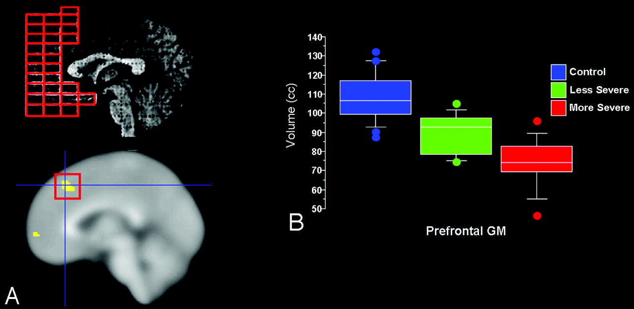
  • Fig 2.
    • Download figure
    • Open in new tab
    • Download powerpoint
    Fig 2.

    Concordance of Talairach and voxel-based findings in the prefrontal GM in more severe versus less severe RTT subjects. Girls with less severe clinical presentation, according to gait abnormality criterion, showed relative preservation of prefrontal GM volumes, corresponding roughly with Brodmann areas 10/46. A, Top left depicts Talairach-derived data, whereas bottom left shows VBM-derived data. B, Boxplots (percentiles) of Talairach-based prefrontal GM volumes illustrate that girls with less severe clinical presentation show volumes intermediate between control subjects and those with more severe phenotype.

  • Fig 3.
    • Download figure
    • Open in new tab
    • Download powerpoint
    Fig 3.

    Cerebral lobar GM volumes in monozygotic twins discordant for the RTT phenotype. Note that the unaffected twin showed frontal, parietal, and temporal GM volumes within the normal control range (depicted as mean +1 SD), whereas the affected twin had smaller volumes in these regions, with the exception of the occipital GM volume, which was at nearly identical levels in both twins and only slightly below the normal control range.

Tables

  • Figures
    • View popup
    Table 1:

    Demographic and clinical characteristics of the study cohort

    VariablenAge, Mean ± SD, yHead Circumference, Mean ± SD, cmClinical Severity Scores, Mean ± SD
    TotalHead GrowthSeizuresRespiratory IrregularitiesScoliosisGait Abnormalities
    All RTT subjects228.6 ± 1.849.5 ± 2.17.8 ± 2.42.5 ± 0.81.2 ± 1.01.7 ± 0.80.7 ± 1.01.6 ± 1.0
    Affected MZ twin8.450.5831202
    More severe (gait criterion)128.8 ± 1.649.3 ± 2.49.6 ± 1.3*2.6 ± 0.71.4 ± 1.01.8 ± 0.81.7 ± 1.2*2.4 ± 0.7*
    Less severe (gait criterion)108.3 ± 2.149.4 ± 1.85.9 ± 1.52.5 ± 1.01.0 ± 1.11.6 ± 0.80.1 ± 0.30.7 ± 0.7
    Control subjects258.9 ± 1.9
    • Note:—RTT indicates Rett syndrome; MZ, monozygotic. Age represents age at time of MR imaging scan. The maximum total clinical severity score is 15. The “more severe” group included the following mutations: R106W (n = 1), T158M (n = 3), R255X (n = 1), R270X (n = 2), R294X (n = 2), and C-terminal deletions (n = 3). The “less severe” group included the following mutations: R133C (n = 1), R168X (n = 1), R255X (n = 1), R294X (n = 4), R306C (n = 2), and G451T/D151Y (n = 1 each).

    • * P < .05 when comparing more severe versus less severe gait.

PreviousNext
Back to top

In this issue

American Journal of Neuroradiology: 29 (3)
American Journal of Neuroradiology
Vol. 29, Issue 3
March 2008
  • Table of Contents
  • Index by author
Advertisement
Print
Download PDF
Email Article

Thank you for your interest in spreading the word on American Journal of Neuroradiology.

NOTE: We only request your email address so that the person you are recommending the page to knows that you wanted them to see it, and that it is not junk mail. We do not capture any email address.

Enter multiple addresses on separate lines or separate them with commas.
Selective Cerebral Volume Reduction in Rett Syndrome: A Multiple-Approach MR Imaging Study
(Your Name) has sent you a message from American Journal of Neuroradiology
(Your Name) thought you would like to see the American Journal of Neuroradiology web site.
CAPTCHA
This question is for testing whether or not you are a human visitor and to prevent automated spam submissions.
Cite this article
J.C. Carter, D.C. Lanham, D. Pham, G. Bibat, S. Naidu, W.E. Kaufmann
Selective Cerebral Volume Reduction in Rett Syndrome: A Multiple-Approach MR Imaging Study
American Journal of Neuroradiology Mar 2008, 29 (3) 436-441; DOI: 10.3174/ajnr.A0857

Citation Manager Formats

  • BibTeX
  • Bookends
  • EasyBib
  • EndNote (tagged)
  • EndNote 8 (xml)
  • Medlars
  • Mendeley
  • Papers
  • RefWorks Tagged
  • Ref Manager
  • RIS
  • Zotero
0 Responses
Respond to this article
Share
Bookmark this article
Selective Cerebral Volume Reduction in Rett Syndrome: A Multiple-Approach MR Imaging Study
J.C. Carter, D.C. Lanham, D. Pham, G. Bibat, S. Naidu, W.E. Kaufmann
American Journal of Neuroradiology Mar 2008, 29 (3) 436-441; DOI: 10.3174/ajnr.A0857
del.icio.us logo Twitter logo Facebook logo Mendeley logo
  • Tweet Widget
  • Facebook Like
  • Google Plus One
Purchase

Jump to section

  • Article
    • Abstract
    • Methods
    • Results
    • Discussion
    • Acknowledgments
    • Footnotes
    • References
  • Figures & Data
  • Supplemental
  • Info & Metrics
  • Responses
  • References
  • PDF

Related Articles

  • PubMed
  • Google Scholar

Cited By...

  • The enhancement of activity rescues the establishment of Mecp2 null neuronal phenotypes
  • Hand stereotypies: Lessons from the Rett Syndrome Natural History Study
  • Development of the Tailored Rett Intervention and Assessment Longitudinal (TRIAL) database and the Rett Evaluation of Symptoms and Treatments (REST) Questionnaire
  • White Matter Impairment in Rett Syndrome: Diffusion Tensor Imaging Study with Clinical Correlations
  • A common MECP2 haplotype associates with reduced cortical surface area in humans in two independent populations
  • Crossref
  • Google Scholar

This article has not yet been cited by articles in journals that are participating in Crossref Cited-by Linking.

More in this TOC Section

  • Comparing MRI Perfusion in Pediatric Brain Tumors
  • Sodium MRI in Pediatric Brain Tumors
  • FRACTURE MR in Congenital Vertebral Anomalies
Show more Pediatric Neuroimaging

Similar Articles

Advertisement

Indexed Content

  • Current Issue
  • Accepted Manuscripts
  • Article Preview
  • Past Issues
  • Editorials
  • Editor's Choice
  • Fellows' Journal Club
  • Letters to the Editor
  • Video Articles

Cases

  • Case Collection
  • Archive - Case of the Week
  • Archive - Case of the Month
  • Archive - Classic Case

More from AJNR

  • Trainee Corner
  • Imaging Protocols
  • MRI Safety Corner
  • Book Reviews

Multimedia

  • AJNR Podcasts
  • AJNR Scantastics

Resources

  • Turnaround Time
  • Submit a Manuscript
  • Submit a Video Article
  • Submit an eLetter to the Editor/Response
  • Manuscript Submission Guidelines
  • Statistical Tips
  • Fast Publishing of Accepted Manuscripts
  • Graphical Abstract Preparation
  • Imaging Protocol Submission
  • Evidence-Based Medicine Level Guide
  • Publishing Checklists
  • Author Policies
  • Become a Reviewer/Academy of Reviewers
  • News and Updates

About Us

  • About AJNR
  • Editorial Board
  • Editorial Board Alumni
  • Alerts
  • Permissions
  • Not an AJNR Subscriber? Join Now
  • Advertise with Us
  • Librarian Resources
  • Feedback
  • Terms and Conditions
  • AJNR Editorial Board Alumni

American Society of Neuroradiology

  • Not an ASNR Member? Join Now

© 2025 by the American Society of Neuroradiology All rights, including for text and data mining, AI training, and similar technologies, are reserved.
Print ISSN: 0195-6108 Online ISSN: 1936-959X

Powered by HighWire