Advertisement

Main menu

  • Home
  • Content
    • Current Issue
    • Accepted Manuscripts
    • Article Preview
    • Past Issue Archive
    • AJNR Case Collection
    • Case of the Week Archive
    • Classic Case Archive
    • Case of the Month Archive
  • Special Collections
    • Spinal CSF Leak Articles (Jan 2020-June 2024)
    • 2024 AJNR Journal Awards
    • Most Impactful AJNR Articles
  • Multimedia
    • AJNR Podcast
    • AJNR Scantastics
    • Video Articles
  • For Authors
    • Submit a Manuscript
    • Author Policies
    • Fast publishing of Accepted Manuscripts
    • Graphical Abstract Preparation
    • Manuscript Submission Guidelines
    • Imaging Protocol Submission
    • Submit a Case for the Case Collection
  • About Us
    • About AJNR
    • Editorial Board
  • More
    • Become a Reviewer/Academy of Reviewers
    • Subscribers
    • Permissions
    • Alerts
    • Feedback
    • Advertisers
    • ASNR Home
  • Other Publications
    • ajnr

User menu

  • Alerts
  • Log in

Search

  • Advanced search
American Journal of Neuroradiology

American Journal of Neuroradiology

ASHNR American Society of Functional Neuroradiology ASHNR American Society of Pediatric Neuroradiology ASSR
  • Alerts
  • Log in

Advanced Search

  • Home
  • Content
    • Current Issue
    • Accepted Manuscripts
    • Article Preview
    • Past Issue Archive
    • AJNR Case Collection
    • Case of the Week Archive
    • Classic Case Archive
    • Case of the Month Archive
  • Special Collections
    • Spinal CSF Leak Articles (Jan 2020-June 2024)
    • 2024 AJNR Journal Awards
    • Most Impactful AJNR Articles
  • Multimedia
    • AJNR Podcast
    • AJNR Scantastics
    • Video Articles
  • For Authors
    • Submit a Manuscript
    • Author Policies
    • Fast publishing of Accepted Manuscripts
    • Graphical Abstract Preparation
    • Manuscript Submission Guidelines
    • Imaging Protocol Submission
    • Submit a Case for the Case Collection
  • About Us
    • About AJNR
    • Editorial Board
  • More
    • Become a Reviewer/Academy of Reviewers
    • Subscribers
    • Permissions
    • Alerts
    • Feedback
    • Advertisers
    • ASNR Home
  • Follow AJNR on Twitter
  • Visit AJNR on Facebook
  • Follow AJNR on Instagram
  • Join AJNR on LinkedIn
  • RSS Feeds

Welcome to the new AJNR, Updated Hall of Fame, and more. Read the full announcements.


AJNR is seeking candidates for the position of Associate Section Editor, AJNR Case Collection. Read the full announcement.


Getting new auth cookie, if you see this message a lot, tell someone!
Research ArticleHEAD & NECK

CSF Leaks: Correlation of High-Resolution CT and Multiplanar Reformations with Intraoperative Endoscopic Findings

V. La Fata, N. McLean, S.K. Wise, J.M. DelGaudio and P.A. Hudgins
American Journal of Neuroradiology March 2008, 29 (3) 536-541; DOI: https://doi.org/10.3174/ajnr.A0885
V. La Fata
  • Find this author on Google Scholar
  • Find this author on PubMed
  • Search for this author on this site
N. McLean
  • Find this author on Google Scholar
  • Find this author on PubMed
  • Search for this author on this site
S.K. Wise
  • Find this author on Google Scholar
  • Find this author on PubMed
  • Search for this author on this site
J.M. DelGaudio
  • Find this author on Google Scholar
  • Find this author on PubMed
  • Search for this author on this site
P.A. Hudgins
  • Find this author on Google Scholar
  • Find this author on PubMed
  • Search for this author on this site
  • Article
  • Figures & Data
  • Info & Metrics
  • Responses
  • References
  • PDF
Loading

Article Figures & Data

Figures

  • Tables
  • Fig 1.
    • Download figure
    • Open in new tab
    • Download powerpoint
    Fig 1.

    Patient 5. CT and endoscopy are concordant. Coronal (A) and oblique sagittal (B) MPR images generated from a 0.625-mm axial dataset demonstrate a 6-mm defect in a pneumatized pterygoid recess of the left sphenoid bone. There is mucosal thickening and an air-fluid level was present (other images) in the sphenoid sinus. At endoscopy, CSF was actively leaking at this site and the maximum size of the skull base defect (anteroposterior dimension), was identical to the CT measurement (6 mm).
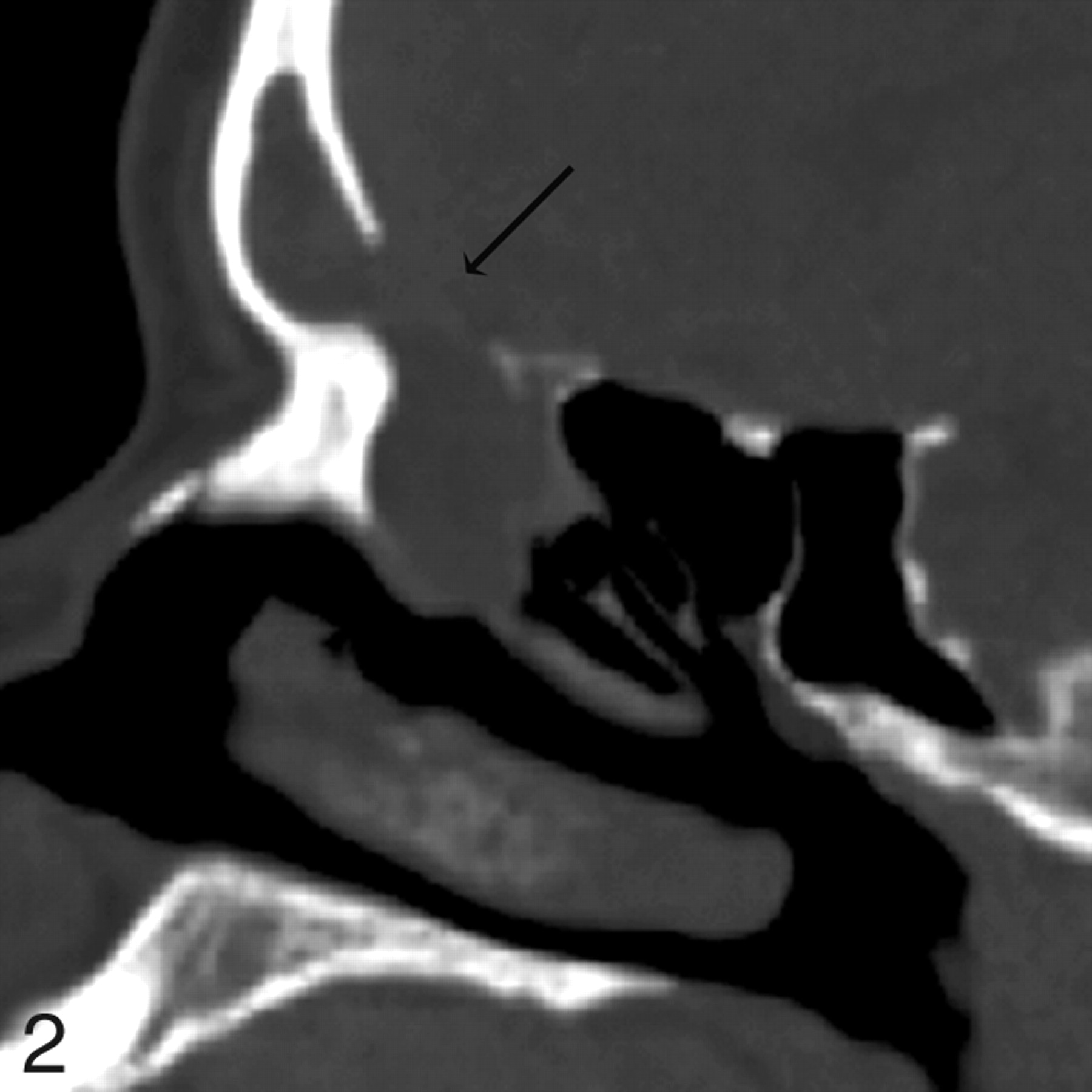
  • Fig 2.
    • Download figure
    • Open in new tab
    • Download powerpoint
    Fig 2.

    Patient 4. CT underestimates defect size by 2 mm. Sagittal MPR image from 1.25-mm axial dataset demonstrates a 13-mm defect in the posterior wall of the right frontal sinus with soft tissue suggestive of encephalocele protruding through the defect. At endoscopy, this was a 15-mm defect. MR was performed preoperatively and showed no encephalocele.
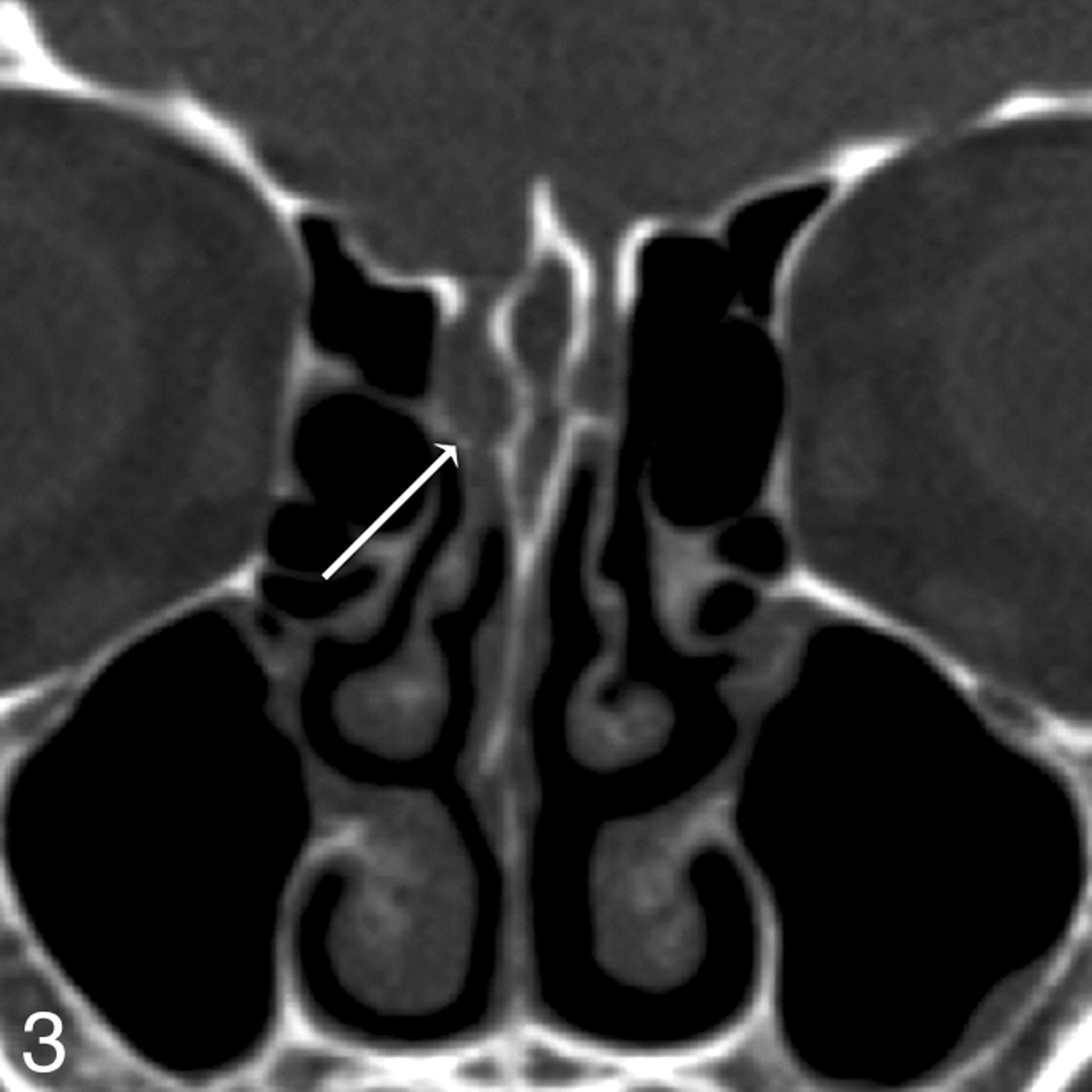
  • Fig 3.
    • Download figure
    • Open in new tab
    • Download powerpoint
    Fig 3.

    Patient 12. Both readers missed a defect found at endoscopy. Coronal MPR image from 0.625-mm axial dataset missed a subtle defect of the right cribriform plate, measuring 3 mm endoscopically and 2 mm by imaging. Two defects were present on the right in this patient; readers detected the first, but not the second, defect. In retrospect, a small amount of soft tissue or fluid attenuation extends medially along the horizontal insertion of the middle turbinate, or olfactory recess, through the skull base defect.

  • Fig 4.
    • Download figure
    • Open in new tab
    • Download powerpoint
    Fig 4.

    Patient 3. CT shows a defect but no CSF leak at endoscopy. Axial image from 1.25-mm axial dataset demonstrates a 3-mm defect in the right sphenoid bone. During endoscopy, after mucosa was elevated off the defect, no egress of CSF was seen from the defect.
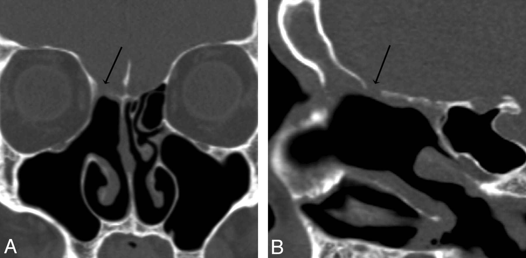
  • Fig 5.
    • Download figure
    • Open in new tab
    • Download powerpoint
    Fig 5.

    Patient 6. CT overestimated the size of the defect. Coronal (A) and sagittal (B) MPR images generated from a 0.625-mm axial dataset demonstrate a 5-mm defect in the right ethmoid roof. This patient has had a total ethmoidectomy, and the right middle turbinate has been resected at the skull base. There is mild mucosal thickening below the defect. At endoscopy, the CSF leak localized to this defect but measured only 2 mm.
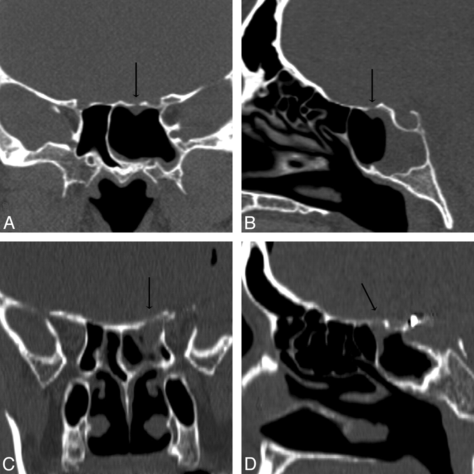
  • Fig 6.
    • Download figure
    • Open in new tab
    • Download powerpoint
    Fig 6.

    Patients 13 (A and B) and 19 (C and D). Coronal and sagittal MPR images in A1 and A2 and B1 and B2 are generated from 0.625-mm and 1.25-mm axial datasets, respectively. Although both of these MPR images resulted in accurate measurements of the CSF leaks, a 2.0-mm defect of the left sphenoid roof in patient 13 is better appreciated than the larger, 5.0-mm defect of the left sphenoid roof in patient 19 because of the overall improved resolution of submillimeter axial collimation.

Tables

  • Figures
  • Imaging and clinical data on CSF leaks

    Patient No.CauseSC (mm)CT Defect (mm)Endo Defect (mm)Accurate (± 2 mm)Discrepancy (mm, Over/Under)
    1SP1.2R eth (6)R eth (5)Y−
    L eth (6)−−−
    2S0.6L eth (6)L eth (6)Y−
    3SP1.2R sph (3)−−−
    L eth (3)−−−
    4SP1.2R fr p wall (13)R fr p wall (15)Y−
    5SP0.6L sph (6)L sph (6)Y−
    6S0.6R eth (5)R eth (2)N3 over
    7S0.6Rt sph (3)R sph (3)Y−
    8S0.6L eth (9)L eth (10)Y−
    9S1.2L crib (7)L crib (7)Y−
    10S1.2L sph (7)L sph (14)N7 under
    R sph (3)R sph (2)Y−
    −R sph (2)−−
    11SP1.2L crib (12)L crib (10)Y−
    12SP0.6R crib (5)R crib (4)Y−
    −R crib (3)−−
    13SP0.6L sph (2)L sph (2)Y−
    14T0.6L eth (11)L eth (10)Y−
    L fr p wall (3)L fr p wall (3)Y−
    15S0.6R eth (6)R eth (3)N3 over
    16S1.2R eth (18)R eth (5)N13 over
    17SP0.6R fr p wall (6)R fr p wall (8)Y−
    18I1.2L crib (8)L crib (3)N5 over
    19S1.2L sph (5)L sph (4)Y−
    • Note:—Endo indicates endoscopy; SP, spontaneous; S, surgical; T, traumatic; I, iatrogenic (other); p, posterior; eth, ethmoid; sph, sphenoid; fr, frontal; crib, cribriform; SC, slice collimation; L, left; R, right.

PreviousNext
Back to top

In this issue

American Journal of Neuroradiology: 29 (3)
American Journal of Neuroradiology
Vol. 29, Issue 3
March 2008
  • Table of Contents
  • Index by author
Advertisement
Print
Download PDF
Email Article

Thank you for your interest in spreading the word on American Journal of Neuroradiology.

NOTE: We only request your email address so that the person you are recommending the page to knows that you wanted them to see it, and that it is not junk mail. We do not capture any email address.

Enter multiple addresses on separate lines or separate them with commas.
CSF Leaks: Correlation of High-Resolution CT and Multiplanar Reformations with Intraoperative Endoscopic Findings
(Your Name) has sent you a message from American Journal of Neuroradiology
(Your Name) thought you would like to see the American Journal of Neuroradiology web site.
CAPTCHA
This question is for testing whether or not you are a human visitor and to prevent automated spam submissions.
Cite this article
V. La Fata, N. McLean, S.K. Wise, J.M. DelGaudio, P.A. Hudgins
CSF Leaks: Correlation of High-Resolution CT and Multiplanar Reformations with Intraoperative Endoscopic Findings
American Journal of Neuroradiology Mar 2008, 29 (3) 536-541; DOI: 10.3174/ajnr.A0885

Citation Manager Formats

  • BibTeX
  • Bookends
  • EasyBib
  • EndNote (tagged)
  • EndNote 8 (xml)
  • Medlars
  • Mendeley
  • Papers
  • RefWorks Tagged
  • Ref Manager
  • RIS
  • Zotero
0 Responses
Respond to this article
Share
Bookmark this article
CSF Leaks: Correlation of High-Resolution CT and Multiplanar Reformations with Intraoperative Endoscopic Findings
V. La Fata, N. McLean, S.K. Wise, J.M. DelGaudio, P.A. Hudgins
American Journal of Neuroradiology Mar 2008, 29 (3) 536-541; DOI: 10.3174/ajnr.A0885
del.icio.us logo Twitter logo Facebook logo Mendeley logo
  • Tweet Widget
  • Facebook Like
  • Google Plus One
Purchase

Jump to section

  • Article
    • Abstract
    • Methods
    • Results
    • Discussion
    • Conclusion
    • Footnotes
    • References
  • Figures & Data
  • Info & Metrics
  • Responses
  • References
  • PDF

Related Articles

  • No related articles found.
  • PubMed
  • Google Scholar

Cited By...

  • Spontaneous Lateral Sphenoid Cephaloceles: Anatomic Factors Contributing to Pathogenesis and Proposed Classification
  • Crossref (76)
  • Google Scholar

This article has been cited by the following articles in journals that are participating in Crossref Cited-by Linking.

  • Imaging of Skull Base Cerebrospinal Fluid Leaks in Adults
    Kristen M. Lloyd, John M. DelGaudio, Patricia A. Hudgins
    Radiology 2008 248 3
  • Diagnosis and management of rhinosinusitis: a practice parameter update
    Anju T. Peters, Sheldon Spector, Joy Hsu, Daniel L. Hamilos, Fuad M. Baroody, Rakesh K. Chandra, Leslie C. Grammer, David W. Kennedy, Noam A. Cohen, Michael A. Kaliner, Ellen R. Wald, Achilles Karagianis, Raymond G. Slavin
    Annals of Allergy, Asthma & Immunology 2014 113 4
  • Diagnosis of cerebrospinal fluid rhinorrhea: an evidence‐based review with recommendations
    Gretchen M. Oakley, Jeremiah A. Alt, Rodney J. Schlosser, Richard J. Harvey, Richard R. Orlandi
    International Forum of Allergy & Rhinology 2016 6 1
  • Spontaneous Skull Base Meningoencephaloceles and Cerebrospinal Fluid Fistulas
    Raquel Cano Alonso, Mar Jimenez de la Peña, Anne Gomez Caicoya, Manuel Recio Rodriguez, Elena Alvarez Moreno, Vicente Martinez de Vega Fernandez
    RadioGraphics 2013 33 2
  • Traumatic Cerebrospinal Fluid Leak: Diagnosis and Management
    Ji-Woong Oh, So-Hyun Kim, Kum Whang
    Korean Journal of Neurotrauma 2017 13 2
  • Spontaneous Lateral Sphenoid Cephaloceles: Anatomic Factors Contributing to Pathogenesis and Proposed Classification
    F. Settecase, H. R. Harnsberger, M. A. Michel, P. Chapman, C. M. Glastonbury
    American Journal of Neuroradiology 2014 35 4
  • Imaging of skull-base cephalocoeles and cerebrospinal fluid leaks
    S.E.J. Connor
    Clinical Radiology 2010 65 10
  • Imaging of Cerebrospinal Fluid Rhinorrhea and Otorrhea
    Mahati Reddy, Kristen Baugnon
    Radiologic Clinics of North America 2017 55 1
  • Detection of Cerebrospinal Fluid Leaks: Is There a Radiologic Standard of Care? A Systematic Review
    Rachelle Eljazzar, Joshua Loewenstern, Jennifer B. Dai, Raj K. Shrivastava, Alfred M. Iloreta
    World Neurosurgery 2019 127
  • Imaging of the Anterior Skull Base
    Hemant Parmar, Sachin Gujar, Gaurang Shah, Suresh K. Mukherji
    Neuroimaging Clinics of North America 2009 19 3

More in this TOC Section

  • WHO Classification Update: Nasal&Skull Base Tumors
  • Peritumoral Signal in Vestibular Schwannomas
  • Chondrosarcoma vs Synovial Chondromatosis: Imaging
Show more HEAD & NECK

Similar Articles

Advertisement

Indexed Content

  • Current Issue
  • Accepted Manuscripts
  • Article Preview
  • Past Issues
  • Editorials
  • Editors Choice
  • Fellow Journal Club
  • Letters to the Editor

Cases

  • Case Collection
  • Archive - Case of the Week
  • Archive - Case of the Month
  • Archive - Classic Case

Special Collections

  • Special Collections

Resources

  • News and Updates
  • Turn around Times
  • Submit a Manuscript
  • Author Policies
  • Manuscript Submission Guidelines
  • Evidence-Based Medicine Level Guide
  • Publishing Checklists
  • Graphical Abstract Preparation
  • Imaging Protocol Submission
  • Submit a Case
  • Become a Reviewer/Academy of Reviewers
  • Get Peer Review Credit from Publons

Multimedia

  • AJNR Podcast
  • AJNR SCANtastic
  • Video Articles

About Us

  • About AJNR
  • Editorial Board
  • Not an AJNR Subscriber? Join Now
  • Alerts
  • Feedback
  • Advertise with us
  • Librarian Resources
  • Permissions
  • Terms and Conditions

American Society of Neuroradiology

  • Not an ASNR Member? Join Now

© 2025 by the American Society of Neuroradiology All rights, including for text and data mining, AI training, and similar technologies, are reserved.
Print ISSN: 0195-6108 Online ISSN: 1936-959X

Powered by HighWire