Advertisement

Main menu

  • Home
  • Content
    • Current Issue
    • Accepted Manuscripts
    • Article Preview
    • Past Issue Archive
    • Video Articles
    • AJNR Case Collection
    • Case of the Week Archive
    • Case of the Month Archive
    • Classic Case Archive
  • Special Collections
    • AJNR Awards
    • Low-Field MRI
    • Alzheimer Disease
    • ASNR Foundation Special Collection
    • Photon-Counting CT
    • View All
  • Multimedia
    • AJNR Podcasts
    • AJNR SCANtastic
    • Trainee Corner
    • MRI Safety Corner
    • Imaging Protocols
  • For Authors
    • Submit a Manuscript
    • Submit a Video Article
    • Submit an eLetter to the Editor/Response
    • Manuscript Submission Guidelines
    • Statistical Tips
    • Fast Publishing of Accepted Manuscripts
    • Graphical Abstract Preparation
    • Imaging Protocol Submission
    • Author Policies
  • About Us
    • About AJNR
    • Editorial Board
    • Editorial Board Alumni
  • More
    • Become a Reviewer/Academy of Reviewers
    • Subscribers
    • Permissions
    • Alerts
    • Feedback
    • Advertisers
    • ASNR Home

User menu

  • Alerts
  • Log in

Search

  • Advanced search
American Journal of Neuroradiology

American Journal of Neuroradiology

ASHNR American Society of Functional Neuroradiology ASHNR American Society of Pediatric Neuroradiology ASSR
  • Alerts
  • Log in

Advanced Search

  • Home
  • Content
    • Current Issue
    • Accepted Manuscripts
    • Article Preview
    • Past Issue Archive
    • Video Articles
    • AJNR Case Collection
    • Case of the Week Archive
    • Case of the Month Archive
    • Classic Case Archive
  • Special Collections
    • AJNR Awards
    • Low-Field MRI
    • Alzheimer Disease
    • ASNR Foundation Special Collection
    • Photon-Counting CT
    • View All
  • Multimedia
    • AJNR Podcasts
    • AJNR SCANtastic
    • Trainee Corner
    • MRI Safety Corner
    • Imaging Protocols
  • For Authors
    • Submit a Manuscript
    • Submit a Video Article
    • Submit an eLetter to the Editor/Response
    • Manuscript Submission Guidelines
    • Statistical Tips
    • Fast Publishing of Accepted Manuscripts
    • Graphical Abstract Preparation
    • Imaging Protocol Submission
    • Author Policies
  • About Us
    • About AJNR
    • Editorial Board
    • Editorial Board Alumni
  • More
    • Become a Reviewer/Academy of Reviewers
    • Subscribers
    • Permissions
    • Alerts
    • Feedback
    • Advertisers
    • ASNR Home
  • Follow AJNR on Twitter
  • Visit AJNR on Facebook
  • Follow AJNR on Instagram
  • Join AJNR on LinkedIn
  • RSS Feeds

AJNR Awards, New Junior Editors, and more. Read the latest AJNR updates

Research ArticleHead and Neck Imaging

Benign Ulceration as a Manifestation of Soft Tissue Radiation Necrosis: Imaging Findings

J.M. Debnam, A.S. Garden and L.E. Ginsberg
American Journal of Neuroradiology March 2008, 29 (3) 558-562; DOI: https://doi.org/10.3174/ajnr.A0886
J.M. Debnam
  • Find this author on Google Scholar
  • Find this author on PubMed
  • Search for this author on this site
A.S. Garden
  • Find this author on Google Scholar
  • Find this author on PubMed
  • Search for this author on this site
L.E. Ginsberg
  • Find this author on Google Scholar
  • Find this author on PubMed
  • Search for this author on this site
  • Article
  • Figures & Data
  • Info & Metrics
  • Responses
  • References
  • PDF
Loading

Article Figures & Data

Figures

  • Tables
  • Fig 1.
    • Download figure
    • Open in new tab
    • Download powerpoint
    Fig 1.

    Patient 3, a 46-year-old man status post 7 months of radiation therapy (72 Gy) for treatment of a squamous cell carcinoma of the left oropharynx, with a nonenhancing ulceration with negative biopsy results (category 1) in the left oropharynx (arrows). This patient has been followed for 11.5 months since initial diagnosis without evidence of tumor recurrence.
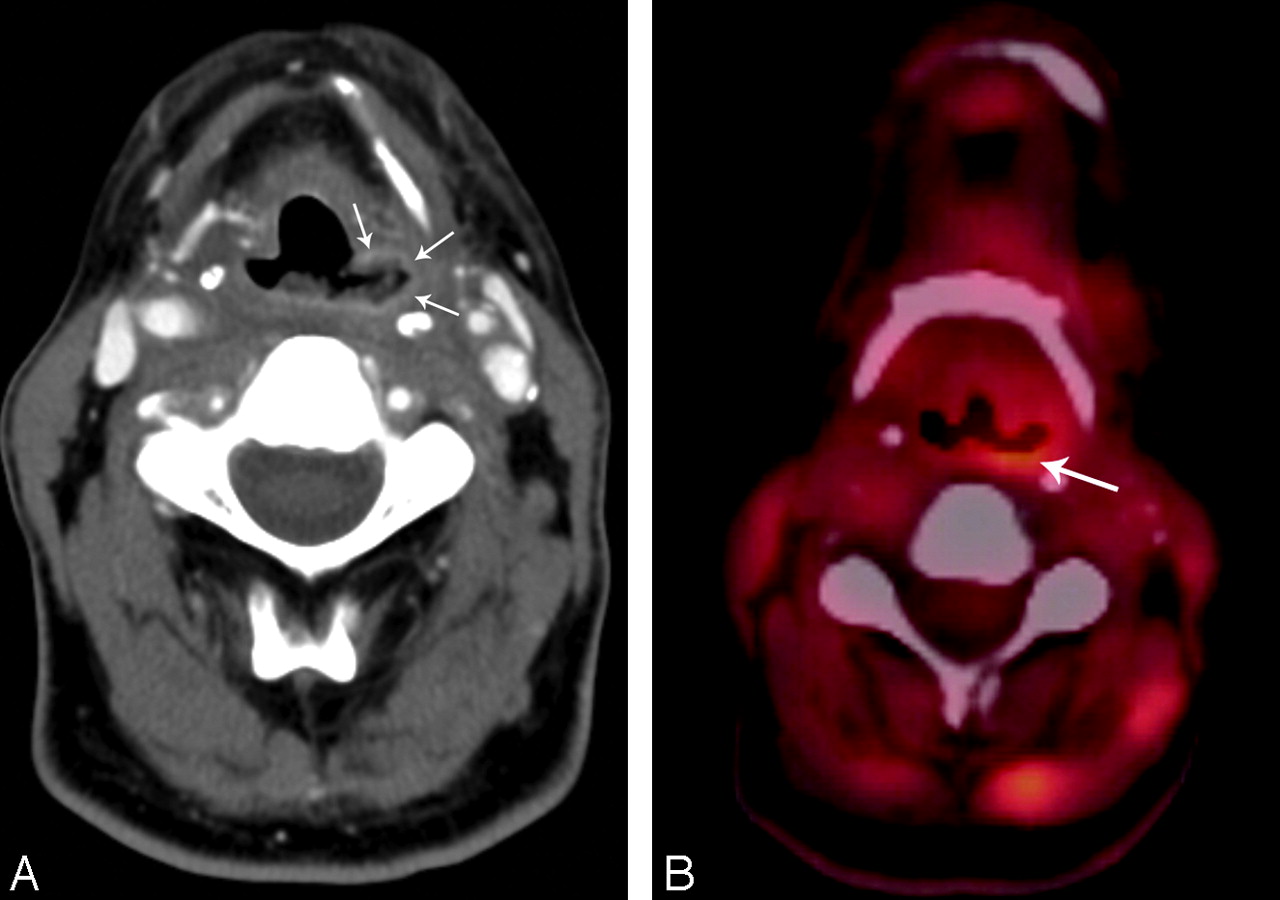
  • Fig 2.
    • Download figure
    • Open in new tab
    • Download powerpoint
    Fig 2.

    Patient 5, a 59-year-old woman status post 3.5 months of radiation therapy (60 Gy) for treatment of squamous cell carcinoma of the oral tongue, with a nonenhancing ulceration with negative biopsy results (category 1). This patient has been followed for 16 months since initial diagnosis without evidence of tumor recurrence. A, Contrast-enhanced CT with ulceration in the right lateral part of the oral tongue (arrows). B, PET/CT with 18F-FDG activity (SUV, 12 mg/mL) along the posterior border (arrows) on the ulceration, suspicious for a recurrent tumor.
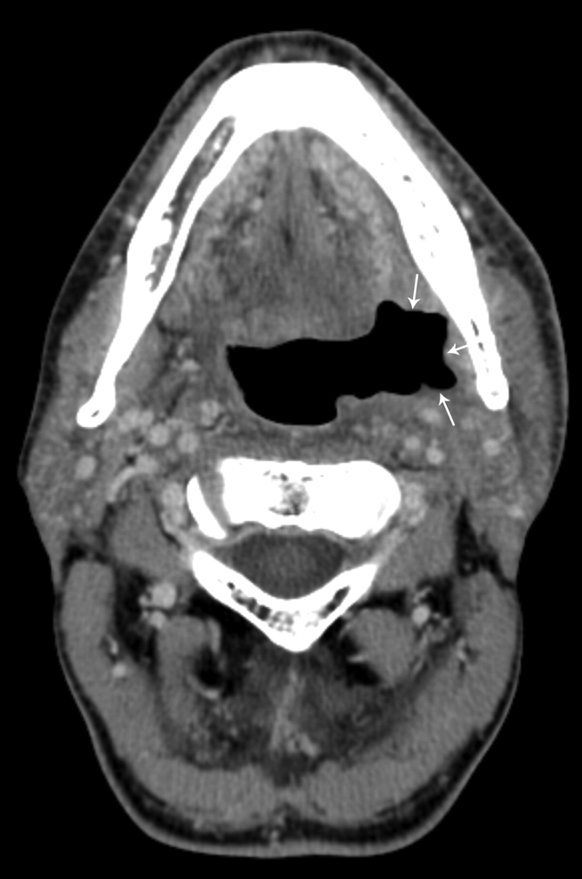
  • Fig 3.
    • Download figure
    • Open in new tab
    • Download powerpoint
    Fig 3.

    Patient 11, a 75-year-old woman status post 5 months of radiation therapy (70 Gy) for treatment of squamous cell carcinoma of the left oropharyngeal wall, with a nonenhancing ulceration of the left oropharynx (arrow) in which a biopsy was not performed (category 2). This patient has been followed for 30 months since initial diagnosis without evidence of tumor recurrence.
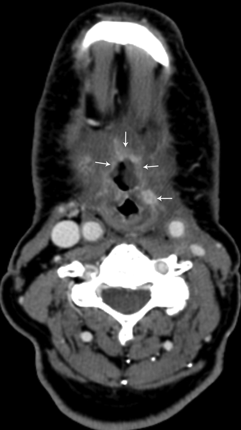
  • Fig 4.
    • Download figure
    • Open in new tab
    • Download powerpoint
    Fig 4.

    Patient 13, a 46-year-old man status post 7 months of radiation therapy (70 Gy) for treatment of a squamous cell carcinoma of the left oropharynx and hypopharynx, with an enhancing ulceration with positive biopsy results (category 3) of the left pyriform sinus. A, Contrast-enhanced CT with an enhancing ulceration of the pyriform sinus (arrows). B, PET/CT with 18F-FDG activity (SUV, 4.6 mg/mL) along the posterior border of the ulceration (arrow).

  • Fig 5.
    • Download figure
    • Open in new tab
    • Download powerpoint
    Fig 5.

    Patient 18, a 70-year-old men status post 61 months of radiation therapy for treatment of a squamous cell carcinoma of the larynx, followed by total laryngectomy with an enhancing ulceration with negative biopsy results (category 4) of the base of the tongue. There is adjacent peripheral and nodular enhancement (arrows). This patient has been followed for 19.5 months since initial diagnosis without evidence of tumor recurrence.

  • Fig 6.
    • Download figure
    • Open in new tab
    • Download powerpoint
    Fig 6.

    Patient 19, a 64-year-old man status post 5 months of radiation therapy (70 Gy) for treatment of a squamous cell carcinoma of the left tonsil, with an enhancing ulceration in the left oropharyngeal wall (arrows) in which biopsy was not performed (category 5). This patient has been followed for 20 months since initial diagnosis without evidence or tumor recurrence.

Tables

  • Figures
  • Summary of clinical data of patients with soft tissue ulceration

    Patient No.CategoryAge/SexRad Dose (Gy)Months After XRTLocation of UlcerationEnhancementBiopsy Results
    1155/M664.5Left palatine tonsilNoneNegative
    2153/M668.5Left palatine tonsilNoneNegative
    3146/M727Left oropharyngeal wallNoneNegative
    4158/M706.5Left vocal cordsNoneNegative
    5159/F603.5Right oral tongueNoneNegative
    6150/MOSH5Left base of tongueNoneNegative
    7161/FOSH12EpiglottisNoneNegative
    8159/M703.5EpiglottisNoneNegative
    9151/F703Right base of tongueNoneNegative
    10270/M603.5Left lateral nasopharynxNoneN/A
    11275/F705Left oropharyngeal wallNoneN/A
    12264/M705Right floor of mouthNoneN/A
    13346/M707Left pyriform sinusSubmucosalPositive
    14363/F769Midline oral tongueSubmucosalPositive
    15370/M666.5Right oral cavityNodularPositive
    16354/M665Left palatine tonsilSubmucosalPositive
    17478/F7210.5Right retromolar trigoneHeterogeneousNegative
    18470/MOSH61Base of tonguePeripheral/NodularNegative
    19564/M705Left oropharyngeal wallPeripheralN/A
    20574/F706Left oropharynxPeripheralN/A
    • Note:—Rad indicates radiation; OSH, outside hospital; XRT, external beam radiation therapy; N/A, biopsy not performed.

    • N.B.—Categoric description of ulcerations is author's terminology.

PreviousNext
Back to top

In this issue

American Journal of Neuroradiology: 29 (3)
American Journal of Neuroradiology
Vol. 29, Issue 3
March 2008
  • Table of Contents
  • Index by author
Advertisement
Print
Download PDF
Email Article

Thank you for your interest in spreading the word on American Journal of Neuroradiology.

NOTE: We only request your email address so that the person you are recommending the page to knows that you wanted them to see it, and that it is not junk mail. We do not capture any email address.

Enter multiple addresses on separate lines or separate them with commas.
Benign Ulceration as a Manifestation of Soft Tissue Radiation Necrosis: Imaging Findings
(Your Name) has sent you a message from American Journal of Neuroradiology
(Your Name) thought you would like to see the American Journal of Neuroradiology web site.
CAPTCHA
This question is for testing whether or not you are a human visitor and to prevent automated spam submissions.
Cite this article
J.M. Debnam, A.S. Garden, L.E. Ginsberg
Benign Ulceration as a Manifestation of Soft Tissue Radiation Necrosis: Imaging Findings
American Journal of Neuroradiology Mar 2008, 29 (3) 558-562; DOI: 10.3174/ajnr.A0886

Citation Manager Formats

  • BibTeX
  • Bookends
  • EasyBib
  • EndNote (tagged)
  • EndNote 8 (xml)
  • Medlars
  • Mendeley
  • Papers
  • RefWorks Tagged
  • Ref Manager
  • RIS
  • Zotero
0 Responses
Respond to this article
Share
Bookmark this article
Benign Ulceration as a Manifestation of Soft Tissue Radiation Necrosis: Imaging Findings
J.M. Debnam, A.S. Garden, L.E. Ginsberg
American Journal of Neuroradiology Mar 2008, 29 (3) 558-562; DOI: 10.3174/ajnr.A0886
del.icio.us logo Twitter logo Facebook logo Mendeley logo
  • Tweet Widget
  • Facebook Like
  • Google Plus One
Purchase

Jump to section

  • Article
    • Abstract
    • Materials and Methods
    • Results
    • Discussion
    • Conclusion
    • Footnotes
    • References
  • Figures & Data
  • Info & Metrics
  • Responses
  • References
  • PDF

Related Articles

  • No related articles found.
  • PubMed
  • Google Scholar

Cited By...

  • Osteoradionecrosis of the Hyoid Bone: Imaging Findings
  • Crossref
  • Google Scholar

This article has not yet been cited by articles in journals that are participating in Crossref Cited-by Linking.

More in this TOC Section

  • Hydrops Herniation into the Semicircular Canals
  • ASL Sensitivity for Head and Neck Paraganglioma
  • Post SRS Peritumoral Hyperintense Signal of VSs
Show more HEAD AND NECK IMAGING

Similar Articles

Advertisement

Indexed Content

  • Current Issue
  • Accepted Manuscripts
  • Article Preview
  • Past Issues
  • Editorials
  • Editor's Choice
  • Fellows' Journal Club
  • Letters to the Editor
  • Video Articles

Cases

  • Case Collection
  • Archive - Case of the Week
  • Archive - Case of the Month
  • Archive - Classic Case

More from AJNR

  • Trainee Corner
  • Imaging Protocols
  • MRI Safety Corner
  • Book Reviews

Multimedia

  • AJNR Podcasts
  • AJNR Scantastics

Resources

  • Turnaround Time
  • Submit a Manuscript
  • Submit a Video Article
  • Submit an eLetter to the Editor/Response
  • Manuscript Submission Guidelines
  • Statistical Tips
  • Fast Publishing of Accepted Manuscripts
  • Graphical Abstract Preparation
  • Imaging Protocol Submission
  • Evidence-Based Medicine Level Guide
  • Publishing Checklists
  • Author Policies
  • Become a Reviewer/Academy of Reviewers
  • News and Updates

About Us

  • About AJNR
  • Editorial Board
  • Editorial Board Alumni
  • Alerts
  • Permissions
  • Not an AJNR Subscriber? Join Now
  • Advertise with Us
  • Librarian Resources
  • Feedback
  • Terms and Conditions
  • AJNR Editorial Board Alumni

American Society of Neuroradiology

  • Not an ASNR Member? Join Now

© 2025 by the American Society of Neuroradiology All rights, including for text and data mining, AI training, and similar technologies, are reserved.
Print ISSN: 0195-6108 Online ISSN: 1936-959X

Powered by HighWire