
Article Figures & Data
Figures
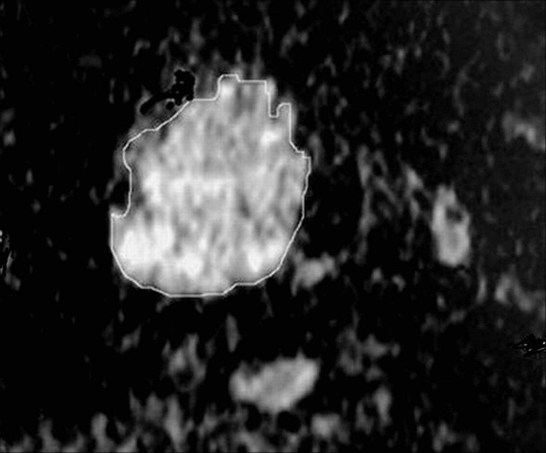
- Fig 1.
Measurement of the ADC value of a thyroid nodule in an ADC map image. The region of interest is drawn around the thyroid nodule, and the ADC value is measured.
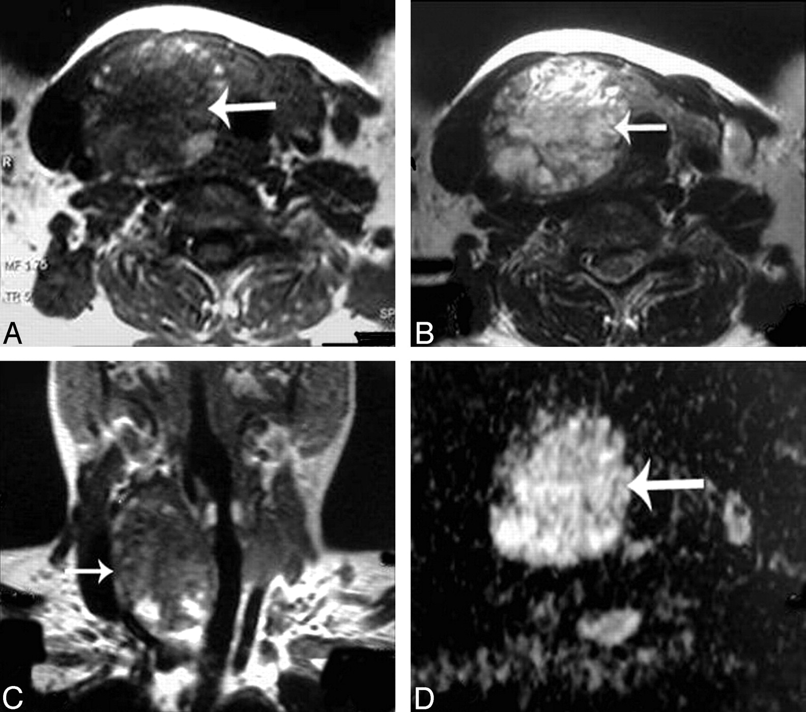
- Fig 2.
Adenomatous nodule. A–C, Axial T1- and T2-weighted and coronal T1-weighted MR images of the neck, respectively, showing a well-defined oval mainly solid solitary nodule (arrow) affecting the right thyroid lobe with contralateral tracheal displacement. D, An ADC map image with hyperintensity of the nodule (arrow) denoting increased diffusion and a measured ADC value of 1.57 ± 0.11 × 10⁻³ mm2/s.
- Fig 3.
Follicular adenoma. A and B, Axial T1- and T2-weighted MR images, respectively, showing a well-defined more or less oval solitary nodule affecting the right thyroid lobe with contralateral tracheal displacement. The nodule has an anterior cystic part (arrow) and another posterior solid one (arrowhead). C, An ADC map image with marked hyperintensity of the anterior cystic portion of the nodule (arrow), denotes increased diffusion with a measured ADC value of 2.25 ± 0.18 × 10⁻³ mm2/s and a relatively hypointense posterior solid portion (arrowhead), denotes relatively restricted diffusion with a measured ADC value of 1.2 ± 0.08 × 10⁻³ mm2/s.
- Fig 4.
Thyroid cyst. A–C, Axial T1- and T2-weighted and coronal T1-weighted MR images, respectively, showing a well-defined more or less oval solitary nodule (arrow) affecting the right thyroid lobe. D, An ADC map image with marked hyperintensity of the nodule (arrow) denoting increased diffusion, and the measured ADC value is 2.05 ± 0.13 × 10⁻³ mm2/s.
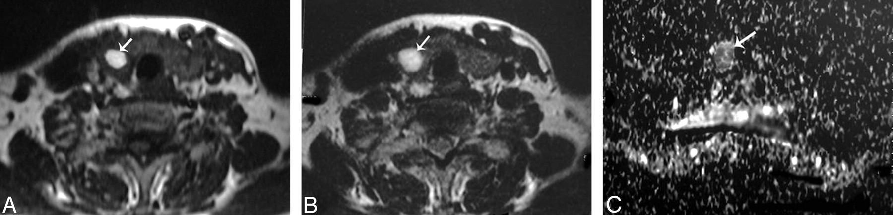
- Fig 5.
Hemorrhagic thyroid cyst. A and B, Axial T1- and T2-weighted MR images, respectively, showing a small well-defined more or less rounded solitary nodule (arrow) affecting the right thyroid lobe. The nodule is hyperintense on both T1- and T2-weighted images. C, An ADC map image shows low signal intensity of the nodule. The measured ADC value is 0.5 ± 0.07 × 10⁻³ mm2/s.
- Fig 6.
Papillary carcinoma of the thyroid. A and B, Axial T1- and T2-weighted MR images, respectively, showing a well-defined irregular mainly solid solitary nodule (arrow) involving all the right thyroid lobe with contralateral tracheal displacement. C, ADC map image shows hypointensity of the nodule (arrow). The measured ADC value is 0.97 ± 0.1 × 10⁻³ mm2/s.
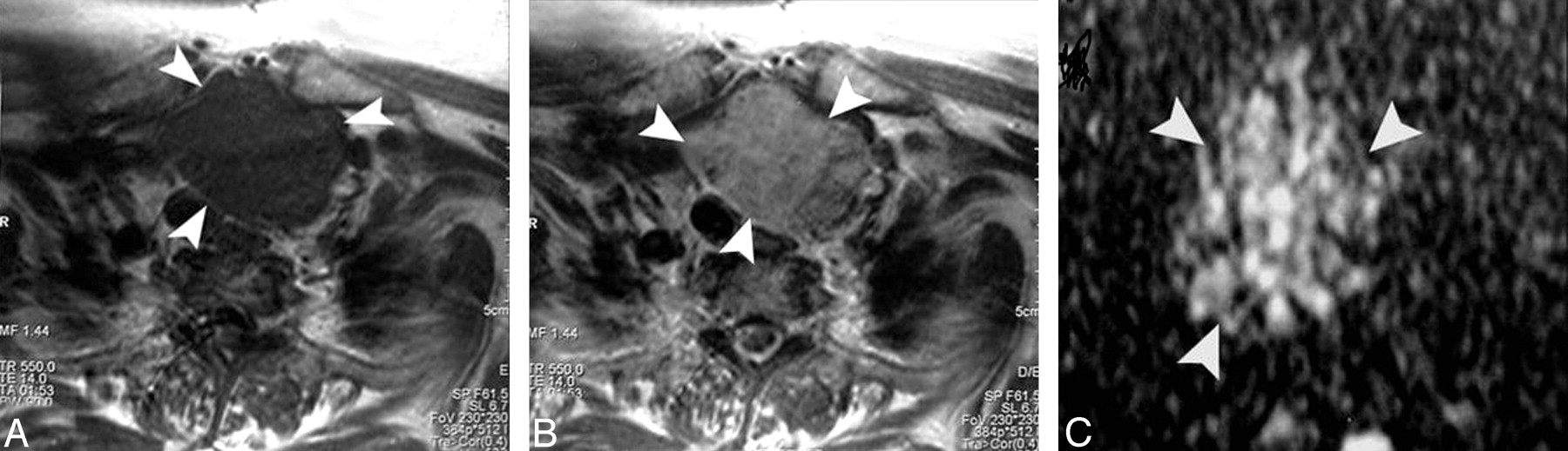
- Fig 7.
Follicular carcinoma of the thyroid. A and B, Axial T1- and T2-weighted MR images, respectively, showing a well-defined more or less oval mainly solid solitary nodule (arrowheads) affecting the right thyroid lobe with contralateral tracheal displacement. C, ADC map image shows a low ADC value (0.92 ± 0.06 × 10⁻³ mm2/s) of the thyroid nodule (arrowhead).
- Fig 8.
ROC curve of the ADC value used for differentiation of benign from malignant solitary thyroid nodules. The area under the curve measures 97%.


Tables
Type of Thyroid Nodules No. (%) of Thyroid Nodules Range of ADC Values (mm2/s) Mean ADC Values (mm2/s) Benign 56 (88.9 ) 0.5–2.3 × 10⁻³ 1.8 ± 0.27 × 10⁻³ Adenomatous nodule 42 (66.7) 1.1–1.9 × 10⁻³ 1.8 ± 0.14 × 10⁻³ Follicular adenoma 6 (9.5) 1.2–2 × 10⁻³ 1.7 ± 0.17 × 10⁻³ Thyroid cyst 8 (12.7) 0.5–2.3 × 10⁻³ 1.9 ± 0.38 × 10⁻³ Malignant 7 (11.1) 0.5–1.1 × 10⁻³ 0.73 ± 0.19 × 10⁻³ Papillary carcinoma 4 (6.3) 0.5–1.1 × 10⁻³ 0.68 ± 0.23 × 10⁻³ Follicular carcinoma 3 (4.8) 0.6–1 × 10⁻³ 0.77 ± 0.17 × 10⁻³ - Table 2:
Comparison between the ADC values of benign and malignant solitary thyroid nodules
Pathology of Thyroid Nodule No. (%) of Thyroid Nodules Mean ADC Values (mm2/s) P Value* Benign 56 (88.9) 1.8 ± 0.27 × 10⁻³ .0001 Malignant 7 (11.1) 0.73 ± 0.19 × 10⁻³ * Significance was considered when P <.05.
- Table 3:
Comparison between the ADC values in the different subtypes of benign and malignant solitary thyroid nodules
Type of Thyroid Nodule No. (%) of Thyroid Nodules Mean ADC Values (mm2/s) P Value* Benign Adenomatous nodule 42 (66.7) 1.8 ± 0.14 × 10⁻³ Follicular adenoma 6 (9.5) 1.7 ± 0.17 × 10⁻³ .0001 Thyroid cyst 8 (12.7) 1.9 ± 0.38 × 10⁻³ Malignant Papillary carcinoma 4 (6.3) 0.68 ± 0.23 × 10⁻³ .464 Follicular carcinoma 3 (4.8) 0.77 ± 0.17 × 10⁻³ * Significance was considered when P < .05.
In this issue


Jump to section
Related Articles
Cited By...
- Diffusion-weighted MRI in differentiating malignant from benign thyroid nodules: a meta-analysis
- 3T diffusion-weighted MRI of the thyroid gland with reduced distortion: preliminary results
- Diffusion MR Imaging Features of Skull Base Osteomyelitis Compared with Skull Base Malignancy
- Non-Gaussian Analysis of Diffusion-Weighted MR Imaging in Head and Neck Squamous Cell Carcinoma: A Feasibility Study
- Can Quantitative Diffusion-Weighted MR Imaging Differentiate Benign and Malignant Cold Thyroid Nodules? Initial Results in 25 Patients