Advertisement

Main menu

  • Home
  • Content
    • Current Issue
    • Accepted Manuscripts
    • Article Preview
    • Past Issue Archive
    • AJNR Case Collection
    • Case of the Week Archive
    • Classic Case Archive
    • Case of the Month Archive
  • Special Collections
    • Spinal CSF Leak Articles (Jan 2020-June 2024)
    • 2024 AJNR Journal Awards
    • Most Impactful AJNR Articles
  • Multimedia
    • AJNR Podcast
    • AJNR Scantastics
    • Video Articles
  • For Authors
    • Submit a Manuscript
    • Author Policies
    • Fast publishing of Accepted Manuscripts
    • Graphical Abstract Preparation
    • Manuscript Submission Guidelines
    • Imaging Protocol Submission
    • Submit a Case for the Case Collection
  • About Us
    • About AJNR
    • Editorial Board
  • More
    • Become a Reviewer/Academy of Reviewers
    • Subscribers
    • Permissions
    • Alerts
    • Feedback
    • Advertisers
    • ASNR Home
  • Other Publications
    • ajnr

User menu

  • Alerts
  • Log in

Search

  • Advanced search
American Journal of Neuroradiology

American Journal of Neuroradiology

ASHNR American Society of Functional Neuroradiology ASHNR American Society of Pediatric Neuroradiology ASSR
  • Alerts
  • Log in

Advanced Search

  • Home
  • Content
    • Current Issue
    • Accepted Manuscripts
    • Article Preview
    • Past Issue Archive
    • AJNR Case Collection
    • Case of the Week Archive
    • Classic Case Archive
    • Case of the Month Archive
  • Special Collections
    • Spinal CSF Leak Articles (Jan 2020-June 2024)
    • 2024 AJNR Journal Awards
    • Most Impactful AJNR Articles
  • Multimedia
    • AJNR Podcast
    • AJNR Scantastics
    • Video Articles
  • For Authors
    • Submit a Manuscript
    • Author Policies
    • Fast publishing of Accepted Manuscripts
    • Graphical Abstract Preparation
    • Manuscript Submission Guidelines
    • Imaging Protocol Submission
    • Submit a Case for the Case Collection
  • About Us
    • About AJNR
    • Editorial Board
  • More
    • Become a Reviewer/Academy of Reviewers
    • Subscribers
    • Permissions
    • Alerts
    • Feedback
    • Advertisers
    • ASNR Home
  • Follow AJNR on Twitter
  • Visit AJNR on Facebook
  • Follow AJNR on Instagram
  • Join AJNR on LinkedIn
  • RSS Feeds

Welcome to the new AJNR, Updated Hall of Fame, and more. Read the full announcements.


AJNR is seeking candidates for the position of Associate Section Editor, AJNR Case Collection. Read the full announcement.


OtherHEAD & NECK

Identification of the Normal Jugular Foramen and Lower Cranial Nerve Anatomy: Contrast-Enhanced 3D Fast Imaging Employing Steady-State Acquisition MR Imaging

I. Davagnanam and S.V. Chavda
American Journal of Neuroradiology March 2008, 29 (3) 574-576; DOI: https://doi.org/10.3174/ajnr.A0860
I. Davagnanam
  • Find this author on Google Scholar
  • Find this author on PubMed
  • Search for this author on this site
S.V. Chavda
  • Find this author on Google Scholar
  • Find this author on PubMed
  • Search for this author on this site
  • Article
  • Figures & Data
  • Info & Metrics
  • Responses
  • References
  • PDF
Loading

Abstract

SUMMARY: Conventional imaging protocols are unable to visualize the intraforaminal/canalicular segments of the lower cranial nerves (IX–XII). On the basis of previous successful demonstration of individual cranial nerves within the cavernous sinus by constructive interference in steady-state MR imaging, we describe the use of contrast-enhanced 3D fast imaging employing steady-state acquisition MR imaging to demonstrate normal in vivo intraforaminal and canalicular segments of cranial nerves IX–XII in 10 patients by using a standardized imaging protocol.

The detection of individual cranial nerves in the intraforaminal portion of the jugular foramen and hypoglossal canal is useful in diagnostic imaging1; however, visualization by using conventional MR imaging protocols does not provide adequate detail of individual nerves.2,3 Recently, contrast-enhanced 3D constructive interference in steady state (CISS) MR imaging has successfully demonstrated the trigeminal ganglion and its divisions and individual cranial nerves within the cavernous sinus.4,5 3D fast imaging employing steady-state acquisition (FIESTA) is a similar form of steady-state sequence MR imaging. Evidence has shown a proportional increase in contrast between the background structures and the cranial nerves within the cisterns as the concentration of gadolinium-based contrast agent increases with 3D steady-state imaging sequences.4,6

This Technical Note describes the use of 3D-FIESTA imaging after intravenous administration of gadodiamide hydrate contrast to demonstrate the normal anatomy of the intraforaminal and canalicular portions of cranial nerves IX–XII.

Description of the Technique

Detectability of the intraforaminal/canalicular segments of cranial nerves IX–XII was evaluated in 10 consecutive patients undergoing routine follow-up imaging for vestibular schwannomas. Patients included 6 men and 4 women (mean age, 56.8; range, 36–68 years).

MR Imaging

The examinations were performed on a 1.5T unit (Signa Excite HD; GE Healthcare, Bucks, UK) with a standard head coil and pre- and postcontrast enhancement with a standardized FIESTA MR imaging protocol. Gadodiamide hydrate (Omniscan; GE Healthcare) was administered at 0.1-mmol/kg body weight as an intravenous contrast agent. The pulse sequence used was the following: 3D FIESTA-C (TR/TE/NEX, 5.5/1.7/3.00; 180 × 180 mm [read x phase encode] FOV; 0.4-mm effective section thickness; 448 × 448 matrix; and imaging time of 8 minutes 21 seconds).

Image Analysis

The datasets obtained were reconstructed in the 3 orthogonal/oblique planes in relation to the cisternal course of the cranial nerves. Images were viewed on high-resolution monitors with a multiplanar reconstruction program (Advantage Workstations; GE Healthcare).

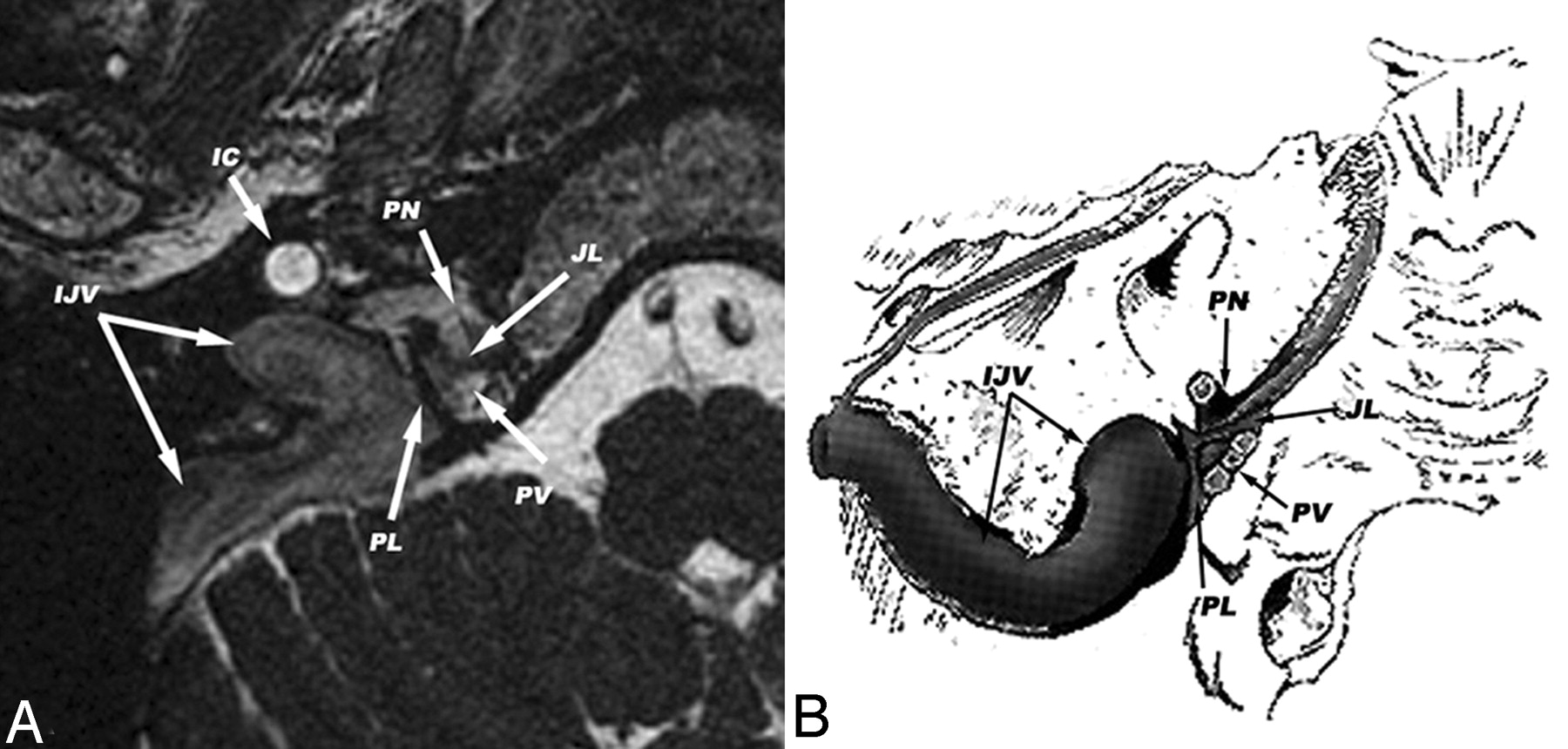
The initial evaluation involved an assessment of the division of the jugular foramen into the pars nervosa and pars vascularis, separated by the fibrous or bony septum (jugular ligament) represented by a dark line (Fig 1). A further attempt was then made to identify cranial nerves IX–XII within the jugular foramen and hypoglossal canal, appearing as hypointense bands or spots. The continuity of nerves was confirmed by tracking their courses proximal and distal to the skull base on >3 consecutive sections in the reconstructed planes to distinguish these from other structures and artifacts, thereby reducing false-positives. Comparison with the precontrast images was then undertaken after identification of cranial nerves on the postcontrast images. This process averaged 26 minutes (range, 18–35) per patient to reconstruct, analyze, and correlate the imaging with the anatomic course of lower cranial nerves.

Fig 1.
  • Download figure
  • Open in new tab
  • Download powerpoint
Fig 1.

Axial postcontrast FIESTA acquisition through the jugular foramen (A) and a diagrammatic representation of the corresponding axial section (B) (adapted from Leblanc A. The Cranial Nerves: Anatomy-Imaging-Vascularization. 2nd ed. Berlin, Germany: Springer; 1996) showing the following: opacified internal jugular vein (IJV), internal carotid artery (IC), jugular ligament (JL), petro-occipital ligament (PL), and division of the pars nervosa (PN) from the pars vascularis (PV).

The images were evaluated by 2 neuroradiologists, making initial evaluations independently but resolving any inconsistencies by collaborative review and consensus agreement.

Results

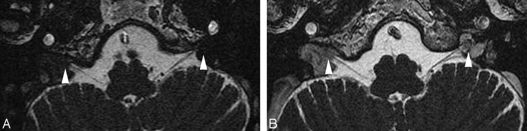
There was consensus agreement that enhancement of the jugular vein and associated venous plexus surrounding the cranial nerves provided excellent contrast of the small structures within the jugular foramen on FIESTA images, unlike on precontrast FIESTA (Fig 2) and standard imaging sequences.

Fig 2.
  • Download figure
  • Open in new tab
  • Download powerpoint
Fig 2.

Axial pre- (A) and postgadolinium (B) FIESTA acquisitions showing the length of the vagus nerve (X) bilaterally. The images were windowed at comparable levels (C:3886, W:12,502) demonstrating the effect of enhancement of the venous plexus adjacent to the intraforaminal segment of the cranial nerves (arrowheads).

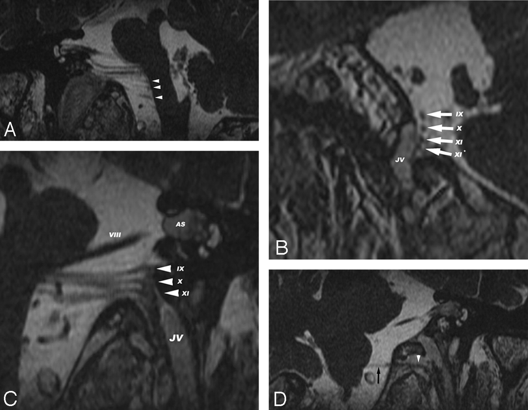
The jugular ligament was identified on the contrast-enhanced sequence in all 20 jugular foramina. The intraforaminal and canalicular segments of cranial nerves IX–XII were localized in all cases consistent with the known anatomic courses of cranial nerves IX–XII (Fig 3). Disagreement between observers relating to the identification of the spinal roots of the accessory nerve (XI) occurred in 2 cases but was resolved by consensus review and agreement.

Fig 3.
  • Download figure
  • Open in new tab
  • Download powerpoint
Fig 3.

Postgadolinium FIESTA acquisitions reformatted in the following planes: the coronal-oblique plane demonstrating the spinal roots of the accessory nerve (XI) (A, white arrowheads); the sagittal-oblique plane demonstrating the intraforaminal segments (B, white arrows) of the glossopharyngeal (IX), vagus (X), cranial accessory (XI), and spinal accessory (XI*) nerves adjacent to the opacified jugular vein (JV); the coronal-oblique plane demonstrating a left vestibular schwannoma (AS) of the vestibulocochlear nerve (VIII) and the intraforaminal segments (C, white arrowheads) of the glossopharyngeal (IX), vagus (X), and cranial accessory (XI) nerves adjacent to the opacified jugular vein (JV); and the coronal-oblique plane demonstrating the cisternal course (D, black arrow) and intracanalicular segment (D, white arrowhead) of the hypoglossal nerve (XII).

Discussion

As early as 1985, Daniels et al7 attempted to correlate the course of cranial nerves IX through XI in cadaveric specimens with standard 1.5T MR imaging. With the advent of steady-state imaging, the use of uncontrasted CISS/FIESTA sequences have become an integral part of the assessment of pathology involving the cranial nerves VII and VIII (particularly vestibular schwannomas).6,8 In 2001, Seitz et al2 suggested a combination of 3D CISS and magnetization-preparation rapid gradient-echo imaging, where the 2D technique had failed to demonstrate lower cranial nerves. Its utility had been extended in the successful demonstration of the visualization of microvascular compression of cranial nerve IX in glossopharyngeal neuralgia9 and the cochlear nerve in presurgical cochlear-implant candidates (high-field 3T).10

The steady-state sequence depicts small structures surrounded by CSF with high contrast and spatial resolution, using a refocused gradient-echo sequence. This uses a phase-cycling technique and a steady-state contrast mechanism to provide high signal intensity-to-noise ratio images with heavy T2-weighted signal intensity while suppressing background tissue. The sequences are extremely short (TR) between radio-frequency pulses, such that high-resolution 3D volume images can be acquired rapidly.11 The jugular ligament and intraforaminal/canalicular segments of cranial nerves IX–XII were consistently and distinctly demonstrated in all our patients with this technique. This was further facilitated by enhancement of the adjacent jugular vein and venous plexus, resulting in an increase in contrast between cranial nerves and the background, similar to that previously described within the cisterns.4–6

The application of contrast-enhanced steady-state imaging has recently successfully delineated cranial nerves in the cavernous sinus4 as well as the trigeminal ganglion and its motor and sensory divisions.5 To our knowledge, assessment by contrast-enhanced steady-state MR imaging has, however, never been performed to visualize the intraforaminal/canalicular segments of the lower cranial nerves before this study.

The identification of normal cranial nerves may have implications in determining the involvement by pathologic processes as well as for neural preservation surgery of tumors in this region.12 However, the authors acknowledge that the variability in the course of the cranial nerves within the jugular foramen may hinder the full utility of this technique.

Conclusion

The reproducibility and consistency in demonstrating the distinct anatomic courses of cranial nerves IX–XII of contrast-enhanced 3D-FIESTA MR imaging in vivo makes this technique potentially useful in the evaluation of the extent of cranial nerve involvement by tumors. It can be more accurately compared with traditional combinations of clinical symptoms, standard CT, and MR imaging and is useful in planning before biopsy and neural preserving surgical procedures.

Footnotes

  • Paper previously presented at: Annual Meeting of the American Society of Head and Neck Radiology, September 27, 2006, Phoenix, Ariz; Annual Meeting of the International Congress of Head and Neck Radiology, September 29, 2006, Budapest, Hungary; and Royal Society of Medicine, London, UK, for the Finzi Prize, November 24, 2006.

References

  1. ↵
    Eldevik OP, Gabrielsen TO, Jacobsen EA. Imaging findings in schwannomas of the jugular foramen. AJNR Am J Neuroradiol 2000;21:1139–44
    Abstract/FREE Full Text
  2. ↵
    Seitz J, Held P, Fründ R, et al. Visualization of the IXth to XIIth cranial nerves using 3-dimensional constructive interference in steady state, 3-dimensional magnetization-prepared rapid gradient echo and T2-weighted 2-dimensional turbo spin echo magnetic resonance imaging sequences. J Neuroimaging 2001;11:160–64
    PubMed
  3. ↵
    Suzuki H, Maki H, Maeda M, et al. Visualization of the intracisternal angioarchitecture at the posterior fossa by use of image fusion. Neurosurgery 2005;56:335–42
    CrossRefPubMed
  4. ↵
    Yagi A, Sato N, Taketomi A, et al. Normal cranial nerves in the cavernous sinuses: contrast-enhanced three-dimensional constructive interference in the steady state MR imaging. AJNR Am J Neuroradiol 2005;26:946–50
    Abstract/FREE Full Text
  5. ↵
    Yousry I, Moriggl B, Schmid UD, et al. Trigeminal ganglion and its divisions: detailed anatomic MR imaging with contrast-enhanced 3D constructive interference in the steady state sequences. AJNR Am J Neuroradiol 2005;26:1128–35
    Abstract/FREE Full Text
  6. ↵
    Shigematsu Y, Korogi Y, Hirai T, et al. Contrast-enhanced CISS MRI of vestibular schwannomas: phantom and clinical studies. J Comput Assist Tomogr 1999;23:224–31
    CrossRefPubMed
  7. ↵
    Daniels DL, Schenck JF, Foster T, et al. Magnetic resonance imaging of the jugular foramen. AJNR Am J Neuroradiol 1985;6:699–703
    Abstract/FREE Full Text
  8. ↵
    Casselman JW, Kuhweide R, Deimling M, et al. Constructive interference in steady state-3DFT MR imaging of the inner ear and cerebellopontine angle. AJNR Am J Neuroradiol 1993;14:47–57
    Abstract/FREE Full Text
  9. ↵
    Karibe H, Shirane R, Yoshimoto T. Preoperative visualization of microvascular compression of cranial nerve IX using constructive interference in steady state magnetic resonance imaging in glossopharyngeal neuralgia. J Clin Neurosci 2004;11:679–81
    CrossRefPubMed
  10. ↵
    Lane JI, Ward H, Witte RJ, et al. 3-T imaging of the cochlear nerve and labyrinth in cochlear-implant candidates: 3D fast recovery fast spin-echo versus 3D constructive interference in the steady state techniques. AJNR Am J Neuroradiol 2004;25:618–22
    Abstract/FREE Full Text
  11. ↵
    Nitz WR. Fast and ultrafast non-echo-planar MR imaging techniques. Eur Radiol 2002;12:2866–82
    PubMed
  12. ↵
    Lustig LR, Jackler RK. The variable relationship between the lower cranial nerves and jugular foramen tumors: implications for neural preservation. Am J Otol 1996;17:658–68
    PubMed
  • Received August 1, 2007.
  • Accepted after revision August 2, 2007.
  • Copyright © American Society of Neuroradiology
View Abstract
PreviousNext
Back to top

In this issue

American Journal of Neuroradiology: 29 (3)
American Journal of Neuroradiology
Vol. 29, Issue 3
March 2008
  • Table of Contents
  • Index by author
Advertisement
Print
Download PDF
Email Article

Thank you for your interest in spreading the word on American Journal of Neuroradiology.

NOTE: We only request your email address so that the person you are recommending the page to knows that you wanted them to see it, and that it is not junk mail. We do not capture any email address.

Enter multiple addresses on separate lines or separate them with commas.
Identification of the Normal Jugular Foramen and Lower Cranial Nerve Anatomy: Contrast-Enhanced 3D Fast Imaging Employing Steady-State Acquisition MR Imaging
(Your Name) has sent you a message from American Journal of Neuroradiology
(Your Name) thought you would like to see the American Journal of Neuroradiology web site.
CAPTCHA
This question is for testing whether or not you are a human visitor and to prevent automated spam submissions.
Cite this article
I. Davagnanam, S.V. Chavda
Identification of the Normal Jugular Foramen and Lower Cranial Nerve Anatomy: Contrast-Enhanced 3D Fast Imaging Employing Steady-State Acquisition MR Imaging
American Journal of Neuroradiology Mar 2008, 29 (3) 574-576; DOI: 10.3174/ajnr.A0860

Citation Manager Formats

  • BibTeX
  • Bookends
  • EasyBib
  • EndNote (tagged)
  • EndNote 8 (xml)
  • Medlars
  • Mendeley
  • Papers
  • RefWorks Tagged
  • Ref Manager
  • RIS
  • Zotero
0 Responses
Respond to this article
Share
Bookmark this article
Identification of the Normal Jugular Foramen and Lower Cranial Nerve Anatomy: Contrast-Enhanced 3D Fast Imaging Employing Steady-State Acquisition MR Imaging
I. Davagnanam, S.V. Chavda
American Journal of Neuroradiology Mar 2008, 29 (3) 574-576; DOI: 10.3174/ajnr.A0860
del.icio.us logo Twitter logo Facebook logo Mendeley logo
  • Tweet Widget
  • Facebook Like
  • Google Plus One
Purchase

Jump to section

  • Article
    • Abstract
    • Description of the Technique
    • Results
    • Discussion
    • Conclusion
    • Footnotes
    • References
  • Figures & Data
  • Info & Metrics
  • Responses
  • References
  • PDF

Related Articles

  • No related articles found.
  • PubMed
  • Google Scholar

Cited By...

  • Pituitary Macroadenoma and Visual Impairment: Postoperative Outcome Prediction with Contrast-Enhanced FIESTA
  • Is All "Communicating" Hydrocephalus Really Communicating? Prospective Study on the Value of 3D-Constructive Interference in Steady State Sequence at 3T
  • Detailed MR Imaging Anatomy of the Cisternal Segments of the Glossopharyngeal, Vagus, and Spinal Accessory Nerves in the Posterior Fossa: The Use of 3D Balanced Fast-Field Echo MR Imaging
  • The Jugular Foramen: Imaging Strategy and Detailed Anatomy at 3T
  • Crossref (32)
  • Google Scholar

This article has been cited by the following articles in journals that are participating in Crossref Cited-by Linking.

  • Is All “Communicating” Hydrocephalus Really Communicating? Prospective Study on the Value of 3D-Constructive Interference in Steady State Sequence at 3T
    A. Dinçer, S. Kohan, M.M. Özek
    American Journal of Neuroradiology 2009 30 10
  • Anatomic Considerations, Nomenclature, and Advanced Cross-sectional Imaging Techniques for Visualization of the Cranial Nerve Segments by MR Imaging
    Ari M. Blitz, Asim F. Choudhri, Zachary D. Chonka, Ahmet T. Ilica, Leonardo L. Macedo, Avneesh Chhabra, Gary L. Gallia, Nafi Aygun
    Neuroimaging Clinics of North America 2014 24 1
  • Prediction of hard meningiomas: quantitative evaluation based on the magnetic resonance signal intensity
    Keita Watanabe, Shingo Kakeda, Junkoh Yamamoto, Satoru Ide, Norihiro Ohnari, Shigeru Nishizawa, Yukunori Korogi
    Acta Radiologica 2016 57 3
  • The Jugular Foramen: Imaging Strategy and Detailed Anatomy at 3T
    J. Linn, F. Peters, B. Moriggl, T.P. Naidich, H. Brückmann, I. Yousry
    American Journal of Neuroradiology 2009 30 1
  • Delineation of Optic Nerves and Chiasm in Close Proximity to Large Suprasellar Tumors with Contrast-enhanced FIESTA MR Imaging
    Keita Watanabe, Shingo Kakeda, Junkoh Yamamoto, Rieko Watanabe, Joji Nishimura, Norihiro Ohnari, Shigeru Nishizawa, Yukunori Korogi
    Radiology 2012 264 3
  • The lower cranial nerves: IX, X, XI, XII
    J.-L. Sarrazin, F. Toulgoat, F. Benoudiba
    Diagnostic and Interventional Imaging 2013 94 10
  • Detailed MR Imaging Anatomy of the Cisternal Segments of the Glossopharyngeal, Vagus, and Spinal Accessory Nerves in the Posterior Fossa: The Use of 3D Balanced Fast-Field Echo MR Imaging
    W.-J. Moon, H.G. Roh, E.C. Chung
    American Journal of Neuroradiology 2009 30 6
  • The glossopharyngeal, vagus and spinal accessory nerves
    Cheng Kang Ong, Vincent Fook Hin Chong
    European Journal of Radiology 2010 74 2
  • Cost-effectiveness analysis of a non-contrast screening MRI protocol for vestibular schwannoma in patients with asymmetric sensorineural hearing loss
    Matthew G. Crowson, Daniel J. Rocke, Jenny K. Hoang, Jane L. Weissman, David M. Kaylie
    Neuroradiology 2017 59 8
  • Fast imaging employing steady-state acquisition (FIESTA) MRI to investigate cerebrospinal fluid (CSF) within dural reflections of posterior fossa cranial nerves
    David J Noble, Daniel Scoffings, Thankamma Ajithkumar, Michael V Williams, Sarah J Jefferies
    The British Journal of Radiology 2016 89 1067

More in this TOC Section

  • WHO Classification Update: Nasal&Skull Base Tumors
  • Peritumoral Signal in Vestibular Schwannomas
  • Chondrosarcoma vs Synovial Chondromatosis: Imaging
Show more HEAD & NECK

Similar Articles

Advertisement

Indexed Content

  • Current Issue
  • Accepted Manuscripts
  • Article Preview
  • Past Issues
  • Editorials
  • Editors Choice
  • Fellow Journal Club
  • Letters to the Editor

Cases

  • Case Collection
  • Archive - Case of the Week
  • Archive - Case of the Month
  • Archive - Classic Case

Special Collections

  • Special Collections

Resources

  • News and Updates
  • Turn around Times
  • Submit a Manuscript
  • Author Policies
  • Manuscript Submission Guidelines
  • Evidence-Based Medicine Level Guide
  • Publishing Checklists
  • Graphical Abstract Preparation
  • Imaging Protocol Submission
  • Submit a Case
  • Become a Reviewer/Academy of Reviewers
  • Get Peer Review Credit from Publons

Multimedia

  • AJNR Podcast
  • AJNR SCANtastic
  • Video Articles

About Us

  • About AJNR
  • Editorial Board
  • Not an AJNR Subscriber? Join Now
  • Alerts
  • Feedback
  • Advertise with us
  • Librarian Resources
  • Permissions
  • Terms and Conditions

American Society of Neuroradiology

  • Not an ASNR Member? Join Now

© 2025 by the American Society of Neuroradiology All rights, including for text and data mining, AI training, and similar technologies, are reserved.
Print ISSN: 0195-6108 Online ISSN: 1936-959X

Powered by HighWire