Advertisement

Main menu

  • Home
  • Content
    • Current Issue
    • Accepted Manuscripts
    • Article Preview
    • Past Issue Archive
    • AJNR Case Collection
    • Case of the Week Archive
    • Classic Case Archive
    • Case of the Month Archive
  • Special Collections
    • Spinal CSF Leak Articles (Jan 2020-June 2024)
    • 2024 AJNR Journal Awards
    • Most Impactful AJNR Articles
  • Multimedia
    • AJNR Podcast
    • AJNR Scantastics
    • Video Articles
  • For Authors
    • Submit a Manuscript
    • Author Policies
    • Fast publishing of Accepted Manuscripts
    • Graphical Abstract Preparation
    • Manuscript Submission Guidelines
    • Imaging Protocol Submission
    • Submit a Case for the Case Collection
  • About Us
    • About AJNR
    • Editorial Board
  • More
    • Become a Reviewer/Academy of Reviewers
    • Subscribers
    • Permissions
    • Alerts
    • Feedback
    • Advertisers
    • ASNR Home
  • Other Publications
    • ajnr

User menu

  • Alerts
  • Log in

Search

  • Advanced search
American Journal of Neuroradiology

American Journal of Neuroradiology

ASHNR American Society of Functional Neuroradiology ASHNR American Society of Pediatric Neuroradiology ASSR
  • Alerts
  • Log in

Advanced Search

  • Home
  • Content
    • Current Issue
    • Accepted Manuscripts
    • Article Preview
    • Past Issue Archive
    • AJNR Case Collection
    • Case of the Week Archive
    • Classic Case Archive
    • Case of the Month Archive
  • Special Collections
    • Spinal CSF Leak Articles (Jan 2020-June 2024)
    • 2024 AJNR Journal Awards
    • Most Impactful AJNR Articles
  • Multimedia
    • AJNR Podcast
    • AJNR Scantastics
    • Video Articles
  • For Authors
    • Submit a Manuscript
    • Author Policies
    • Fast publishing of Accepted Manuscripts
    • Graphical Abstract Preparation
    • Manuscript Submission Guidelines
    • Imaging Protocol Submission
    • Submit a Case for the Case Collection
  • About Us
    • About AJNR
    • Editorial Board
  • More
    • Become a Reviewer/Academy of Reviewers
    • Subscribers
    • Permissions
    • Alerts
    • Feedback
    • Advertisers
    • ASNR Home
  • Follow AJNR on Twitter
  • Visit AJNR on Facebook
  • Follow AJNR on Instagram
  • Join AJNR on LinkedIn
  • RSS Feeds

Welcome to the new AJNR, Updated Hall of Fame, and more. Read the full announcements.


AJNR is seeking candidates for the position of Associate Section Editor, AJNR Case Collection. Read the full announcement.


Getting new auth cookie, if you see this message a lot, tell someone!
Research ArticleINTERVENTIONAL

Detection of Aneurysms by 64-Section Multidetector CT Angiography in Patients Acutely Suspected of Having an Intracranial Aneurysm and Comparison with Digital Subtraction and 3D Rotational Angiography

A.M. McKinney, C.S. Palmer, C.L. Truwit, A. Karagulle and M. Teksam
American Journal of Neuroradiology March 2008, 29 (3) 594-602; DOI: https://doi.org/10.3174/ajnr.A0848
A.M. McKinney
  • Find this author on Google Scholar
  • Find this author on PubMed
  • Search for this author on this site
C.S. Palmer
  • Find this author on Google Scholar
  • Find this author on PubMed
  • Search for this author on this site
C.L. Truwit
  • Find this author on Google Scholar
  • Find this author on PubMed
  • Search for this author on this site
A. Karagulle
  • Find this author on Google Scholar
  • Find this author on PubMed
  • Search for this author on this site
M. Teksam
  • Find this author on Google Scholar
  • Find this author on PubMed
  • Search for this author on this site
  • Article
  • Figures & Data
  • Info & Metrics
  • Responses
  • References
  • PDF
Loading

Article Figures & Data

Figures

  • Tables
  • Fig 1.
    • Download figure
    • Open in new tab
    • Download powerpoint
    Fig 1.

    An 83-year-old woman with SAH and an aneurysm less than 4 mm. Emergent CTA showed a 2-mm MCA bifurcation aneurysm. This was difficult to visualize on conventional DSA (not shown) but was confirmed on 3DRA (B) and surgically.

  • Fig 2.
    • Download figure
    • Open in new tab
    • Download powerpoint
    Fig 2.

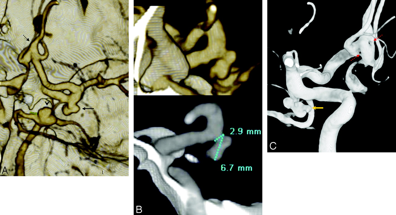
    A 47-year-old woman with SAH and multiple 4- to 10-mm aneurysms; aneurysm measurement technique is demonstrated. MSCTA showed 3 aneurysms: a 7 mm basilar tip aneurysm (arrowhead, A), a right posterior communicating artery (PcomA) segment aneurysm (solid arrow, A), and a fenestrated, fusiform anterior communicating artery (AcomA, dashed arrow, A). Sectioned 3D (B, top) and MPR (B, bottom) images were used to measure the PcomA aneurysm. 3DRA demonstrated the PcomA (gold arrow denotes the site of hemorrhage, C) and the double-fenestration AcomA (dashed red arrows, C).

  • Fig 3.
    • Download figure
    • Open in new tab
    • Download powerpoint
    Fig 3.

    A 78-year-old woman with SAH from an aneurysm more than 10 mm in size. MSCTA showed a 12-mm aneurysm in the periophthalmic ICA segment on 3D-VR images (data not shown), with peripheral calcifications, best seen on MPR (short white arrows, A). The aneurysm was noted to be separate from the ophthalmic artery origin on 3DRA (black arrow, B).

  • Fig 4.
    • Download figure
    • Open in new tab
    • Download powerpoint
    Fig 4.

    The only false-negative CTA, in a 72-year-old man with severe headache, lacking hemorrhage on CT. However, the symptoms prompted an MR imaging/MR angiography (data not shown), with tiny infarcts and a questionable left supraclinoid ICA outpouching. Thereafter, the patient underwent catheter DSA to exclude vasculitis (which was negative), which showed a 2-mm periophthalmic aneurysm on 3DRA (dashed arrow, A). Closer, repeat review of the CTA showed the lesion projecting medially over the bony sella (arrow, B).

  • Fig 5.
    • Download figure
    • Open in new tab
    • Download powerpoint
    Fig 5.

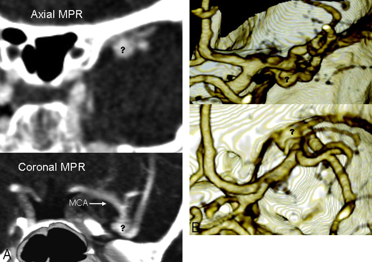
    The only false-positive CTA. A 38-year-old woman with confusion had a head CT negative for hemorrhage but with an isoattenuated structure in the region of the left MCA (data not shown) and corresponding flow void on T2-weighted MR imaging (data not shown), suspicious for aneurysm. Both 16-section (data not shown) and repeat 64-section CTA were performed, which showed a bizarre 6- to 7-mm outpouching (question marks, A and B) overlying the left MCA bifurcation on MPR axial (A, top) and coronal (A, bottom) and 3D MIP/VR posterior (B, top) and superior (B, bottom) views. This was considered a prominent middle cerebral venous plexus, because the catheter DSA and 3DRA (data not shown) were completely negative.

  • Fig 6.
    • Download figure
    • Open in new tab
    • Download powerpoint
    Fig 6.

    Blisterlike lesion in a 46-year-old man with SAH from a 3.5-mm MCA aneurysm, noted on CTA and 3DRA (asterisk, A). The sessile lesion was noted on the undersurface of the ICA and not noted on CTA 3D-VR (data not shown) or MPR (B) views. This was not changed on 2-week repeat catheter 3DRA.

  • Fig 7.
    • Download figure
    • Open in new tab
    • Download powerpoint
    Fig 7.

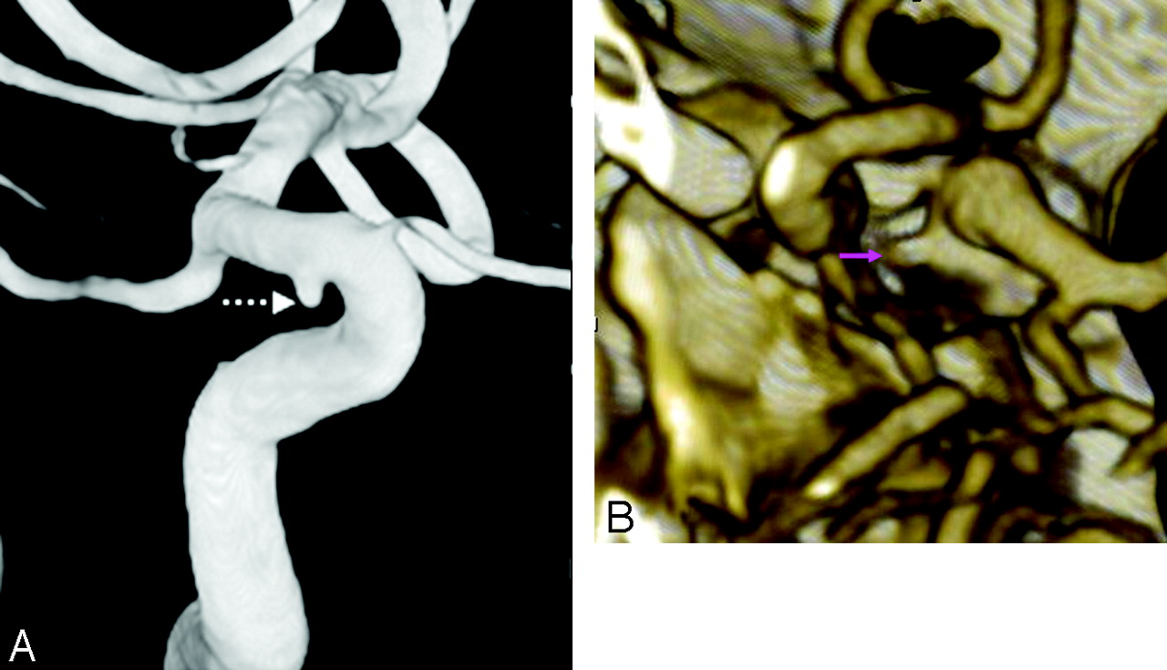
    Atherosclerosis simulating a blister-like lesion. A 52-year-old woman with SAH and focal parenchymal hematoma on CT. CTA showed a 2.9-mm M3 mycotic aneurysm adjacent to the hematoma, also present on 3DRA (dashed arrows, A and B). However, a sessile outpouching was noted on the cavernous ICA undersurface (solid arrows, A and B). Further review of the CTA MPR images revealed this to be an atherosclerotic calcification (pink arrow, C).

Tables

  • Figures
    • View popup
    Table 1:

    Aneurysm detection by MSCTA, confirmation by DSA/3DRA, and locations

    Aneurysm Size, mmCTA: No. of Aneurysms*DSA: No. of CTA True-PositivesDSA: No. of CTA False-NegativesDSA: No. of CTA False-PositivesAneurysm Locations (No.) of Each Size Range as on DSA
    <4151310AcomA (1)
    ACA: A1/A2 Bif (2)
    ICA: cavernous (1)
    ICA: periophthalmic (2)
    ICA: PcomA segment/origin (3)
    MCA: Bif/Trif (2)
    MCA: M2 segment (2)
    4–10201801AcomA (4)
    ICA: PcomA segment/origin (7)
    ICA: terminus (1)
    MCA: Bif/Trif (4)
    Basilar: tip (1)
    Basilar: midportion (1)
    >106600AcomA (2)
    ICA: periophthalmic (1)
    ICA: PcomA segment/origin (1)
    ICA: terminus (1)
    MCA: Bif/Trif (1)
    Total413711AcomA (7)
    ACA: A1/A2 Bif (2)
    ICA: cavernous (1)
    ICA: periophthalmic (3)
    ICA: PcomA segment/origin (11)
    ICA: terminus (2)
    MCA: Bif/Trif (7)
    MCA: M2 segment (2)
    Basilar: (2)
    • Note:—PcomA indicates posterior communicating artery; AcomA, anterior communicating artery; Bif/Trif, bifurcation/trifurcation; ICA, internal carotid artery; MSCTA, multisection CT angiography; DSA, digital subtraction angiography; 3DRA, 3D rotational angiography; CTA, CT angiography; ACA, anterior cerebral artery.

    • * Note: Number of aneurysms on CTA includes 2 patients with aneurysms who did not undergo DSA/3DRA.

    • View popup
    Table 2:

    Accuracy of 64MSCTA on a per-aneurysm/per-patient basis, with κ and ρ per size

    Size on DSA/RASensitivity of MSCTA, %Specificity of MSCTA, %PPV of MSCTA, %NPV of MSCTA, %Accuracy of MSCTA, %κ/ρ (CTA versus DSA) Aneurysm Size
    Overall97.4/96.090.0/90.097.4/96.090.0/90.095.8/94.30.965/0.967
    <4 mm92.3/90.9100.0/100.0100.0/100.090.9/90.995.2/95.60.711/0.753
    4–10 mm100.0/100.090.0/90.994.7/92.3100.0/100.095.8/96.40.841/0.861
    >10 mm100.0/100.0100.0/100.0100.0/100.0100.0/100.0100.0/100.00.476/0.888
    • Note:—MSCTA indicates multisection CT angiography; DSA, digital subtraction angiography; RA, rotational angiography; κ = κ reliability coefficient, ρ = ρ correlation coefficient; CTA, CT angiography. Data show a per-aneurysm/per-patient percentages.

PreviousNext
Back to top

In this issue

American Journal of Neuroradiology: 29 (3)
American Journal of Neuroradiology
Vol. 29, Issue 3
March 2008
  • Table of Contents
  • Index by author
Advertisement
Print
Download PDF
Email Article

Thank you for your interest in spreading the word on American Journal of Neuroradiology.

NOTE: We only request your email address so that the person you are recommending the page to knows that you wanted them to see it, and that it is not junk mail. We do not capture any email address.

Enter multiple addresses on separate lines or separate them with commas.
Detection of Aneurysms by 64-Section Multidetector CT Angiography in Patients Acutely Suspected of Having an Intracranial Aneurysm and Comparison with Digital Subtraction and 3D Rotational Angiography
(Your Name) has sent you a message from American Journal of Neuroradiology
(Your Name) thought you would like to see the American Journal of Neuroradiology web site.
CAPTCHA
This question is for testing whether or not you are a human visitor and to prevent automated spam submissions.
Cite this article
A.M. McKinney, C.S. Palmer, C.L. Truwit, A. Karagulle, M. Teksam
Detection of Aneurysms by 64-Section Multidetector CT Angiography in Patients Acutely Suspected of Having an Intracranial Aneurysm and Comparison with Digital Subtraction and 3D Rotational Angiography
American Journal of Neuroradiology Mar 2008, 29 (3) 594-602; DOI: 10.3174/ajnr.A0848

Citation Manager Formats

  • BibTeX
  • Bookends
  • EasyBib
  • EndNote (tagged)
  • EndNote 8 (xml)
  • Medlars
  • Mendeley
  • Papers
  • RefWorks Tagged
  • Ref Manager
  • RIS
  • Zotero
0 Responses
Respond to this article
Share
Bookmark this article
Detection of Aneurysms by 64-Section Multidetector CT Angiography in Patients Acutely Suspected of Having an Intracranial Aneurysm and Comparison with Digital Subtraction and 3D Rotational Angiography
A.M. McKinney, C.S. Palmer, C.L. Truwit, A. Karagulle, M. Teksam
American Journal of Neuroradiology Mar 2008, 29 (3) 594-602; DOI: 10.3174/ajnr.A0848
del.icio.us logo Twitter logo Facebook logo Mendeley logo
  • Tweet Widget
  • Facebook Like
  • Google Plus One
Purchase

Jump to section

  • Article
    • Abstract
    • Methods
    • Results
    • Discussion
    • Conclusion
    • Footnotes
    • References
  • Figures & Data
  • Info & Metrics
  • Responses
  • References
  • PDF

Related Articles

  • No related articles found.
  • PubMed
  • Google Scholar

Cited By...

  • Ruptured lenticulostriate artery aneurysm: a report of a case treated with endovascular embolisation
  • Comparison of eye-lens doses imparted during interventional and non-interventional neuroimaging techniques for assessment of intracranial aneurysms
  • Sidewall cerebral aneurysms: effect of an outflow angle-assisted approach on diagnosis
  • Aneurysm outflow angle at MRA as discriminant for accurate diagnosis and differentiation between small sidewall cerebral aneurysms and infundibula
  • Predicting intraprocedural rupture and thrombus formation during coiling of ruptured anterior communicating artery aneurysms
  • Geometric Parameter Analysis of Ruptured and Unruptured Aneurysms in Patients with Symmetric Bilateral Intracranial Aneurysms: A Multicenter CT Angiography Study
  • Diagnostic Impact of Bone-Subtraction CT Angiography for Patients with Acute Subarachnoid Hemorrhage
  • Guidelines for the Management of Patients With Unruptured Intracranial Aneurysms: A Guideline for Healthcare Professionals From the American Heart Association/American Stroke Association
  • Use of Follow-Up Imaging in Isolated Perimesencephalic Subarachnoid Hemorrhage: A Meta-Analysis
  • Pearls and Oy-sters: Small but consequential: Intracerebral hemorrhage caused by lenticulostriate artery aneurysm
  • Guidelines for the Management of Aneurysmal Subarachnoid Hemorrhage: A Guideline for Healthcare Professionals From the American Heart Association/American Stroke Association
  • Bilateral mirror posterior inferior cerebellar artery aneurysms: diagnostic caveat on catheter angiography
  • Contrast-free MRA at 3.0 T for the detection of intracranial aneurysms
  • Investigating suspected subarachnoid haemorrhage in adults
  • Patient-Specific Computational Hemodynamics of Intracranial Aneurysms from 3D Rotational Angiography and CT Angiography: An In Vivo Reproducibility Study
  • Utility of CT Angiography in the Identification and Characterization of Supraclinoid Internal Carotid Artery Blister Aneurysms
  • The clinical conundrum of convexal subarachnoid hemorrhage
  • Crossref (189)
  • Google Scholar

This article has been cited by the following articles in journals that are participating in Crossref Cited-by Linking.

  • Guidelines for the Management of Aneurysmal Subarachnoid Hemorrhage
    E. Sander Connolly, Alejandro A. Rabinstein, J. Ricardo Carhuapoma, Colin P. Derdeyn, Jacques Dion, Randall T. Higashida, Brian L. Hoh, Catherine J. Kirkness, Andrew M. Naidech, Christopher S. Ogilvy, Aman B. Patel, B. Gregory Thompson, Paul Vespa
    Stroke 2012 43 6
  • Guidelines for the Management of Patients With Unruptured Intracranial Aneurysms
    B. Gregory Thompson, Robert D. Brown, Sepideh Amin-Hanjani, Joseph P. Broderick, Kevin M. Cockroft, E. Sander Connolly, Gary R. Duckwiler, Catherine C. Harris, Virginia J. Howard, S. Claiborne (Clay) Johnston, Philip M. Meyers, Andrew Molyneux, Christopher S. Ogilvy, Andrew J. Ringer, James Torner
    Stroke 2015 46 8
  • Prevalence of Unruptured Cerebral Aneurysms in Chinese Adults Aged 35 to 75 Years
    Ming-Hua Li, Shi-Wen Chen, Yong-Dong Li, Yuan-Chang Chen, Ying-Sheng Cheng, Ding-Jun Hu, Hua-Qiao Tan, Qian Wu, Wu Wang, Zhen-Kui Sun, Xiao-Er Wei, Jia-Yin Zhang, Rui-Hua Qiao, Wen-Hong Zong, Yin Zhang, Wei Lou, Zhi-Yuan Chen, Yu Zhu, De-Rong Peng, Sui-Xin Ding, Xue-Fan Xu, Xu-Hong Hou, Wei-Ping Jia
    Annals of Internal Medicine 2013 159 8
  • Multidisciplinary management and emerging therapeutic strategies in aneurysmal subarachnoid haemorrhage
    Alejandro A Rabinstein, Giuseppe Lanzino, Eelco FM Wijdicks
    The Lancet Neurology 2010 9 5
  • Diagnosis and Treatment of Unruptured Intracranial Aneurysms and Aneurysmal Subarachnoid Hemorrhage
    Rabih G. Tawk, Tasneem F. Hasan, Caitlin E. D’Souza, Jeffrey B. Peel, William D. Freeman
    Mayo Clinic Proceedings 2021 96 7
  • Diagnosing cerebral aneurysms by computed tomographic angiography: Meta‐analysis
    Jan Menke, Jörg Larsen, Kai Kallenberg
    Annals of Neurology 2011 69 4
  • Dual-Energy CT Angiography in the Evaluation of Intracranial Aneurysms: Image Quality, Radiation Dose, and Comparison With 3D Rotational Digital Subtraction Angiography
    Long-Jiang Zhang, Sheng-Yong Wu, Jing-Bo Niu, Zhuo-Li Zhang, Henry Z. Wang, Yan-E Zhao, Xue Chai, Chang-Sheng Zhou, Guang-Ming Lu
    American Journal of Roentgenology 2010 194 1
  • Aneurysmal Subarachnoid Hemorrhage: Review of the Pathophysiology and Management Strategies
    Marcey L. Osgood
    Current Neurology and Neuroscience Reports 2021 21 9
  • Current Imaging Assessment and Treatment of Intracranial Aneurysms
    Lotfi Hacein-Bey, James M. Provenzale
    American Journal of Roentgenology 2011 196 1
  • ACR Appropriateness Criteria ® Cerebrovascular Disease
    Michael B. Salmela, Shabnam Mortazavi, Bharathi D. Jagadeesan, Daniel F. Broderick, Judah Burns, Tejaswini K. Deshmukh, H. Benjamin Harvey, Jenny Hoang, Christopher H. Hunt, Tabassum A. Kennedy, Alexander A. Khalessi, William Mack, Nandini D. Patel, Joel S. Perlmutter, Bruno Policeni, Jason W. Schroeder, Gavin Setzen, Matthew T. Whitehead, Rebecca S. Cornelius, Amanda S. Corey
    Journal of the American College of Radiology 2017 14 5

More in this TOC Section

  • SAVE vs. Solumbra Techniques for Thrombectomy
  • Delayed Reperfusion Post-Thrombectomy&Thrombolysis
  • Contrast-Induced Encephalopathy after NeuroIR
Show more Interventional

Similar Articles

Advertisement

Indexed Content

  • Current Issue
  • Accepted Manuscripts
  • Article Preview
  • Past Issues
  • Editorials
  • Editors Choice
  • Fellow Journal Club
  • Letters to the Editor

Cases

  • Case Collection
  • Archive - Case of the Week
  • Archive - Case of the Month
  • Archive - Classic Case

Special Collections

  • Special Collections

Resources

  • News and Updates
  • Turn around Times
  • Submit a Manuscript
  • Author Policies
  • Manuscript Submission Guidelines
  • Evidence-Based Medicine Level Guide
  • Publishing Checklists
  • Graphical Abstract Preparation
  • Imaging Protocol Submission
  • Submit a Case
  • Become a Reviewer/Academy of Reviewers
  • Get Peer Review Credit from Publons

Multimedia

  • AJNR Podcast
  • AJNR SCANtastic
  • Video Articles

About Us

  • About AJNR
  • Editorial Board
  • Not an AJNR Subscriber? Join Now
  • Alerts
  • Feedback
  • Advertise with us
  • Librarian Resources
  • Permissions
  • Terms and Conditions

American Society of Neuroradiology

  • Not an ASNR Member? Join Now

© 2025 by the American Society of Neuroradiology All rights, including for text and data mining, AI training, and similar technologies, are reserved.
Print ISSN: 0195-6108 Online ISSN: 1936-959X

Powered by HighWire