Advertisement

Main menu

  • Home
  • Content
    • Current Issue
    • Accepted Manuscripts
    • Article Preview
    • Past Issue Archive
    • AJNR Case Collection
    • Case of the Week Archive
    • Classic Case Archive
    • Case of the Month Archive
  • Special Collections
    • Spinal CSF Leak Articles (Jan 2020-June 2024)
    • 2024 AJNR Journal Awards
    • Most Impactful AJNR Articles
  • Multimedia
    • AJNR Podcast
    • AJNR Scantastics
    • Video Articles
  • For Authors
    • Submit a Manuscript
    • Author Policies
    • Fast publishing of Accepted Manuscripts
    • Graphical Abstract Preparation
    • Manuscript Submission Guidelines
    • Imaging Protocol Submission
    • Submit a Case for the Case Collection
  • About Us
    • About AJNR
    • Editorial Board
  • More
    • Become a Reviewer/Academy of Reviewers
    • Subscribers
    • Permissions
    • Alerts
    • Feedback
    • Advertisers
    • ASNR Home
  • Other Publications
    • ajnr

User menu

  • Alerts
  • Log in

Search

  • Advanced search
American Journal of Neuroradiology

American Journal of Neuroradiology

ASHNR American Society of Functional Neuroradiology ASHNR American Society of Pediatric Neuroradiology ASSR
  • Alerts
  • Log in

Advanced Search

  • Home
  • Content
    • Current Issue
    • Accepted Manuscripts
    • Article Preview
    • Past Issue Archive
    • AJNR Case Collection
    • Case of the Week Archive
    • Classic Case Archive
    • Case of the Month Archive
  • Special Collections
    • Spinal CSF Leak Articles (Jan 2020-June 2024)
    • 2024 AJNR Journal Awards
    • Most Impactful AJNR Articles
  • Multimedia
    • AJNR Podcast
    • AJNR Scantastics
    • Video Articles
  • For Authors
    • Submit a Manuscript
    • Author Policies
    • Fast publishing of Accepted Manuscripts
    • Graphical Abstract Preparation
    • Manuscript Submission Guidelines
    • Imaging Protocol Submission
    • Submit a Case for the Case Collection
  • About Us
    • About AJNR
    • Editorial Board
  • More
    • Become a Reviewer/Academy of Reviewers
    • Subscribers
    • Permissions
    • Alerts
    • Feedback
    • Advertisers
    • ASNR Home
  • Follow AJNR on Twitter
  • Visit AJNR on Facebook
  • Follow AJNR on Instagram
  • Join AJNR on LinkedIn
  • RSS Feeds

Welcome to the new AJNR, Updated Hall of Fame, and more. Read the full announcements.


AJNR is seeking candidates for the position of Associate Section Editor, AJNR Case Collection. Read the full announcement.


LetterLetter

MR Imaging in Biphasic Japanese Encephalitis

S.K. Handique and N. Barkataky
American Journal of Neuroradiology March 2008, 29 (3) e3; DOI: https://doi.org/10.3174/ajnr.A0862
S.K. Handique
  • Find this author on Google Scholar
  • Find this author on PubMed
  • Search for this author on this site
N. Barkataky
  • Find this author on Google Scholar
  • Find this author on PubMed
  • Search for this author on this site
  • Article
  • Figures & Data
  • Info & Metrics
  • Responses
  • References
  • PDF
Loading

Japanese encephalitis (JE) is usually a monophasic disease characterized by fever, altered sensorium with or without seizures, and other focal neurologic symptoms. If the patient survives, there is gradual recovery with or without persistent signs of central nervous system (CNS) injury. MR imaging helps in establishing an early diagnosis and shows lesions in the thalami, substantia nigra, basal ganglia, hippocampi, cerebral cortex, brain stem, and cerebellum. A biphasic form of JE has been reported, with early relapse of symptoms within days or a few weeks after recovery from the first phase. To our knowledge, this is the only report in literature of biphasic JE.1 We report the MR imaging findings of a patient with JE and biphasic illness.

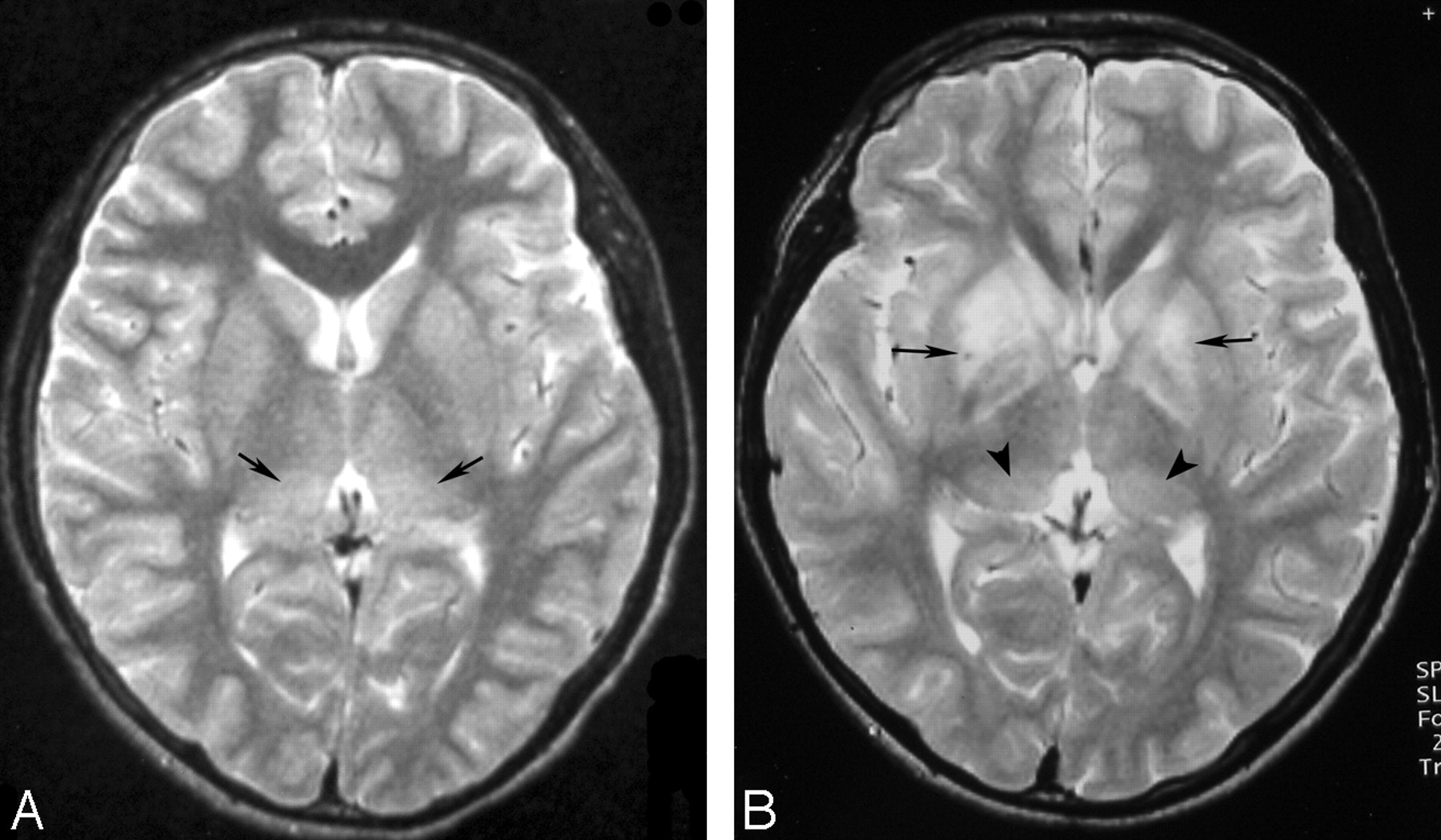
A 16-year-old boy presented to our institute with a history of fever, headache, body ache, and occasional vomiting for 14 days and altered sensorium for a day before admission. On examination, he had meningeal irritation, spasticity, mutism, tremors, and perioral dyskinesia. Findings of routine blood examination were normal. CSF examination results showed lymphocytic pleocytosis and raised protein. CSF immunoglobulin capture enzyme-linked immunosorbent assay (MAC-ELISA) result was 250 U, establishing a diagnosis of JE. MR imaging showed T2-hyperintense and mildly T1-hypointense lesions in both thalami (Fig 1A) and substantia nigra. On treatment, there was gradual recovery, and the patient was discharged a week later.

Twenty-five days later he was readmitted with a history of low-grade fever, headache, and vomiting for 3 days and altered sensorium for 1 day. He had signs of meningeal irritation, limb spasticity, hypokinesia, tremors, perioral dyskinesia, and behavioral changes in the form of a violent agitational state. CSF examination was repeated on days 47 and 62 after the onset of the first phase. There was mild increase in cells, mainly lymphocytes and protein on day 47, with normal cells and raised protein on day 62. CSF MAC-ELISA results were 247 and 261 U, respectively. On MR imaging, fresh lesions resembling the acute lesions seen earlier were now noted in both basal ganglia. The earlier noted thalamic and substantia nigra lesions had partially resolved (Fig 1B). He recovered gradually with treatment and had completely recovered without any deficit on follow-up in 3 months.

Biphasic JE is characterized by early relapse of meningoencephalitic symptoms, which may be somewhat different from those of the first phase but still conform to known features of JE. Patients usually recover well. MR imaging shows fresh JE lesions in the second phase, in addition to JE lesions in the first.1 Our patient showed similar clinical and imaging features. Lesions in the substantia nigra and thalami were seen on MR imaging in the first phase, followed by fresh lesions in the basal ganglia in the second phase, all known sites of involvement of JE. Behavioral changes and extrapyramidal features were more prominent in the second phase in comparison with the first. Our patient also showed good recovery. Biphasic JE is not common in our experience, and this is the only case we saw among 87 consecutive confirmed cases of JE in the last 7 years.

Persistence of the JE virus (JEV) in the CNS is suggested by the raised JEV-specific immunoglobulin M (IgM) levels in the CSF in our patient. Detection of JEV-specific IgM in the CSF long after the acute phase of the illness has been considered as immunologic evidence of virus persistence in the CNS.2 Whether persistence of JEV may be the reason for the biphasic illness in our patient is obscure. Alteration in the host immune mechanisms and a unique strain of JEV have been implicated. The former is a more likely mechanism because reactivation of latent JE infection has been experimentally demonstrated in immunologic-poor states.3

In conclusion, JE may rarely present as a biphasic illness. MR imaging shows typical JE lesions in the first phase, with fresh areas of involvement in the second phase.

  Fig 1.
  • Download figure
  • Open in new tab
  • Download powerpoint
Fig 1.

A, Axial T2-weighted MR image obtained in the first phase (day 17 of onset of JE) of the disease shows bilateral thalamic lesions (arrows). Substantia nigra lesions were also seen at this stage (not shown). B, Axial T2-weighted MR image obtained in the second phase (45 days after onset of first phase) shows fresh lesions in the basal ganglia (arrows). Residual lesions (arrowheads) are noted in the thalami. These have partially resolved. Residual lesions were also seen in the substantia nigra at this stage (not shown).

References

  1. ↵
    Pradhan S, Gupta RK, Singh MB, et al. Biphasic illness pattern due to early relapse in Japanese-B virus encephalitis. J Neurol Sci 2001;183:13–18
    CrossRefPubMed
  2. ↵
    Ravi V, Desai AS, Shenoy PK, et al. Persistence of Japanese encephalitis virus in the human nervous system. J Med Virol 1993;40:326–29
    PubMed
  3. ↵
    Mathur A, Arora KL, Rawat S, et al. Persistence, latency and reactivation of Japanese encephalitis virus infection in mice. J Gen Virol 1986;67 (Pt 2):381–85
    Abstract/FREE Full Text
  • Copyright © American Society of Neuroradiology
PreviousNext
Back to top

In this issue

American Journal of Neuroradiology: 29 (3)
American Journal of Neuroradiology
Vol. 29, Issue 3
March 2008
  • Table of Contents
  • Index by author
Advertisement
Print
Download PDF
Email Article

Thank you for your interest in spreading the word on American Journal of Neuroradiology.

NOTE: We only request your email address so that the person you are recommending the page to knows that you wanted them to see it, and that it is not junk mail. We do not capture any email address.

Enter multiple addresses on separate lines or separate them with commas.
MR Imaging in Biphasic Japanese Encephalitis
(Your Name) has sent you a message from American Journal of Neuroradiology
(Your Name) thought you would like to see the American Journal of Neuroradiology web site.
CAPTCHA
This question is for testing whether or not you are a human visitor and to prevent automated spam submissions.
Cite this article
S.K. Handique, N. Barkataky
MR Imaging in Biphasic Japanese Encephalitis
American Journal of Neuroradiology Mar 2008, 29 (3) e3; DOI: 10.3174/ajnr.A0862

Citation Manager Formats

  • BibTeX
  • Bookends
  • EasyBib
  • EndNote (tagged)
  • EndNote 8 (xml)
  • Medlars
  • Mendeley
  • Papers
  • RefWorks Tagged
  • Ref Manager
  • RIS
  • Zotero
0 Responses
Respond to this article
Share
Bookmark this article
MR Imaging in Biphasic Japanese Encephalitis
S.K. Handique, N. Barkataky
American Journal of Neuroradiology Mar 2008, 29 (3) e3; DOI: 10.3174/ajnr.A0862
del.icio.us logo Twitter logo Facebook logo Mendeley logo
  • Tweet Widget
  • Facebook Like
  • Google Plus One
Purchase

Jump to section

  • Article
    • References
  • Figures & Data
  • Info & Metrics
  • Responses
  • References
  • PDF

Related Articles

  • No related articles found.
  • PubMed
  • Google Scholar

Cited By...

  • No citing articles found.
  • Crossref (7)
  • Google Scholar

This article has been cited by the following articles in journals that are participating in Crossref Cited-by Linking.

  • Management of Viral Central Nervous System Infections: A Primer for Clinicians
    P Brandon Bookstaver, Phillip L Mohorn, Ansal Shah, Lauren D Tesh, April M Quidley, Ravish Kothari, Christopher M Bland, Sharon Weissman
    Journal of Central Nervous System Disease 2017 9
  • Viral Infections of the Central Nervous System
    Sanjeev Kumar Handique
    Neuroimaging Clinics of North America 2011 21 4
  • Clinical features of adult anti-N-methyl-d-aspartate receptor encephalitis after Japanese encephalitis
    Chun-juan Wang, Zi-ling Zeng, Fu-sheng Zhang, Shou-gang Guo
    Journal of the Neurological Sciences 2020 417
  • Detection of Japanese Encephalitis by Metagenomic Next-Generation Sequencing of Cerebrospinal Fluid: A Case Report and Literature Review
    Xin Li, Jing Li, Guode Wu, Manxia Wang, Zhang Jing
    Frontiers in Cellular Neuroscience 2022 16
  • CNS Infections
    Sanjeev K. Handique, Mausumi Barthakur
    2014
  • Imaging of CNS Infections and Neuroimmunology
    Vivek Yedavalli, Vivek Pandey, Delilah Burrowes
    2019
  • Radiopaedia.org
    Jignesh Modi, Sonam Vadera, Rohit Sharma
    2015

More in this TOC Section

  • Brain AVM’s Nidus: What if We Hadn’t Understood Anything?
  • Letter to the Editor regarding “Automated Volumetric Software in Dementia: Help or Hindrance to the Neuroradiologist?”
  • Reply:
Show more Letters

Similar Articles

Advertisement

Indexed Content

  • Current Issue
  • Accepted Manuscripts
  • Article Preview
  • Past Issues
  • Editorials
  • Editors Choice
  • Fellow Journal Club
  • Letters to the Editor

Cases

  • Case Collection
  • Archive - Case of the Week
  • Archive - Case of the Month
  • Archive - Classic Case

Special Collections

  • Special Collections

Resources

  • News and Updates
  • Turn around Times
  • Submit a Manuscript
  • Author Policies
  • Manuscript Submission Guidelines
  • Evidence-Based Medicine Level Guide
  • Publishing Checklists
  • Graphical Abstract Preparation
  • Imaging Protocol Submission
  • Submit a Case
  • Become a Reviewer/Academy of Reviewers
  • Get Peer Review Credit from Publons

Multimedia

  • AJNR Podcast
  • AJNR SCANtastic
  • Video Articles

About Us

  • About AJNR
  • Editorial Board
  • Not an AJNR Subscriber? Join Now
  • Alerts
  • Feedback
  • Advertise with us
  • Librarian Resources
  • Permissions
  • Terms and Conditions

American Society of Neuroradiology

  • Not an ASNR Member? Join Now

© 2025 by the American Society of Neuroradiology All rights, including for text and data mining, AI training, and similar technologies, are reserved.
Print ISSN: 0195-6108 Online ISSN: 1936-959X

Powered by HighWire