Advertisement

Main menu

  • Home
  • Content
    • Current Issue
    • Accepted Manuscripts
    • Article Preview
    • Past Issue Archive
    • Video Articles
    • AJNR Case Collection
    • Case of the Week Archive
    • Case of the Month Archive
    • Classic Case Archive
  • Special Collections
    • AJNR Awards
    • Low-Field MRI
    • Alzheimer Disease
    • ASNR Foundation Special Collection
    • Photon-Counting CT
    • View All
  • Multimedia
    • AJNR Podcasts
    • AJNR SCANtastic
    • Trainee Corner
    • MRI Safety Corner
    • Imaging Protocols
  • For Authors
    • Submit a Manuscript
    • Submit a Video Article
    • Submit an eLetter to the Editor/Response
    • Manuscript Submission Guidelines
    • Statistical Tips
    • Fast Publishing of Accepted Manuscripts
    • Graphical Abstract Preparation
    • Imaging Protocol Submission
    • Author Policies
  • About Us
    • About AJNR
    • Editorial Board
    • Editorial Board Alumni
  • More
    • Become a Reviewer/Academy of Reviewers
    • Subscribers
    • Permissions
    • Alerts
    • Feedback
    • Advertisers
    • ASNR Home

User menu

  • Alerts
  • Log in

Search

  • Advanced search
American Journal of Neuroradiology

American Journal of Neuroradiology

ASHNR American Society of Functional Neuroradiology ASHNR American Society of Pediatric Neuroradiology ASSR
  • Alerts
  • Log in

Advanced Search

  • Home
  • Content
    • Current Issue
    • Accepted Manuscripts
    • Article Preview
    • Past Issue Archive
    • Video Articles
    • AJNR Case Collection
    • Case of the Week Archive
    • Case of the Month Archive
    • Classic Case Archive
  • Special Collections
    • AJNR Awards
    • Low-Field MRI
    • Alzheimer Disease
    • ASNR Foundation Special Collection
    • Photon-Counting CT
    • View All
  • Multimedia
    • AJNR Podcasts
    • AJNR SCANtastic
    • Trainee Corner
    • MRI Safety Corner
    • Imaging Protocols
  • For Authors
    • Submit a Manuscript
    • Submit a Video Article
    • Submit an eLetter to the Editor/Response
    • Manuscript Submission Guidelines
    • Statistical Tips
    • Fast Publishing of Accepted Manuscripts
    • Graphical Abstract Preparation
    • Imaging Protocol Submission
    • Author Policies
  • About Us
    • About AJNR
    • Editorial Board
    • Editorial Board Alumni
  • More
    • Become a Reviewer/Academy of Reviewers
    • Subscribers
    • Permissions
    • Alerts
    • Feedback
    • Advertisers
    • ASNR Home
  • Follow AJNR on Twitter
  • Visit AJNR on Facebook
  • Follow AJNR on Instagram
  • Join AJNR on LinkedIn
  • RSS Feeds

AJNR Awards, New Junior Editors, and more. Read the latest AJNR updates

Research ArticleBrain

Arteriovenous Shunt Visualization in Arteriovenous Malformations with Arterial Spin-Labeling MR Imaging

R.L. Wolf, J. Wang, J.A. Detre, E.L. Zager and R.W. Hurst
American Journal of Neuroradiology April 2008, 29 (4) 681-687; DOI: https://doi.org/10.3174/ajnr.A0901
R.L. Wolf
  • Find this author on Google Scholar
  • Find this author on PubMed
  • Search for this author on this site
J. Wang
  • Find this author on Google Scholar
  • Find this author on PubMed
  • Search for this author on this site
J.A. Detre
  • Find this author on Google Scholar
  • Find this author on PubMed
  • Search for this author on this site
E.L. Zager
  • Find this author on Google Scholar
  • Find this author on PubMed
  • Search for this author on this site
R.W. Hurst
  • Find this author on Google Scholar
  • Find this author on PubMed
  • Search for this author on this site
  • Article
  • Figures & Data
  • Info & Metrics
  • Responses
  • References
  • PDF
Loading

Article Figures & Data

Figures

  • Tables
  • Fig 1.
    • Download figure
    • Open in new tab
    • Download powerpoint
    Fig 1.

    Image evaluation and shunt approximation for a right hemisphere AVM. A, CASL perfusion ΔM maps show bright signal intensity in the sagittal sinus and right transverse sinus (arrowheads) and in draining veins (arrows) adjacent to the nidus (open arrows). B, A small region of interest (red circle) placed in the left basal ganglia is used for each patient to measure mean ΔM (BGmean) and SD (BGSD). A threshold is generated (empirically chosen as threshold = BGmean + 8 *BGSD) above which voxels are labeled as shunt voxels to create a mask (blue voxels) for the AVM and draining vessels. The AV shunt fraction is estimated by multiplying the mean ΔM in the mask by the number of voxels in the mask, then dividing by the mean ΔM in the entire brain multiplied by all labeled voxels. C, Registered 2D TOF MRA source images (no superior saturation band) are reviewed to verify reasonable selection of AVM and draining vessels (open and closed arrows).

  • Fig 2.
    • Download figure
    • Open in new tab
    • Download powerpoint
    Fig 2.

    AVM before (top) and 2 days after (bottom) partial embolization (patient 1). CASL ΔM images (A, D) show decrease in AV shunt (arrows, D), estimated decrease from 23% to 20%. Superficial venous drainage is identified, including the superior sagittal sinus and sphenobasal sinus on both studies (open arrowheads, A). Of interest, despite intense signal intensity in the sphenobasal sinus, this corresponds to a relatively small venous structure on DSA (black arrowhead, C). DSA lateral projections (C, F) and 3D TOF MRA collapsed maximum intensity projection images (B, E) confirm decreased flow in a portion of AVM, especially the posterior-superior portion (white arrowhead, E).

  • Fig 3.
    • Download figure
    • Open in new tab
    • Download powerpoint
    Fig 3.

    Susceptibility artifact resulting from the presence of embolic material, thrombus, and/or hemorrhage (patient 1). CASL source images from 4 imaging locations are shown.
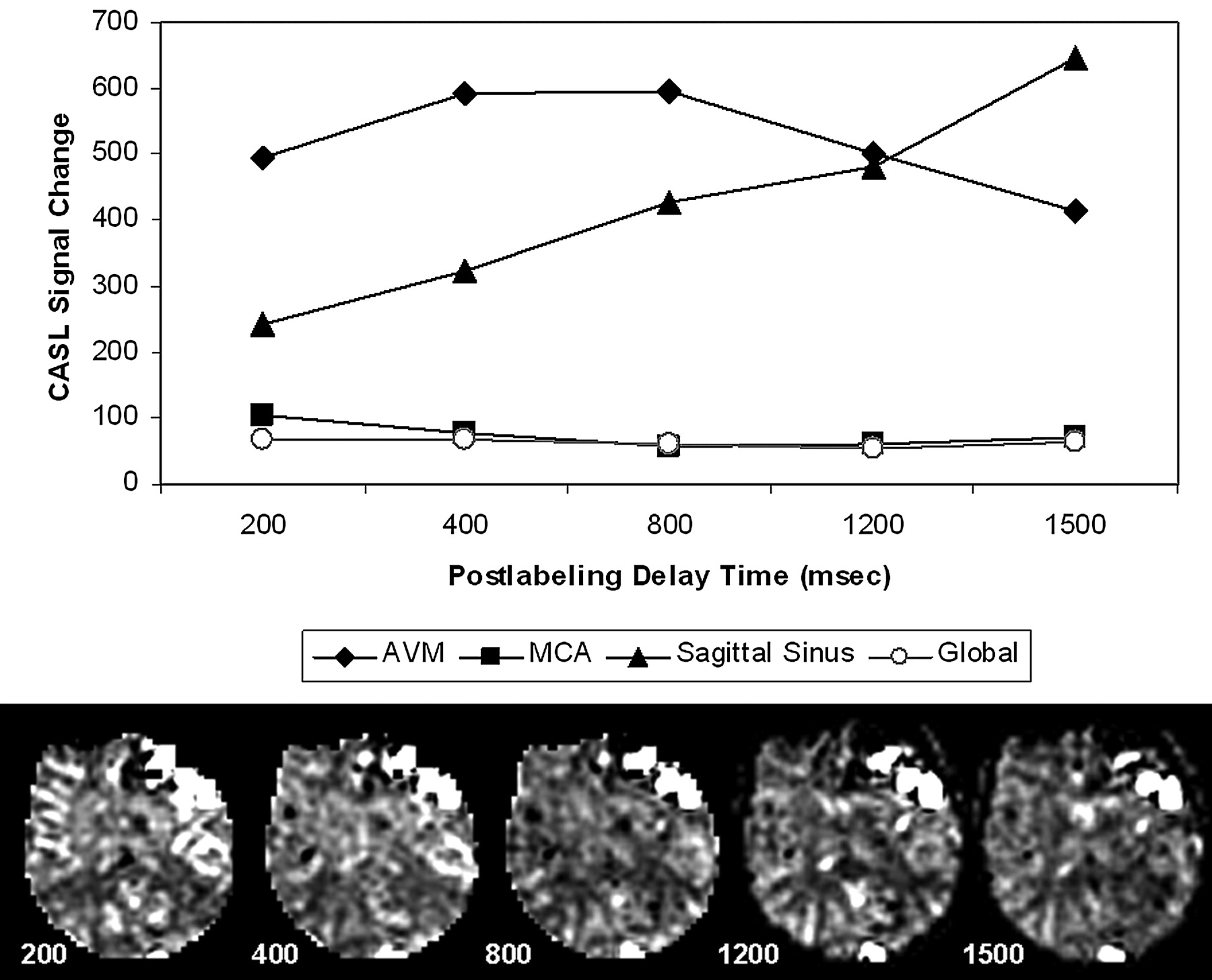
  • Fig 4.
    • Download figure
    • Open in new tab
    • Download powerpoint
    Fig 4.

    Multiple postlabeling delay times (200–1500 ms): effect on ASL signal intensity in the AVM (patient 1). ΔM (T1-corrected) measured in regions of interest for the AVM (diamond), sagittal sinus (triangle), right MCA territory (box), and entire brain (circle) at each delay is plotted (above), with ΔM maps from 1 imaging location at multiple delays for comparison (below). ASL signal intensity is relatively stable in global measures over the entire brain and in the MCA at longer delays as expected. Signal intensity decreases in AVM and increases in sagittal sinus as delay increases.

  • Fig 5.
    • Download figure
    • Open in new tab
    • Download powerpoint
    Fig 5.

    Regional changes in CBF compared with extent of AV shunt. Δ = contralateral − ipsilateral CBF in measured regions of interest in white matter near the AVM, the cortex near the AVM, the thalamus, and the basal ganglia. A, Cortical gray matter CBF Δ values are negatively but weakly correlated with percent AV shunt and are not significant. (B) Δ values in white matter (R2 = 0.70, F = 11.58, P = .02) are negatively correlated (blood flow in adjacent ipsilateral white matter increases as percent shunt increased). Basal ganglia Δ values (C) show little change in CBF with the size of the AV shunt, but thalamic CBF (D) appears to decrease ipsilaterally as percent AV shunt increases (R2 = 0.74, F = 14.19, P = .01).

Tables

  • Figures
    • View popup
    Table 1:

    Clinical data and AVM features

    PatientAge (yr)PresentationLocationEloquent Tissue at RiskGrade (size/eloquent/drainage)*Arterial SupplyVAT AVM/normal (s)
    165HeadacheLeft frontalBroca area2/1/0L M2–3, L ACA0.67/3.87
    244Bleed + seizuresRight frontal-parietalPrimary sensorimotor cortex2/1/0R M3–40.54/3.74
    323Hand/Face numbnessLeft frontal-parietalPrimary sensorimotor cortex1/1/0L M40.27/3.87
    433Bleed + headache, expressive dysphasiaLeft frontalBroca area1/1/0L M4NA
    549HeadacheLeft occipital-temporalVisual cortex, optic radiations1/1/0L M4NA
    623Left face/tongue spasmsRight frontal-parietalPrimary sensorimotor cortex2/1/0R M3–40.4/3.2
    747Headache, blurry visionLeft frontalBroca area2/1/1L M1–4, L ACA0.53/6.13
    • Note:—VAT indicates venous appearance time with contrast appearance in supraclinoid artery as reference point (t = 0 seconds) for both AVM and normal venous structures; NA, not available (DSA performed elsewhere); R, right; L, left; M1–4, middle cerebral artery branches; ACA, anterior cerebral artery.

    • * Spetzler-Martin grade by category (size, involvement of eloquent brain, and drainage pattern) with eloquent regions defined as primary sensorimotor cortex, language and visual cortex, hypothalamus and thalamus, internal capsule, brain stem, and deep cerebellar nuclei.1

    • View popup
    Table 2:

    AVM data and CBF comparisons ipsilateral and contralateral to AVM

    PatientGrade*Shunt (%)CBF (mL/100 g/min)
    CBFmeanBGipsBGcontThalipsThalcontGMipsGMcontWMipsWMcont
    1323.4250.749.637.851.253.961.758.929.823.8
    2319.8054.744.34766.876.685.953.325.415.5
    324.9052.339.049.045.842.967.378.613.616.1
    420.6461.367.863.666.648.584.588.223.834.4
    523.1339.035.036.748.445.149.737.117.422.2
    6310.7239.739.341.436.040.148.062.821.625.0
    7429.7448.234.644.742.658.662.351.239.735.8
    Mean49.444.345.751.152.265.661.524.524.7
    • Note:—CBFmean indicates mean brain cerebral blood flow excluding AVM and associated structures; GM (WM), gray matter (white matter) blood flow averaged from regions of interest anterior and posterior to the AVM; Thal, thalamus; ips, ipsilateral; cont, contralateral.

    • * Grade indicates total Spetzler-Martin grade (sum of scores for size, involvement of eloquent brain, and drainage pattern as listed in Table 1).

PreviousNext
Back to top

In this issue

American Journal of Neuroradiology: 29 (4)
American Journal of Neuroradiology
Vol. 29, Issue 4
April 2008
  • Table of Contents
  • Index by author
Advertisement
Print
Download PDF
Email Article

Thank you for your interest in spreading the word on American Journal of Neuroradiology.

NOTE: We only request your email address so that the person you are recommending the page to knows that you wanted them to see it, and that it is not junk mail. We do not capture any email address.

Enter multiple addresses on separate lines or separate them with commas.
Arteriovenous Shunt Visualization in Arteriovenous Malformations with Arterial Spin-Labeling MR Imaging
(Your Name) has sent you a message from American Journal of Neuroradiology
(Your Name) thought you would like to see the American Journal of Neuroradiology web site.
CAPTCHA
This question is for testing whether or not you are a human visitor and to prevent automated spam submissions.
Cite this article
R.L. Wolf, J. Wang, J.A. Detre, E.L. Zager, R.W. Hurst
Arteriovenous Shunt Visualization in Arteriovenous Malformations with Arterial Spin-Labeling MR Imaging
American Journal of Neuroradiology Apr 2008, 29 (4) 681-687; DOI: 10.3174/ajnr.A0901

Citation Manager Formats

  • BibTeX
  • Bookends
  • EasyBib
  • EndNote (tagged)
  • EndNote 8 (xml)
  • Medlars
  • Mendeley
  • Papers
  • RefWorks Tagged
  • Ref Manager
  • RIS
  • Zotero
0 Responses
Respond to this article
Share
Bookmark this article
Arteriovenous Shunt Visualization in Arteriovenous Malformations with Arterial Spin-Labeling MR Imaging
R.L. Wolf, J. Wang, J.A. Detre, E.L. Zager, R.W. Hurst
American Journal of Neuroradiology Apr 2008, 29 (4) 681-687; DOI: 10.3174/ajnr.A0901
del.icio.us logo Twitter logo Facebook logo Mendeley logo
  • Tweet Widget
  • Facebook Like
  • Google Plus One
Purchase

Jump to section

  • Article
    • Abstract
    • Methods
    • Results
    • Discussion
    • Conclusion
    • Footnotes
    • References
  • Figures & Data
  • Info & Metrics
  • Responses
  • References
  • PDF

Related Articles

  • No related articles found.
  • PubMed
  • Google Scholar

Cited By...

  • Noninvasive Follow-up Imaging of Ruptured Pediatric Brain AVMs Using Arterial Spin-Labeling
  • Follow-Up MRI for Small Brain AVMs Treated by Radiosurgery: Is Gadolinium Really Necessary?
  • Arterial Spin-Labeling Improves Detection of Intracranial Dural Arteriovenous Fistulas with MRI
  • Feasibility of Flat Panel Detector CT in Perfusion Assessment of Brain Arteriovenous Malformations: Initial Clinical Experience
  • Intracranial Arteriovenous Shunting: Detection with Arterial Spin-Labeling and Susceptibility-Weighted Imaging Combined
  • Evaluation of 4D Vascular Flow and Tissue Perfusion in Cerebral Arteriovenous Malformations: Influence of Spetzler-Martin Grade, Clinical Presentation, and AVM Risk Factors
  • Accuracy of Vessel-Encoded Pseudocontinuous Arterial Spin-Labeling in Identification of Feeding Arteries in Patients with Intracranial Arteriovenous Malformations
  • Identification of Venous Signal on Arterial Spin Labeling Improves Diagnosis of Dural Arteriovenous Fistulas and Small Arteriovenous Malformations
  • Crossref
  • Google Scholar

This article has not yet been cited by articles in journals that are participating in Crossref Cited-by Linking.

More in this TOC Section

  • Multimodal CT Provides Improved Performance for Lacunar Infarct Detection
  • Optimal MRI Sequence for Identifying Occlusion Location in Acute Stroke: Which Value of Time-Resolved Contrast-Enhanced MRA?
  • Evaluating the Effects of White Matter Multiple Sclerosis Lesions on the Volume Estimation of 6 Brain Tissue Segmentation Methods
Show more Brain

Similar Articles

Advertisement

Indexed Content

  • Current Issue
  • Accepted Manuscripts
  • Article Preview
  • Past Issues
  • Editorials
  • Editor's Choice
  • Fellows' Journal Club
  • Letters to the Editor
  • Video Articles

Cases

  • Case Collection
  • Archive - Case of the Week
  • Archive - Case of the Month
  • Archive - Classic Case

More from AJNR

  • Trainee Corner
  • Imaging Protocols
  • MRI Safety Corner
  • Book Reviews

Multimedia

  • AJNR Podcasts
  • AJNR Scantastics

Resources

  • Turnaround Time
  • Submit a Manuscript
  • Submit a Video Article
  • Submit an eLetter to the Editor/Response
  • Manuscript Submission Guidelines
  • Statistical Tips
  • Fast Publishing of Accepted Manuscripts
  • Graphical Abstract Preparation
  • Imaging Protocol Submission
  • Evidence-Based Medicine Level Guide
  • Publishing Checklists
  • Author Policies
  • Become a Reviewer/Academy of Reviewers
  • News and Updates

About Us

  • About AJNR
  • Editorial Board
  • Editorial Board Alumni
  • Alerts
  • Permissions
  • Not an AJNR Subscriber? Join Now
  • Advertise with Us
  • Librarian Resources
  • Feedback
  • Terms and Conditions
  • AJNR Editorial Board Alumni

American Society of Neuroradiology

  • Not an ASNR Member? Join Now

© 2025 by the American Society of Neuroradiology All rights, including for text and data mining, AI training, and similar technologies, are reserved.
Print ISSN: 0195-6108 Online ISSN: 1936-959X

Powered by HighWire