Advertisement

Main menu

  • Home
  • Content
    • Current Issue
    • Accepted Manuscripts
    • Article Preview
    • Past Issue Archive
    • AJNR Case Collection
    • Case of the Week Archive
    • Classic Case Archive
    • Case of the Month Archive
  • Special Collections
    • Spinal CSF Leak Articles (Jan 2020-June 2024)
    • 2024 AJNR Journal Awards
    • Most Impactful AJNR Articles
  • Multimedia
    • AJNR Podcast
    • AJNR Scantastics
    • Video Articles
  • For Authors
    • Submit a Manuscript
    • Author Policies
    • Fast publishing of Accepted Manuscripts
    • Graphical Abstract Preparation
    • Manuscript Submission Guidelines
    • Imaging Protocol Submission
    • Submit a Case for the Case Collection
  • About Us
    • About AJNR
    • Editorial Board
  • More
    • Become a Reviewer/Academy of Reviewers
    • Subscribers
    • Permissions
    • Alerts
    • Feedback
    • Advertisers
    • ASNR Home
  • Other Publications
    • ajnr

User menu

  • Alerts
  • Log in

Search

  • Advanced search
American Journal of Neuroradiology

American Journal of Neuroradiology

ASHNR American Society of Functional Neuroradiology ASHNR American Society of Pediatric Neuroradiology ASSR
  • Alerts
  • Log in

Advanced Search

  • Home
  • Content
    • Current Issue
    • Accepted Manuscripts
    • Article Preview
    • Past Issue Archive
    • AJNR Case Collection
    • Case of the Week Archive
    • Classic Case Archive
    • Case of the Month Archive
  • Special Collections
    • Spinal CSF Leak Articles (Jan 2020-June 2024)
    • 2024 AJNR Journal Awards
    • Most Impactful AJNR Articles
  • Multimedia
    • AJNR Podcast
    • AJNR Scantastics
    • Video Articles
  • For Authors
    • Submit a Manuscript
    • Author Policies
    • Fast publishing of Accepted Manuscripts
    • Graphical Abstract Preparation
    • Manuscript Submission Guidelines
    • Imaging Protocol Submission
    • Submit a Case for the Case Collection
  • About Us
    • About AJNR
    • Editorial Board
  • More
    • Become a Reviewer/Academy of Reviewers
    • Subscribers
    • Permissions
    • Alerts
    • Feedback
    • Advertisers
    • ASNR Home
  • Follow AJNR on Twitter
  • Visit AJNR on Facebook
  • Follow AJNR on Instagram
  • Join AJNR on LinkedIn
  • RSS Feeds

Welcome to the new AJNR, Updated Hall of Fame, and more. Read the full announcements.


AJNR is seeking candidates for the position of Associate Section Editor, AJNR Case Collection. Read the full announcement.


OtherFUNCTIONAL

Diffusion Tensor Imaging and Fiber Tractography in Parry-Romberg Syndrome

W.J. Moon, H.J. Kim, H.G. Roh, J. Oh and S.H. Han
American Journal of Neuroradiology April 2008, 29 (4) 714-715; DOI: https://doi.org/10.3174/ajnr.A0967
W.J. Moon
  • Find this author on Google Scholar
  • Find this author on PubMed
  • Search for this author on this site
H.J. Kim
  • Find this author on Google Scholar
  • Find this author on PubMed
  • Search for this author on this site
H.G. Roh
  • Find this author on Google Scholar
  • Find this author on PubMed
  • Search for this author on this site
J. Oh
  • Find this author on Google Scholar
  • Find this author on PubMed
  • Search for this author on this site
S.H. Han
  • Find this author on Google Scholar
  • Find this author on PubMed
  • Search for this author on this site
  • Article
  • Figures & Data
  • Info & Metrics
  • Responses
  • References
  • PDF
Loading

Abstract

SUMMARY: We report a case of Parry-Romberg syndrome in a 32-year-old woman presenting with intermittent headache and mild sensory disturbance. MR imaging revealed minimal asymmetric atrophy of the right hemisphere with a few nonspecific white matter hyperintensities. Diffusion tensor imaging and fiber tractography, however, demonstrated clear fiber derangement, especially in the sensory tract of the right cerebral white matter.

Parry-Romberg syndrome (PRS) is an acquired progressive facial hemiatrophy of the skin and subcutaneous tissue, and sometimes underlying bony structure, with an occasional involvement of the nervous system1–5 The atrophic process commonly appears during the first and second decades of life and affects mainly the face below the forehead.6 The most common neurologic manifestation is epilepsy; however, there are many other neurologic features, such as migraine, facial pain, and structural brain lesions.1–8

We report a case of a patient with PRS presenting with sensory disturbance and subtle white matter changes. We confirmed white matter involvement of PRS by the use of diffusion tensor imaging (DTI) and fiber tractography with 3T MR imaging.

Case Report

A 32-year-old woman presented with intermittent headache, progressive left hemihypoesthesia (lower extremity), and right hemifacial paresthesia for 2 years. The patient recalled that the progressive facial asymmetry started 20 years previously. A physical examination revealed skull depression under the thin skin in the left frontal and right parieto-occipital region, bilateral facial hemiatrophy with alopecia, and bilateral enophthalmos, being more prominent on the left side. On a neurologic examination, the patient showed hyperactive deep tendon reflexes on both knee jerks with a left extensor plantar response, along with a peripheral facial palsy on the right side. Laboratory examinations such as blood counts, renal and hepatic function tests, hypercoagulability screening, and autoantibodies revealed normal findings.

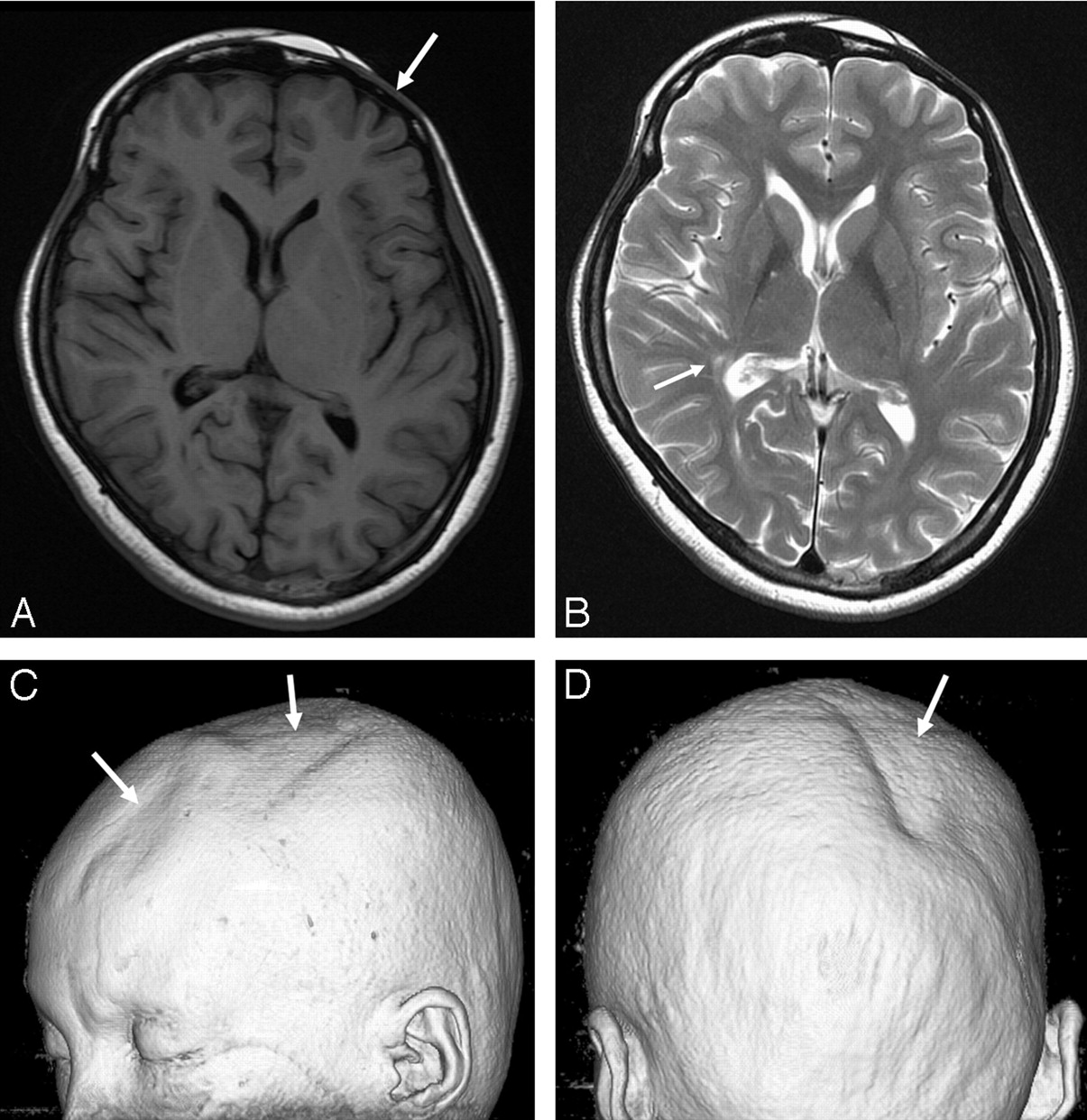
Conventional MR images as well as DTIs were obtained by using a 3T MR imaging scanner (Signa HDx; GE Healthcare, Milwaukee, Wis). T2-weighted images and fluid-attenuated inversion recovery (FLAIR) images showed a few tiny hyperintensities in the right frontoparietal periventricular white matter, right globus pallidus, left lateral thalamus, and right upper pons (Fig 1A, -B). Mild asymmetric atrophy of the right midbrain was noted. A 3D volume rendering image from the T1-weighted images depicted bilateral asymmetric scalp and calvarial atrophy (Fig 1C).

Fig 1.
  • Download figure
  • Open in new tab
  • Download powerpoint
Fig 1.

A, The T1-weighted image (at the level of basal ganglia) shows no abnormal lesion in the brain. However, atrophy of the left frontal skin and calvaria is noted (arrow). B, The T2-weighted image shows a few hyperintense lesions in the right basal ganglia and right posterior periventricular white matter (arrow). The midbrain appears with mild atrophy on the right side (not shown). C and D, A 3D volume rendering image shows focal skin and calvarial atrophy (arrows) over the left frontoparietal and the right parieto-occipital regions.

DTI was performed by using a Stejskal-Tanner sequence with single-shot spin-echo–type echo-planar imaging (TR/TE, 10,000/82 ms; matrix size, 128 × 128; FOV, 240 mm; 66 axial sections; 2.4-mm section thickness). We obtained diffusion-weighted images along 25 different directions with a b-value of 1000 s/mm2, as well as an image without diffusion weighting (b = 0 s/mm2). Brain fiber tracking was performed by using software on a workstation (FuncTool; GE Healthcare). The sensory and pyramidal motor tracts were observed by the method introduced by Yamada et al.9 Fiber tractography demonstrated a markedly decreased fiber tract volume of the sensory and motor pyramidal tract on the right side. Sensory tract derangement was more prominent as compared with motor tract involvement (Fig 2).

Fig 2.
  • Download figure
  • Open in new tab
  • Download powerpoint
Fig 2.

3D fiber tractography of the pyramidal tracts (A) and sensory tract through the dorsal pons and thalamus (B). A, The pyramidal tracts are clearly depicted on the left hemisphere but are sparse on the right hemisphere (arrows). B, The sensory tract on the right side is prominently involved (arrow).

Discussion

Up to 52% of patients with PRS have associated neuropsychiatric abnormalities such as migraine or facial pain, vision problems, depression and anxiety, and epilepsy. Among the described cases, epilepsy is the most common problem requiring patients to seek medical attention.1–3,6

In our patient, scalp and facial atrophy was bilateral but more prominent on the left frontal scalp, right parieto-occipital scalp, and left orbit. Right hemifacial paresthesia can be explained by right periorbital facial atrophy and subsequent trigeminal nerve involvement. In contrast, brain parenchymal involvement was more prominent on the right side. The right brain involvement explains clearly the left hypoesthesia of the extremities by cross-innervation of the sensory tract. In previous reports, abnormal brain lesions have been shown to be ipsilateral to the side of the face with PRS involvement.2–4,6,8

Bilaterality of PRS has been reported in approximately 2%–7.4% of cases.6 Bilateral involvement of facial atrophy and the almost unilateral involvement of the brain abnormality in our patient suggest that PRS might progress sequentially bilaterally for a long time. Bilateral involvement of facial and scalp atrophy can be visualized best by the use of 3D volume rendering images.

White matter and deep gray matter abnormalities on T2-weighted and FLAIR images were subtle in our patient. However, fiber tractography visualized clearly asymmetric involvement of the fiber tracts and larger involvement of the fiber tracts than expected from the conventional MR images. This finding implies that axonal injury in our patient was so profound that the derangement of the fiber tract could occur. Conversely, previous case reports have demonstrated relatively preserved fiber tracts despite widespread white matter abnormalities.2 Although we could not find any cortical and subcortical abnormalities on high-resolution thin-section T1- and T2-weighted images, a microscopic abnormality within the cortex might induce progressive axonal degeneration.

The pathophysiology of PRS remains uncertain. No genetic association has been found so far. A neuroinflammatory process has been suggested as a possible pathogenic mechanism, though no associated serum autoantibodies have been found.1 The disproportionately large involvement of the fiber tracts as compared with the white matter lesion on conventional MR images in our patient suggests a widespread underlying neurodegenerative process.

In conclusion, DTI and fiber tractography are useful in evaluating the neurologic aspects of patients with PRS with the slightest involvement of the brain.

Footnotes

  • This work was supported by the Second-Phase of BK (Brain Korea) 21 Project in 2007.

References

  1. ↵
    Pichiecchio A, Uggetti C, Grazia Egitto M, et al. Parry-Romberg syndrome with migraine and intracranial aneurysm. Neurology 2002;59:606–08, discussion 481
    Abstract/FREE Full Text
  2. ↵
    Okumura A, Ikuta T, Tsuji T, et al. Parry-Romberg syndrome with a clinically silent white matter lesion. AJNR Am J Neuroradiol 2006;27:1729–31
    Abstract/FREE Full Text
  3. ↵
    Paprocka J, Jamroz E, Adamek D, et al. Difficulties in differentiation of Parry-Romberg syndrome, unilateral facial sclerodermia, and Rasmussen syndrome. Childs Nerv Syst 2006;22:409–15. Epub 2005 Oct 25
    CrossRefPubMed
  4. ↵
    Moko SB, Mistry Y, Blandin de Chalain TM. Parry-Romberg syndrome: intracranial MRI appearances. J Craniomaxillofac Surg 2003;31:321–24
    PubMed
  5. ↵
    Blaszczyk M, Krolicki L, Krasu M, et al. Progressive facial hemiatrophy: central nervous system involvement and relationship with scleroderma en coup de sabre. J Rheumatol 2003;30:1997–2004
    Abstract/FREE Full Text
  6. ↵
    Tollefson MM, Witman PM. En coup de sabre morphea and Parry-Romberg syndrome: a retrospective review of 54 patients. J Am Acad Dermatol 2007;56:257–63
    CrossRefPubMed
  7. Shah JR, Juhasz C, Kupsky WJ, et al. Rasmussen encephalitis associated with Parry-Romberg syndrome. Neurology 2003;61:395–97
    Abstract/FREE Full Text
  8. ↵
    Sathornsumetee S, Schanberg L, Rabinovich E, et al. Parry-Romberg syndrome with fatal brain stem involvement. J Pediatr 2005;146:429–31
    CrossRefPubMed
  9. ↵
    Yamada K, Kizu O, Mori S, et al. Brain fiber tracking with clinically feasible diffusion-tensor MR imaging: initial experience. Radiology 2003;227:295–301
    CrossRefPubMed
  • Received November 28, 2007.
  • Accepted after revision December 5, 2007.
  • Copyright © American Society of Neuroradiology
View Abstract
PreviousNext
Back to top

In this issue

American Journal of Neuroradiology: 29 (4)
American Journal of Neuroradiology
Vol. 29, Issue 4
April 2008
  • Table of Contents
  • Index by author
Advertisement
Print
Download PDF
Email Article

Thank you for your interest in spreading the word on American Journal of Neuroradiology.

NOTE: We only request your email address so that the person you are recommending the page to knows that you wanted them to see it, and that it is not junk mail. We do not capture any email address.

Enter multiple addresses on separate lines or separate them with commas.
Diffusion Tensor Imaging and Fiber Tractography in Parry-Romberg Syndrome
(Your Name) has sent you a message from American Journal of Neuroradiology
(Your Name) thought you would like to see the American Journal of Neuroradiology web site.
CAPTCHA
This question is for testing whether or not you are a human visitor and to prevent automated spam submissions.
Cite this article
W.J. Moon, H.J. Kim, H.G. Roh, J. Oh, S.H. Han
Diffusion Tensor Imaging and Fiber Tractography in Parry-Romberg Syndrome
American Journal of Neuroradiology Apr 2008, 29 (4) 714-715; DOI: 10.3174/ajnr.A0967

Citation Manager Formats

  • BibTeX
  • Bookends
  • EasyBib
  • EndNote (tagged)
  • EndNote 8 (xml)
  • Medlars
  • Mendeley
  • Papers
  • RefWorks Tagged
  • Ref Manager
  • RIS
  • Zotero
0 Responses
Respond to this article
Share
Bookmark this article
Diffusion Tensor Imaging and Fiber Tractography in Parry-Romberg Syndrome
W.J. Moon, H.J. Kim, H.G. Roh, J. Oh, S.H. Han
American Journal of Neuroradiology Apr 2008, 29 (4) 714-715; DOI: 10.3174/ajnr.A0967
del.icio.us logo Twitter logo Facebook logo Mendeley logo
  • Tweet Widget
  • Facebook Like
  • Google Plus One
Purchase

Jump to section

  • Article
    • Abstract
    • Case Report
    • Discussion
    • Footnotes
    • References
  • Figures & Data
  • Info & Metrics
  • Responses
  • References
  • PDF

Related Articles

  • No related articles found.
  • PubMed
  • Google Scholar

Cited By...

  • No citing articles found.
  • Crossref (16)
  • Google Scholar

This article has been cited by the following articles in journals that are participating in Crossref Cited-by Linking.

  • A review of Parry-Romberg syndrome
    Jessica El-Kehdy, Ossama Abbas, Nelly Rubeiz
    Journal of the American Academy of Dermatology 2012 67 4
  • Parry-Romberg Syndrome and Rasmussen Encephalitis: Possible Association. Clinical and Neuroimaging Features
    Daniela Longo, Amalia Paonessa, Nicola Specchio, Luciana Nogueira Delfino, Dianela Claps, Lucia Fusco, Francesco Randisi, Elisabetta Genovese, Federico Vigevano, Giuseppe Fariello
    Journal of Neuroimaging 2011 21 2
  • Neurological Manifestations in Parry–Romberg Syndrome
    Justine Vix, Stéphane Mathis, Mathieu Lacoste, Rémy Guillevin, Jean-Philippe Neau
    Medicine 2015 94 28
  • "Scleroderma linearis: hemiatrophia faciei progressiva (Parry-Romberg syndrom) without any changes in CNS and linear scleroderma "en coup de sabre" with CNS tumor
    Beata Bergler-Czop, Anna Lis-Święty, Ligia Brzezińska-Wcisło
    BMC Neurology 2009 9 1
  • Giant Intracranial Aneurysm in a Ten-Year-Old Boy with Parry Romberg Syndrome
    T. Bosman, J. Van Beijnum, M.A.A. Van Walderveen, P.A. Brouwer
    Interventional Neuroradiology 2009 15 2
  • Parry–Romberg syndrome: clinical, electrophysiological and neuroimaging correlations
    Sławomir Budrewicz, Magdalena Koszewicz, Ewa Koziorowska-Gawron, Paweł Szewczyk, Ryszard Podemski, Krzysztof Słotwiński
    Neurological Sciences 2012 33 2
  • Imaging Findings of Various Calvarial Bone Lesions with a Focus on Osteolytic Lesions
    Younghee Yim, Won-Jin Moon, Hyeong Su An, Joon Cho, Myung Ho Rho
    Journal of the Korean Society of Radiology 2016 74 1
  • Síndrome de Parry-Romberg: achados nas sequências avançadas de ressonância magnética - relato de caso
    Rafael Alfenas de Paula, Bruno Niemeyer de Freitas Ribeiro, Paulo Roberto Valle Bahia, Renato Niemeyer de Freitas Ribeiro, Laís Balbi de Carvalho
    Radiologia Brasileira 2014 47 3
  • The Causes of Epilepsy
    2011
  • Parry–Romberg syndrome: conventional and advanced MRI follow-up in a boy
    Paolo Rigamonti, Silvia Squarza, Marco Politi, Roberto Sangermani, Maurizio Cariati, Carla Uggetti
    The Neuroradiology Journal 2017 30 5

More in this TOC Section

  • Glutaric Aciduria Type 1: DK vs. Conventional MRI
  • Kurtosis and Epileptogenic Tubers: A Pilot Study
  • Choroid Plexus Calcification&Microglial Activation
Show more Functional

Similar Articles

Advertisement

Indexed Content

  • Current Issue
  • Accepted Manuscripts
  • Article Preview
  • Past Issues
  • Editorials
  • Editors Choice
  • Fellow Journal Club
  • Letters to the Editor

Cases

  • Case Collection
  • Archive - Case of the Week
  • Archive - Case of the Month
  • Archive - Classic Case

Special Collections

  • Special Collections

Resources

  • News and Updates
  • Turn around Times
  • Submit a Manuscript
  • Author Policies
  • Manuscript Submission Guidelines
  • Evidence-Based Medicine Level Guide
  • Publishing Checklists
  • Graphical Abstract Preparation
  • Imaging Protocol Submission
  • Submit a Case
  • Become a Reviewer/Academy of Reviewers
  • Get Peer Review Credit from Publons

Multimedia

  • AJNR Podcast
  • AJNR SCANtastic
  • Video Articles

About Us

  • About AJNR
  • Editorial Board
  • Not an AJNR Subscriber? Join Now
  • Alerts
  • Feedback
  • Advertise with us
  • Librarian Resources
  • Permissions
  • Terms and Conditions

American Society of Neuroradiology

  • Not an ASNR Member? Join Now

© 2025 by the American Society of Neuroradiology All rights, including for text and data mining, AI training, and similar technologies, are reserved.
Print ISSN: 0195-6108 Online ISSN: 1936-959X

Powered by HighWire