
Article Figures & Data
Figures
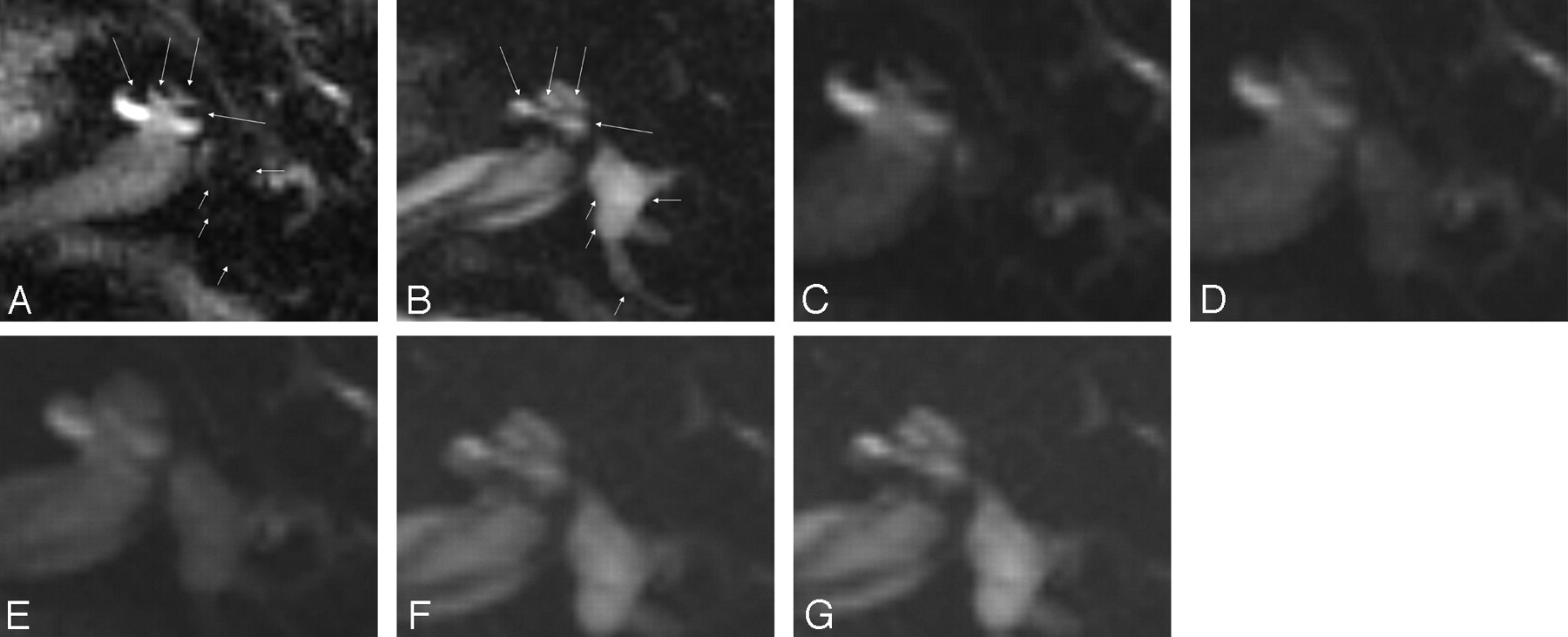
- Fig 1.
A 46-year-old man with Ménière disease. All images were obtained 24 hours after the intratympanic injection of Gd-DTPA. A, Perilymphatic fluid image obtained with 3D-FLAIR (9000/128/2500). Low signal intensity areas (arrows) can be recognized as enlarged endolymphatic space in the cochlea. Note that the vestibule and posterior semicircular canal (short arrows) also showed low signal intensity, so we could not determine whether the vestibule and posterior semicircular canal were filled with endolymphatic fluid. B, Endolymphatic fluid image obtained with a 3D inversion-recovery sequence (9000/128/1000). Endolymphatic space in the cochlea (arrows) shows high signal intensity on this image. This image confirmed that the vestibule and posterior semicircular canal were filled with fluid. C–G, Fusion of a perilymphatic fluid image (C) and an endolymphatic fluid image (G) with transitional images (D–F). By changing the fusion mixture rate on a workstation, the spatial relationship between perilymphatic and endolymphatic space was easily appreciated in both the cochlea and vestibule. In this case, endolymphatic space was enlarged in both the cochlea and vestibule, but the enlargement was especially prominent in the vestibule. Note that CSF in the internal auditory canal is visualized as high signal intensity on the endolymphatic fluid image (G). The signal intensity of perilymphatic space is just suppressed. Thus, the term endolymphatic image is only useful for labyrinthine space.
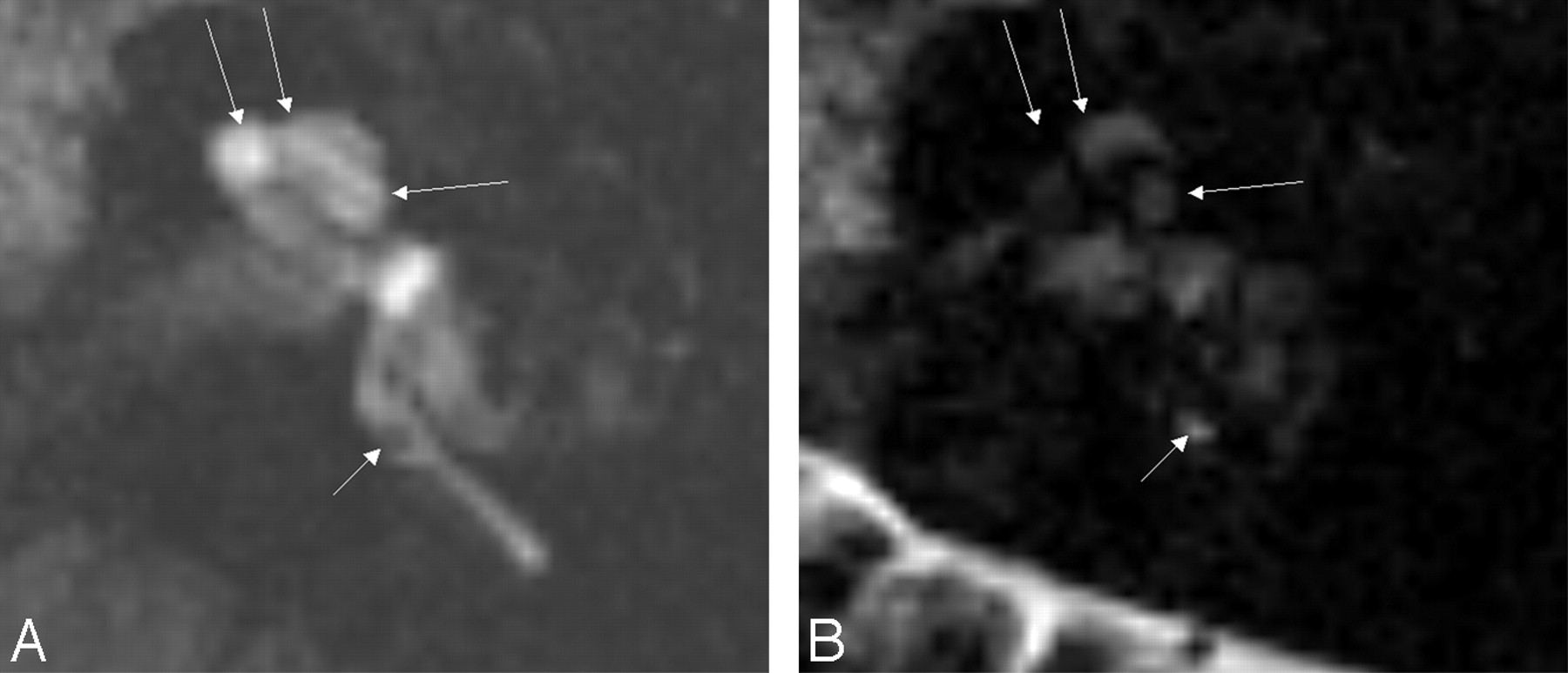
- Fig 2.
A 43-year-old woman with sudden sensorineural hearing loss in the left ear. All images were obtained 24 hours after the intratympanic injection of Gd-DTPA. A, Perilymphatic fluid image obtained with 3D-FLAIR (9000/128/2500). Areas of low signal intensity in the position of the cochlear duct (arrows) cannot be recognized as the endolymphatic space in the cochlea, probably because of their small size. Note that the endolymphatic space of the posterior ampulla (short arrow) showed low signal intensity. B, Endolymphatic fluid image obtained with a 3D inversion-recovery sequence (9000/128/1000). The endolymphatic space in the cochlea (arrows) cannot be recognized as high signal intensity on this image. Note that the endolymphatic space of the posterior ampulla (short arrow) showed high signal intensity alternatively compared with Fig 2A.


In this issue


Jump to section
Related Articles
Cited By...
- Detection and Grading of Endolymphatic Hydrops in Meniere Disease Using MR Imaging
- Meniere's disease: a reappraisal supported by a variable latency of symptoms and the MRI visualisation of endolymphatic hydrops
- Comparison of Contrast Effect on the Cochlear Perilymph after Intratympanic and Intravenous Gadolinium Injection