
Article Figures & Data
Figures
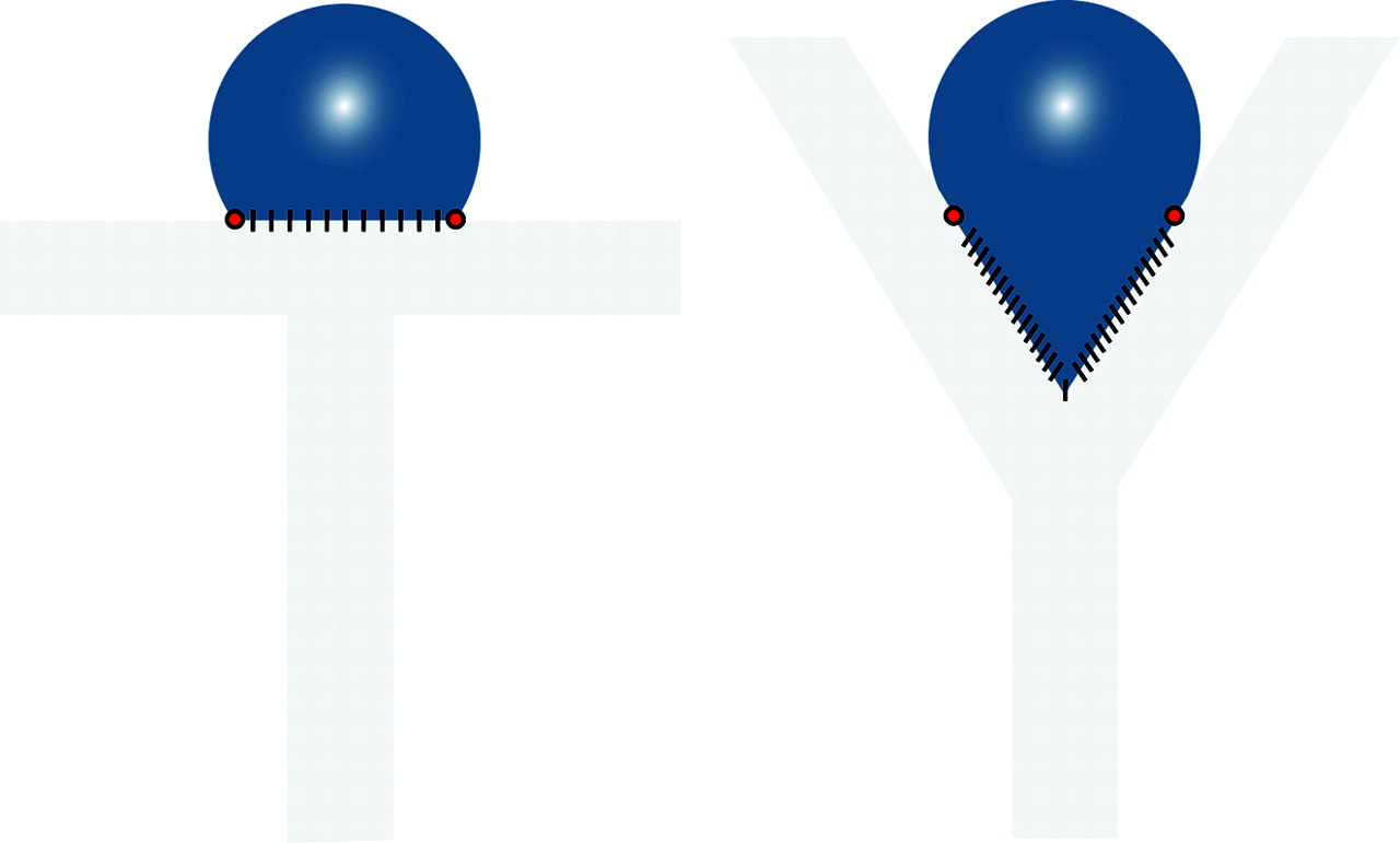
- Fig 1.
Schematic illustration of T- and Y-type carotid bifurcation aneurysm models.
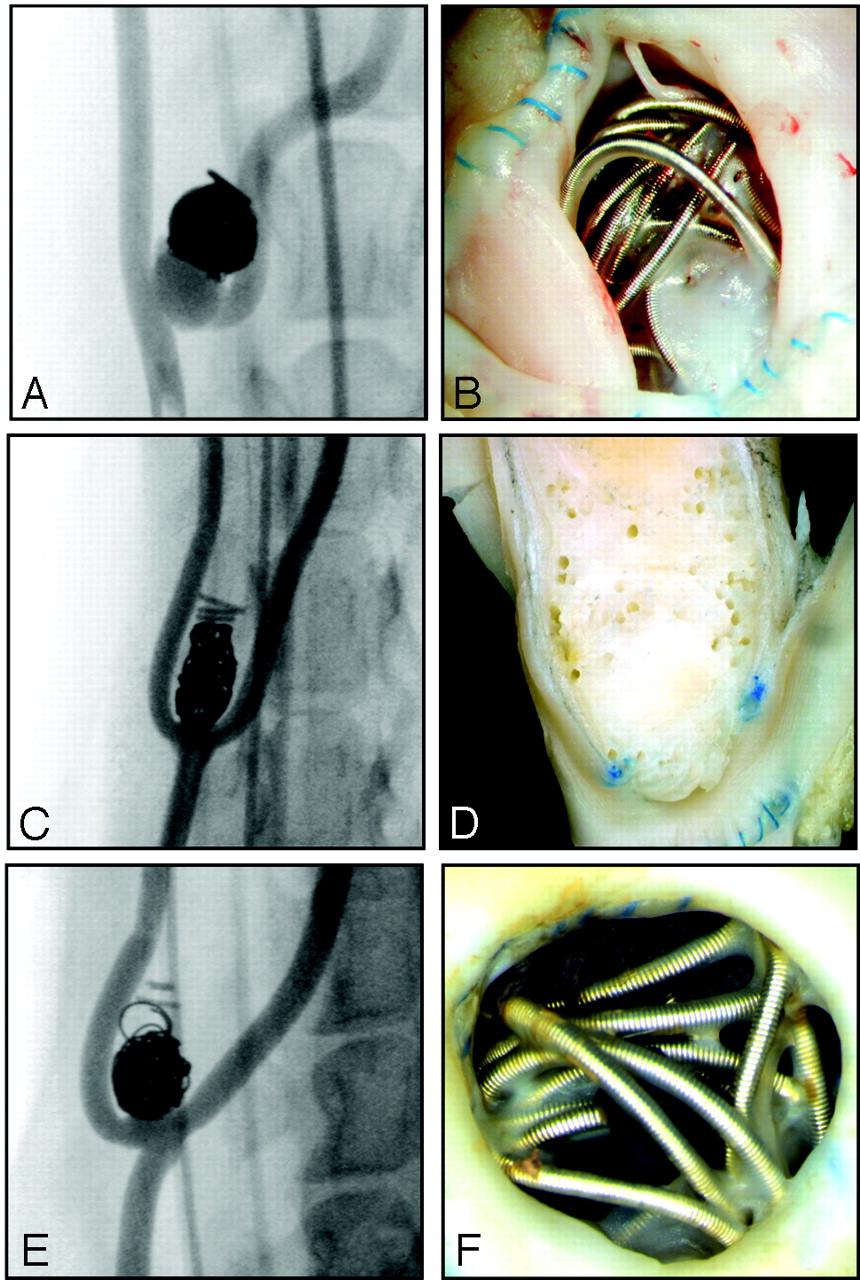
- Fig 2.
Angiographic and macroscopic results. Angiography (A, C, E) and macroscopic photographs (B, D, F) of prototypical cases of recurrence at 3 months and poor neointimal closure (A and B), complete occlusions at 3 months with good neointimal closure of the neck (C and D), and angiographic occlusion but with poor neointimal closure of the neck (E and F). Note that the coils have been removed in D.
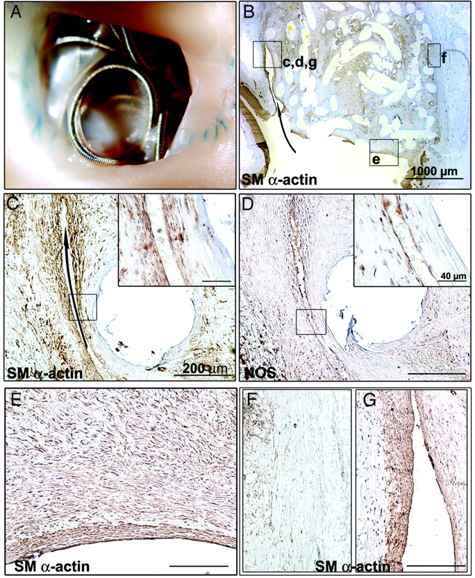
- Fig 3.
Macroscopic and pathologic findings of aneurysm with complete angiographic occlusion at 3 months after coil embolization. A, Photograph of aneurysmal neck en face. Note complete closure of the neck by a translucent layer of membrane tissue. B, Low-magnification overview of an axial aneurysm section showing a continuous layer of smooth muscle (SM) α-actin+ cells completely sealing the neck of the aneurysm. At higher magnification (C and D are magnifications of the inset c,d in B), closed corner (arrows in B and C) shows aligned SM α-actin+ cells covered by a single layer of NOS+ endothelial cells. E, The same neointimal layer is continuous, isolating the aneurysm from the circulation. F, SM α-actin immunostaining of atrophied aneurysmal wall. Sections are hematoxylin-counterstained (scale bar: 1000 μm for B; 200 μm for C–F; 40 μm for insets in C and D).
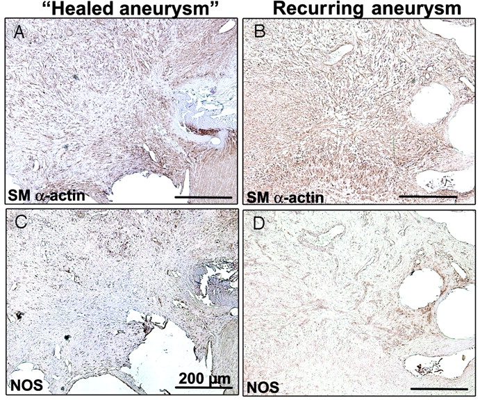
- Fig 4.
Macroscopic and pathologic findings of recurring aneurysm 3 months after coil embolization. A, Macroscopic photograph showing incomplete neck closure. B, Low magnification of overview of axial aneurysm section revealing a recurring lesion extending from the corner of the neck to the body of the aneurysm (arrow). These crescentic spaces are lined by smooth muscle (SM) α–actin+ cells (C) and covered with NOS+ endothelial cells (D). E, Higher magnification showing the same layer of SM α-actin+ cells covered with NOS+ endothelial cells. F, Atrophy of the aneurysmal wall in the occluded zone. G, No atrophy of the aneurysmal wall in the recurring zone. Sections are hematoxylin-counterstained (scale bar: 1000 μm for B; 200 μm for C–G; 40 μm for insets in C and D).
- Fig 5.
Common findings of occluded and recurrent aneurysms. Macroscopic views with coils protruding into the lumen vessel before (A) and after coil retrieval (B). Note that all coils are re-covered with neointima, which consists of smooth muscle (SM) cells (C) embedded in a collagenous matrix surrounded by a unique layer of NOS+ cells (D). Note polypoid filaments (pink arrows in A and E) observed at the neck of aneurysms, with a similar neointima organization (F). Sections are hematoxylin-counterstained (scale bar: 200 μm for C, D, and F; 40 μm for insets in C, D, and F).
- Fig 6.
Vascularized connective tissues are similarly organized in occluded (A and C) and recurrent (B and D) aneurysm with smooth muscle (SM) α–actin+ cells, presumably myofibroblast (A and B) and NOS+ cells for neovascularization (C and D) (scale bar: 200 μm).
- Fig 7.
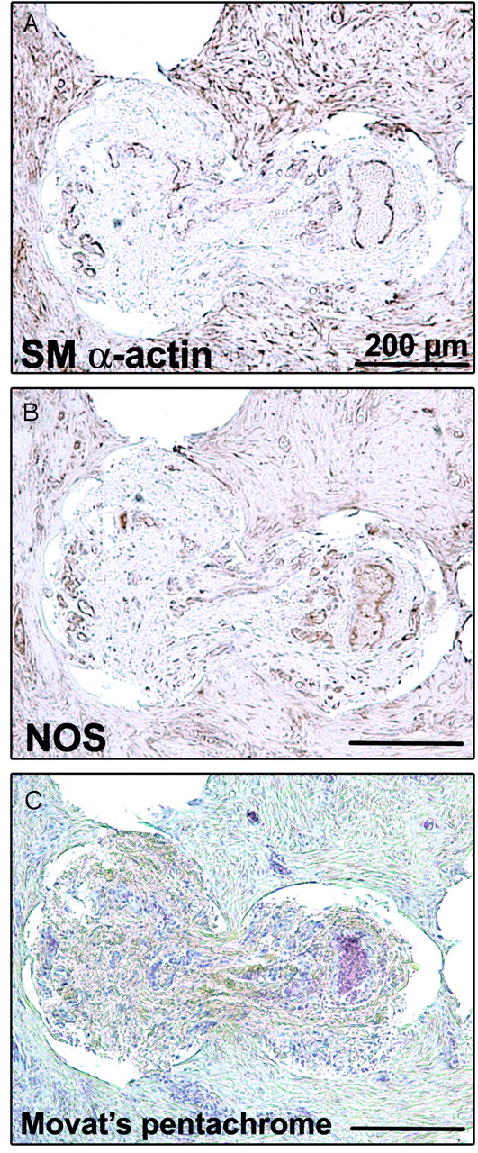
Tissues infiltrating the core of the coils. Immunostaining of smooth muscle (SM) α–actin (A), of NOS (B), and Movat pentachrome stain (C) shows the same structure as the vascularized tissue inside the aneurysm (scale bar: 200 μm).
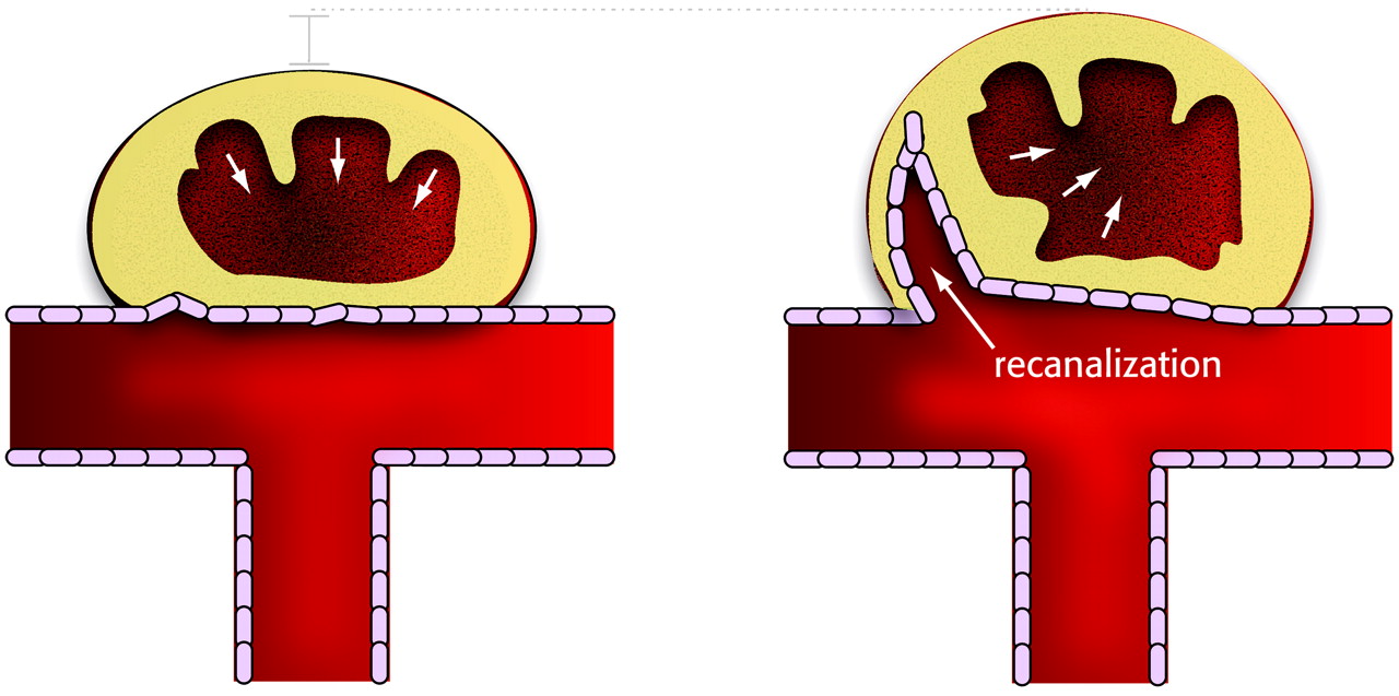
- Fig 8.
Schematic illustration summarizing the differences between occlusion and recanalization.


Tables
In this issue


Jump to section
Related Articles
Cited By...
- Assessment of endothelialization of aneurysm wall over time in a rabbit model through CD31 scoring
- Hemodynamics and coil distribution with changing coil stiffness and length in intracranial aneurysms
- An update to the Raymond-Roy Occlusion Classification of intracranial aneurysms treated with coil embolization
- Creation of Bifurcation-Type Elastase-Induced Aneurysms in Rabbits
- Thrombosis Heralding Aneurysmal Rupture: An Exploration of Potential Mechanisms in a Novel Giant Swine Aneurysm Model
- The impact of coil shape design on angiographic occlusion, packing density and coil mass uniformity in aneurysm embolization: an in vitro study
- Creation of Large Elastase-Induced Aneurysms: Presurgical Arterial Remodeling Using Arteriovenous Fistulas
- A New Canine Carotid Artery Bifurcation Aneurysm Model for the Evaluation of Neurovascular Devices
- In Vivo Experimental Intracranial Aneurysm Models: A Systematic Review
- Minimally invasive neuroradiologic model of preclinical transient middle cerebral artery occlusion in canines