
Article Figures & Data
Figures
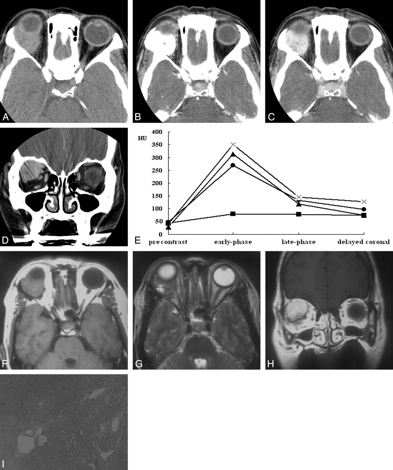
- Fig 1.
A–I, Case 2. Solitary fibrous tumor of the orbit in a 50-year-old woman. Axial precontrast (A), early-phase (B), late-phase (C), and delayed coronal (D) CT scans show a 30-mm well-defined soft tissue mass in the superolateral aspect of the right orbit near the lacrimal gland fossa. Compared with the cerebral gray matter, the mass is slightly hyperattenuated on precontrast CT scan (A). Marked homogeneous enhancement is seen on early-phase CT scan (B), and there is rapid washout of contrast material on late-phase (C) and delayed coronal (D) CT scans. The mass abuts and conforms to the posterior wall of the eyeball without causing significant indentation. E, Time-attenuation curve also reveals rapid enhancement and early washout of contrast material within the tumor (•), which is very similar to those of the internal carotid artery (×). ▴ = internal jugular vein. ▪ = medial rectus muscle. F–H, Compared with the cerebral gray matter, the mass is isointense and mixed isointense and hyperintense on axial T1- (F) and T2-weighted (G) MR images, respectively. Marked homogeneous enhancement is seen on coronal postcontrast T1-weighted MR image (H). Note the small areas of bright signal intensity within the tumor on T2-weighted image (G). I, Photomicrograph shows that the tumor has a patternless histologic architecture with hyalinized collagen bundles and multiple dilated vessels (hematoxylin-eosin, original magnification ×40).
- Fig 2.
A–D, Case 5. Solitary fibrous tumor of the orbit in a 24-year-old woman. Postcontrast (A) axial CT scan and axial T1-weighted (B), T2-weighted (C), and postcontrast fat-suppressed T1-weighted (D) MR images show a 18-mm well-defined ovoid soft tissue mass in the inferomedial aspect of the intraconal space of the left orbit. Like the tumor shown in Fig 1, the mass conforms to the ocular contour without causing significant indentation. Compared with the cerebral gray matter, the mass is slightly hyperattenuated on precontrast CT scan (not shown), isointense on T1-weighted MR image (B), and mixed isointense and hyperintense and T2-weighted MR image (C). There is marked homogeneous enhancement of the tumor on postcontrast CT (A) and MR (D) images. Note the signal intensity void, tubular vascular structures inside the tumor on MR images (arrows).
- Fig 3.
A and B, Case 6. Solitary fibrous tumor of the lower eyelid in a 40-year-old man. Axial early-phase (A) and late-phase (B) CT scans show a 20-mm well-defined ovoid soft tissue mass on the right lower eyelid. Marked homogeneous enhancement is seen on early-phase CT scan (A), followed by rapid washout of contrast material on late-phase CT scan (B).


Tables
Clinical and imaging findings in six patients with solitary fibrous tumor of the orbit
Patient No./Age/Sex Chief Complaint CT Scan/ Dual-Phase CT MR Image Location Size* (mm) Margin Density on Precontrast CT Scan† Signal Intensity on MR Imaging† Enhancement Pattern/Degree T1WI T2WI 1/26 y/M Periocular mass for 10 months Yes/No No Lacrimal sac 30 Well-defined - - - Heterogeneous/marked 2/50 y/F Proptosis for 8 months Yes/Yes Yes Postseptal extraconal 30 Well-defined Slightly hyperdense Isointense Mixed isointense and hyperintense Homogeneous/marked 3/51 y/F Periocular mass for 6 months Yes/Yes No Lacrimal sac 18 Well-defined Isodense - - Homogeneous/marked 4/18 y/F Proptosis for 2 y Yes/No Yes Postseptal extraconal 30 Well-defined Isodense Isointense Mixed isointense and hyperintense Heterogeneous/marked 5/24 y/F Proptosis for 1 y Yes/No Yes Postseptal intraconal 18 Well-defined Slightly hyperdense Isointense Mixed isointense and hyperintense Homogeneous/marked 6/40 y/M Periocular mass for 6 months Yes/Yes No Lower eyelid 20 Well-defined Isodense - - Homogeneous/marked Note:—T1WI indicates T1-weighted image; T2WI, T2-weighted image.
* Size was denoted as greatest diameter.
† Density and signal intensity of the tumor were compared with those of the cerebral gray matter.


