
Article Figures & Data
Figures
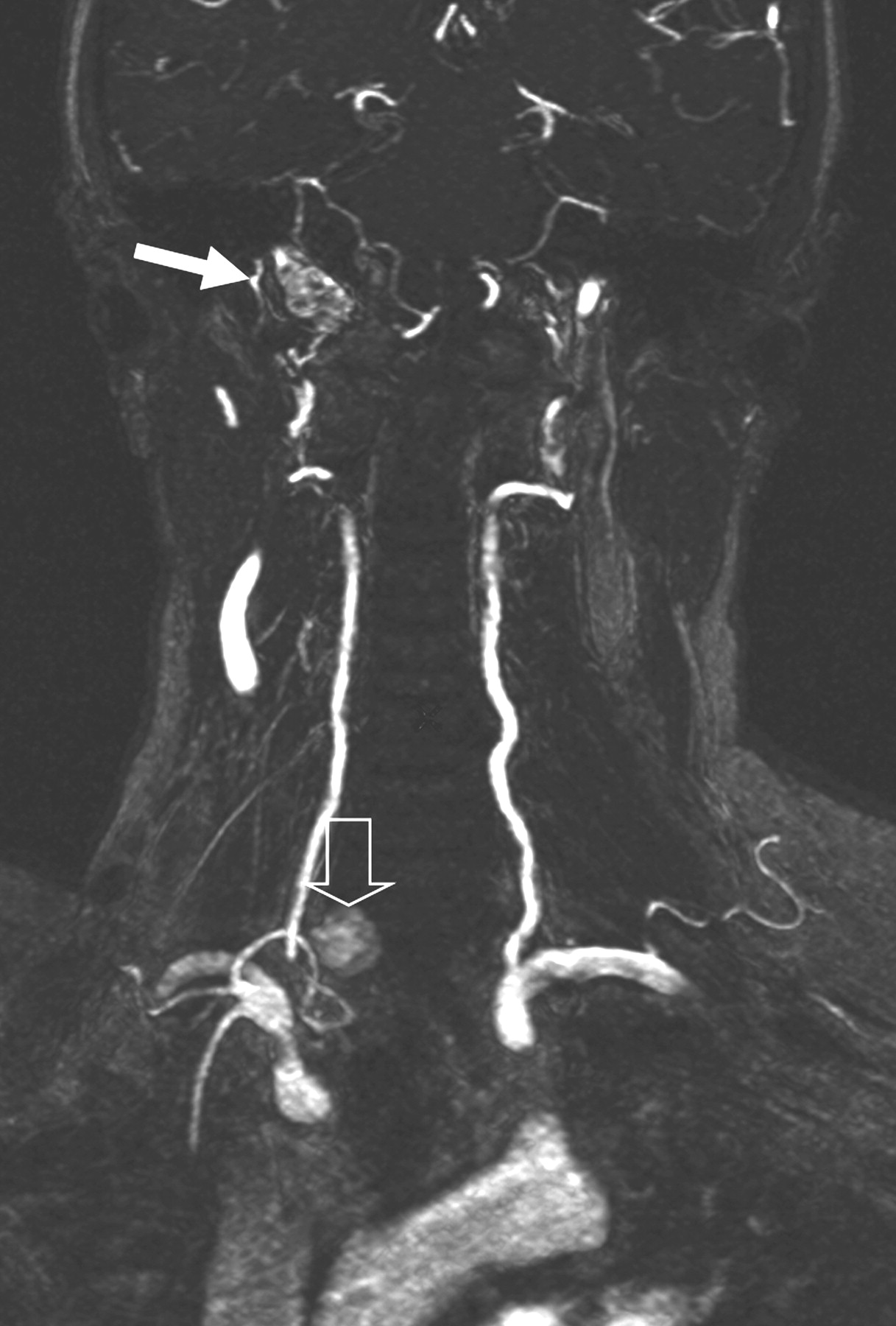
- Fig 1.
A 57-year-old woman with 2 paragangliomas: a residual vagal paraganglioma (solid arrow) and a retrothyroidian paraganglioma (open arrow). The second one was missed by DSA (because the feeding artery was not opacified) and by SE imaging (because it was situated out of the FOV, under the carotid bifurcation). They were depicted by CE-MRA and were both pathologically confirmed. CE-MRA coronal MIP image shows the 2 paragangliomas as intense tumor blush.
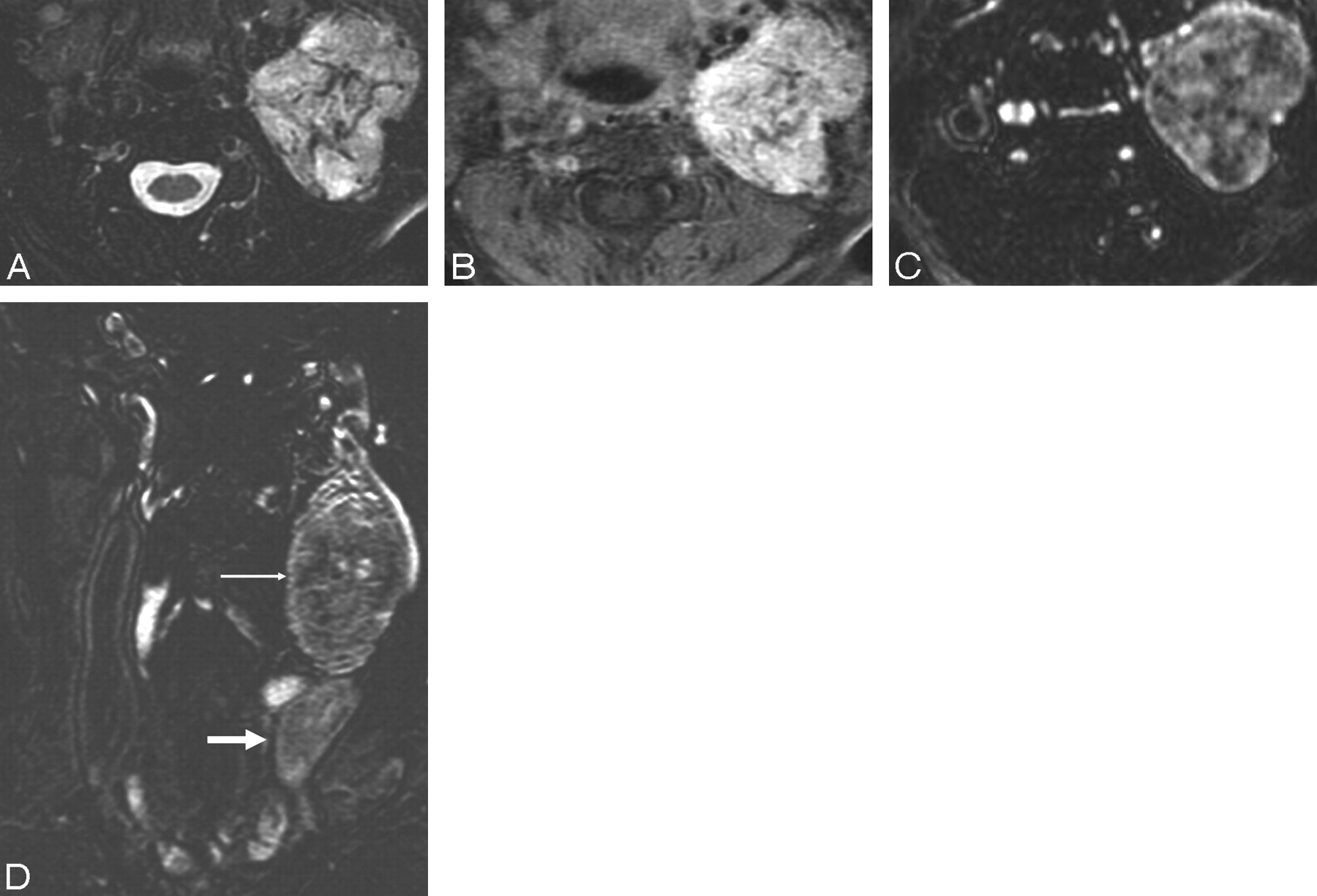
- Fig 2.
A 52-year-old woman with a characteristic vagal paraganglioma. A, Axial T2-weighted fat-saturated image shows a high-signal-intensity left-sided mass with prominent vascular flow voids. B, Axial contrast-enhanced fat-saturated T1-weighted image shows an intensely enhancing mass. C and D, CE-MRA axial MPR image (C) and CE-MRA coronal MIP image (D) show a typical tumor blush in the arterial phase (small arrow). Note the early draining internal jugular vein (large arrow).
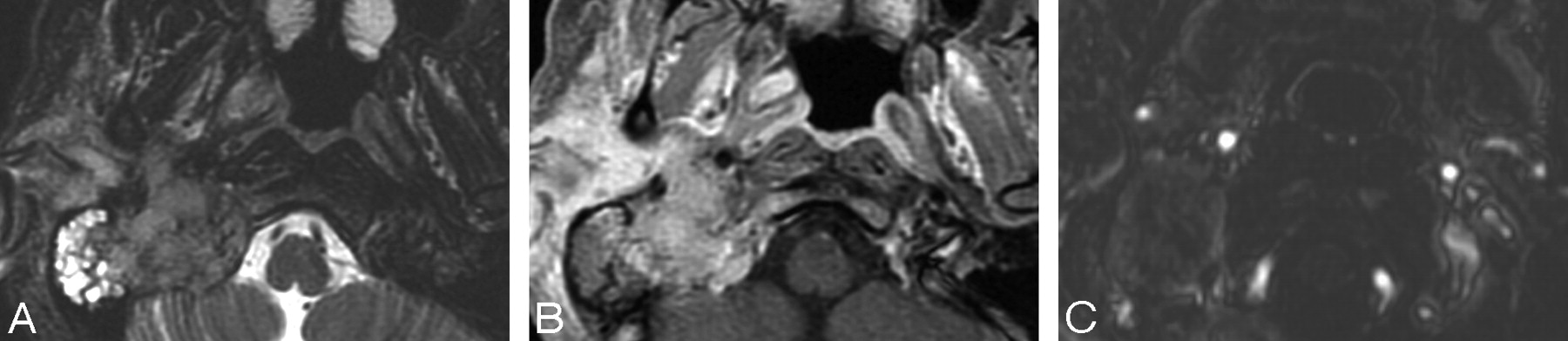
- Fig 3.
A 36-year-old woman with a primary jugular foramen meningioma. A, Axial T2-weighted fat-saturated image shows a mass centered in the right-sided jugular foramen with vascular flow voids mimicking a paraganglioma. B, Axial contrast-enhanced fat-saturated T1-weighted image shows strong enhancement of this mass. The tumor is difficult to differentiate from a paraganglioma on SE sequences. C, CE-MRA axial MPR image does not show the typical tumor blush in the arterial phase, which indicates that this tumor is probably not a paraganglioma.
- Fig 4.
A 37-year-old woman with a cervical schwannoma. A, Axial contrast-enhanced fat-saturated T1-weighted image shows intensely enhancing well-circumscribed right-sided cervical mass. Differentiation with a paraganglioma is difficult. B, CE-MRA axial MPR image does not show the typical tumor blush in the arterial phase, which indicates that this tumor is probably not a paraganglioma. Note the anterior displacement of both the internal and external carotid arteries due to the schwannoma.


Tables
- Table 1:
For the diagnosis of paraganglioma, sensitivity, specificity, PPV, NPV, and accuracy of each reader for both techniques
Reader 1 Reader 2 Reader 3 Consensus Sensitivity, % C MRI 94.1 (80.32–99.28) 94.1 (80.32–99.28) 91.1 (76.32–98.14) 94 (80.32–99.28) CE-MRA 97 (84.67–99.93) 94.1 (80.32–99.28) 97 (84.67–99.93) 100 (89.72–100) Specificity, % C MRI 47 (22.98–72.19) 35.2* (14.21–61.67) 52.9† (27.81–77.02) 41‡ (18.44–67.08) CE-MRA 76.4 (50.1–93.19) 88.2* (63.56–98.54) 88.2† (63.56–98.54) 94‡ (71.31–99.85) PPV, % C MRI 78 (62.39–89.44) 74.4 (58.83–86.48) 79.5 (63.54–90.7) 76.2 (60.55–87.95) CE-MRA 89.2 (74.58–96.97) 94.1 (80.32–99.28) 94.3 (80.84–99.3) 97.1 (85.08–99.93) NPV, % C MRI 80 (44.39–97.48) 75 (34.91–96.81) 75 (42.81–94.51) 77.8 (39.99–97.19) CE-MRA 92.9 (66.13–99.82) 88.2 (63.56–98.54) 93.8 (69.77–99.84) 100 (79.41–100) Accuracy, % C MRI 78.4 (44.39–97.48) 74.5 (34.91–96.81) 78.4 (42.81–94.51) 76.5 (39.99–97.19) CE-MRA 90.2 (66.13–99.82) 92.1 (63.56–98.54) 94.1 (69.77–99.84) 98 (79.41–100) Note:—C MRI indicates conventional MR imaging; in parenthesis, 95% CIs; CE-MRA, contrast-enhanced MR angiography; PPV, positive predictive value; NPV, negative predictive value.
*‡ CE-MRA performed significantly superior compared with conventional MR Imaging.
* P = .012.
† P = .031.
‡ P = .004.
- Table 2:
Patients with suggested recurrence of paraganglioma (surgically treated paragangliomas)
Patient Localization Interval Between Surgery and MRI Maximal Dimension (mm) Probability of PG Final Diagnosis (reference standard) C MRI CE-MRA 1 Vagal 4 years 20 1 2 PG (surgery + pathology) 2 Jugulotympanic 9 months 16 1 2 PG (SD) 3 Vagal 4 years 22 2 2 PG (SD) 4 Jugulotympanic 6 months 41 2 2 PG (SD) CBT 6 months 4 1 0 Postoperative scar (follow-up) Note:—CBT indicates carotid body tumor; C MRI, conventional MR imaging; PG, paraganglioma; SD, surgical data (known tumor residue); 2, probable; 1, uncertain; 0, improbable.
In this issue


Jump to section
Related Articles
Cited By...
- The Sensitivity of Arterial Spin-Labeling Imaging for Detection of Head and Neck Paragangliomas
- 68Ga-DOTATATE PET/CT Versus MRI: Why the Comparison of 68Ga-DOTATATE PET/CT to an Appropriate MRI Protocol Is Essential
- Endocrine tumors associated with the vagus nerve
- 15 YEARS OF PARAGANGLIOMA: Imaging and imaging-based treatment of pheochromocytoma and paraganglioma
- Evaluation of Brain and Head and Neck Tumors with 4D Contrast-Enhanced MR Angiography at 3T