
Article Figures & Data
Figures
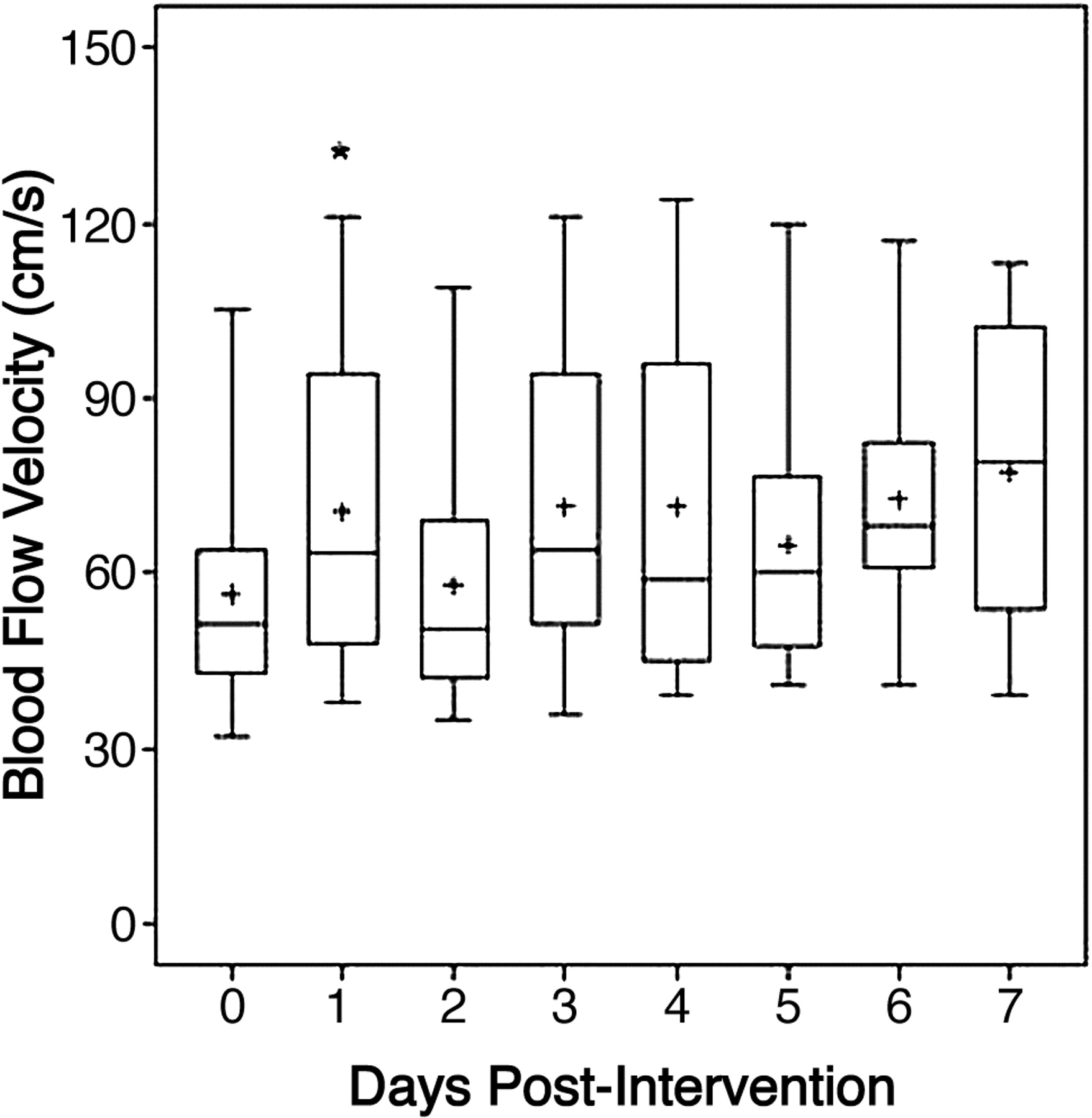
- Fig 1.
The box-plot profiles of blood-flow velocity measured with TCD, from day 0 (preintervention) to the 7 days postintervention. The plus represents the sample mean value. The asterisk indicates P < .05 for the change from baseline.
- Fig 2.
The box-plot profiles of perfusion parameters measured with perfusion CT: MTT (A), Tmax (B), cerebral blood flow (C), and cerebral blood volume (D), by time 0 (preintervention), 1 (first day after intervention), 2 (2–5 days postintervention), to 3 (5–9 days postintervention). The plus signs are the sample mean values. The asterisk indicates P < .05 for the change from baseline.
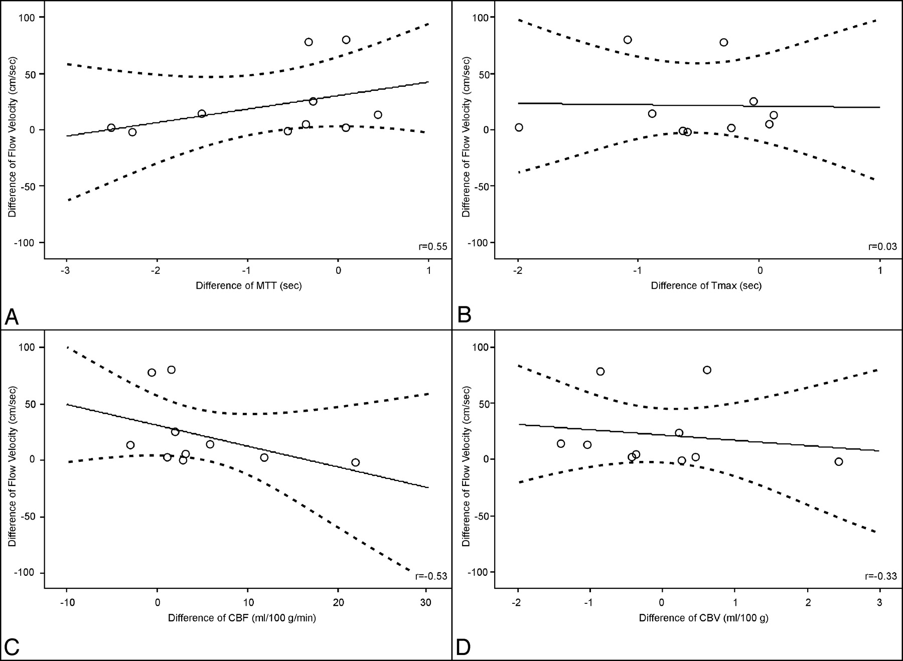
- Fig 3.
Correlation between the first day change of flow velocity measured with TCD and the first day change of perfusion parameters measured with perfusion CT: MTT (A), Tmax (B), cerebral blood flow (C), and cerebral blood volume (D). Data are presented with correlation coefficients (r) and univariate linear regression with 95% confidence intervals.
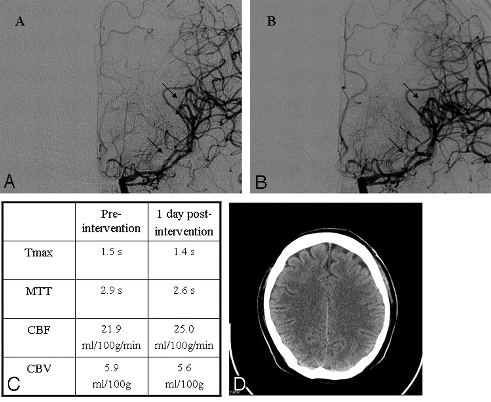
- Fig 4.
Illustrative case 12. A, Baseline angiogram on day 6 documents severe vasospasm in the left M1 and M2 segments (arrows). B, DSA obtained 10 minutes after the injection of 1.6-mg nimodipine reveals reduction of the vasospasm (arrows). C, PCT approximately 24 hours after IAN demonstrates a reduction of MTT and Tmax. D, No ischemic lesions are documented on the CT scan obtained at the time of discharge.


Tables
Patient No. Age (yr)/Sex WFNS Grade Fisher Grade Aneurysm Location Treatment Vasospasm Confirmed by DSA* 1 67/F IV IV PICA left Clipping 15 2 47/M I IV MCA left Clipping 11 3 47/F III IV MCA left Clipping 8 4 59/M III IV Pericallosal left Clipping 6, 9 5 49/F I III BA Coiling 7 6 65/F IV IV AcomA Clipping 16 7 48/M III IV MCA right Clipping 8 8 54/M III IV ICA left Coiling 11 9 36/F III IV AcomA Clipping 9 10 46/F IV IV BA Coiling 5, 6 11 46/F I II AcomA Coiling 6 12 33/M I III MCA left Clipping 7 13 37/M I II AcomA Clipping 3 14 49/F I III AcomA Coiling 6 15 73/F IV IV PcomA left Clipping 4 16 58/M II III PICA right Coiling 10 17 40/M I II AcomA Coiling 5, 6 18 27/M IV IV ICA left Clipping 5, 13 19 48/F IV IV ICA right Clipping 10, 11 20 58/M I II MCA right Clipping 7, 11 21 70/F II III MCA left Clipping 8, 9 22 55/F IV IV PcomA right Coiling 8 23 36/M I IV ICA right Coiling 10 24 65/M I III AcomA Clipping 5, 7, 8, 11 25 49/F IV IV AcomA Coiling 0, 3, 10 26 42/M III IV ICA left Coiling 7, 11, 12, 14, 18 Note:—ICA indicates internal carotid artery; AcomA, anterior communicating artery; MCA, middle cerebral artery; PcomA, posterior communicating artery; BA, basilar artery; PICA, posterior inferior cerebellar artery; WFNS, World Federation of Neurological Surgeons; DSA, digital subtraction angiography.
* In days after the index SAH.
Patient No. Distribution of Vasospasm IAN IAN Dosage per Session (mg) Continuous IAN Former PTA Direct DSA Result 1 ACA/MCA b ICA b 3.2 – – + 2 ACA/MCA l ICA l 1.6 4 mg/h, 2 hours – (+) 3 MCA l ICA l 0.8 – – ++ 4 1) ACA b 1) ICA b 1) 3.2 1) − 1) − 1) ++ 2) ACA b 2) ICA b 2) 3.2 2) – 2) – 2) + 5 MCA b ICA b 3.2 – – ++ 6 ACA/MCA l ICA l 1.6 – – ++ 7 MCA r ICA r 1.6 – Yes ++ 8 MCA l ICA l 1.6 – – (+) 9 MCA l ICA l 0.8 – Yes ++ 10 1) ICA l 1) ICA l 1) 3.2 1) − 1) − 1) + 2) ICA l 2) ICA l 2) 3.2 2) – 2) – 2) + 11 ACA l ICA l 1.6 – Yes ++ 12 MCA l ICA l 1.6 – – ++ 13 ACA r ICA r 1.6 – – (+) 14 ACA b ICA b 3.2 – – + 15 ICA l ICA l 1.6 – – ++ 16 MCA l ICA l 1.6 – (+) PCA l VA l 1.6 4 mg/h, 2 hours 17 1) ICA b 1) ICA b 1) 3.2 1) − 1) − 1) ++ 2) ICA b 2) ICA b 2) 3.2 2) 4 mg/h, 2 hours 2) – 2) (+) 18 1) ICA left 1) ICA left 1) 1.6 1) − 1) Yes 1) + 2) ICA right 2) ICA right 2) 1.6 2) – 2) Yes 2) + Note:—ACA, anterior cerebral artery; MCA, middle cerebral artery; ICA, internal carotid artery; PCA, posterior cerebral artery; VA, vertebral artery; l, left-sided; r, right-sided; b, bilateral; DSA, digital subtraction angiography; PTA, balloon angioplasty; (+), minor changes; +, moderate changes; ++, major changes.
- Table 3:
Change of PCT parameters between immediately before and the day after the IAN intervention*
Preintervention 1 Day Postintervention P† MTT 3.27 (2.99–3.84) 2.72 (2.24–3.38) .17 Tmax 1.66 (1.43–2.29) 1.55 (1.12–1.94) .03 CBF 13.67 (10.86–15.45) 13.90 (9.52–25.96) .15 CBV 4.0 (3.51–5.87) 3.74 (2.97–5.59) .52 Note:—CBF indicates cerebral blood flow; CBV, cerebral blood volume; MTT, mean transit time; Tmax, selective time to peak of brain parenchyma.
* Data are presented as medians (interquartile range).
† With the signed rank test for paired samples.
Patient No. Posttreatment Ischemic Lesions (CT) mRS at Discharge* mRS at 3-Month Follow-Up* Adverse Effects 1 Minor 5 4 None 2 Minor 5 4 Hypotension 3 Minor 5 2 Hypotension 4 Minor 6 – None 5 No 0 0 None 6 No 2 1 None 7 Minor 4 – Hypotension 8 No 0 0 Hypotension 9 No 2 1 None 10 Minor 1 1 None 11 Minor 0 0 None 12 No 0 0 None 13 No 0 0 Hypotension 14 No 0 0 None 15 Major 6 – Hypotension 16 Minor 1 – None 17 Minor 0 0 None 18 Minor 5 5 None Note:—mRS, indicates modified Rankin Scale.
* Scores of 0–2 indicate good outcome; 3–4, moderate outcome; 5–6, poor outcome.
In this issue


Jump to section
Related Articles
Cited By...
- Endovascular treatment of cerebral vasospasm after subarachnoid hemorrhage: More is more
- Feasibility and Safety of Repeat Instant Endovascular Interventions in Patients with Refractory Cerebral Vasospasms
- The detrimental clinical impact of severe angiographic vasospasm may be diminished by maximal medical therapy and intensive endovascular treatment
- Perfusion-diffusion mismatch in MRI to indicate endovascular treatment of cerebral vasospasm after subarachnoid haemorrhage