
Abstract
BACKGROUND AND PURPOSE: The natural history study of experimental aneurysms is important for the evaluation of new endovascular occlusion devices. Our purpose was to evaluate the natural history of experimental venous pouch bifurcation aneurysms in mongrel dogs up to a 10-month period.
MATERIALS AND METHODS: Serial digital subtraction angiography was performed in 5 bifurcation aneurysms 1, 4, 7, and 10 months after surgical creation. Aneurysm dimensions, including height, width, and neck diameter, and animal body weights were measured. Comparisons of each parameter were performed using the Friedman test and the paired Wilcoxon signed-rank test.
RESULTS: Four of 5 aneurysms were patent during a 10-month follow-up period. One aneurysm was regarded as a partially thrombosed aneurysm at 1 month, though the extent of partial thrombosis lessened at 10 months. Bifurcation aneurysms progressively increased in size (aneurysm height, width, and neck diameter) during the first several months.
CONCLUSION: If this experimental model is used to evaluate new endovascular devices for cerebral aneurysm treatment, investigators should be aware of early progressive aneurysm enlargement.
Recently, various modifications have been made to coils used to endovascularly occlude large or giant cerebral aneurysms. These next-generation coils have been evaluated in different types of experimental aneurysms in different species.1–6 However, natural history studies of experimental aneurysms are few.7–9 The rate and timing of spontaneous thrombosis for different types of experimental aneurysms in different species are important to determine. Without these data, evaluations of new endovascular occlusion devices in these animal models may not be valid. In this study, we investigated the natural history of experimental venous pouch bifurcation aneurysms surgically constructed in dogs up to a 10-month period.
Methods
Five male dogs were included in this prospective study. One experienced neurosurgeon (T.T.) constructed all of the experimental bifurcation aneurysms after sufficient training. All of the animal experiments were conducted in accordance with policies set by the St Luke's Roosevelt Hospital Center Animal Care Facility Institutional Animal Care and Use Committee. All of the surgical and endovascular procedures were performed under general anesthesia. Dogs were 3–4 years old, weighted 18–21 kg each, and were maintained on a standard laboratory diet. Anesthesia was induced with thiopental 15–20 mg/kg followed by endotracheal intubation and maintained with isoflurane 1%–3% via the endotracheal tube. The neck was prepped and draped in the usual sterile fashion. During surgery, the heart rate, blood pressure, O2 saturation, electrocardiogram, and depth of anesthesia were monitored.
Aneurysm Construction
The details of bifurcation aneurysm construction have been described previously.10–12 In brief, after a 10-cm midline incision is made in the neck, a 6-cm venous segment is excised from the right external jugular vein. The distal segment of the divided left common carotid artery (CCA) is swung to the right through a tunnel made behind the trachea. A partial end-to-side anastomosis of the left CCA to the proximal arteriotomy of the right CCA is created by using interrupted monofilament 6–0 Prolene sutures (Ethicon, Cincinnati, Ohio). The venous segment is then sutured to the notch formed by the anastomosis of both CCAs. The open end of the grafted vein segment is then ligated with 4–0 Prolene sutures to create a bifurcation aneurysm. After a bifurcation aneurysm was completed, a sidewall aneurysm was created on the right CCA distal to a bifurcation aneurysm for a different experiment.
Angiographic Analysis
Follow-up angiograms were arbitrarily obtained at 1, 4, 7, and 10 months after aneurysm construction because of the cost of animal housing and the difficulty of animal care and handling for a longer time. All of the aneurysms were assessed by digital subtraction angiography (DSA) to document size, configuration changes, and possible spontaneous aneurysm thrombosis. In each session of angiography, almost the same projection was duplicated, comparing the shape of bony structure and aneurysm with those at the previous angiogram.
Image Interpretation
One experienced observer (T.T.) evaluated aneurysm morphology. The DSA images for any given subject were viewed separately. The reader was blinded to measurements obtained from the other reading session.
An aneurysm that had irregular dome filling of contrast material was regarded as a partially thrombosed aneurysm. An aneurysm that showed signs such as irregularity of the parent artery at the neck or no aneurysm filling was regarded as a completely thrombosed aneurysm. The height, width, and neck diameters of the aneurysms were determined by using a nickel coin (diameter, 21 mm) on the skin near the aneurysm as a reference. Under magnified angiograms, the width of the aneurysm was determined at its point of maximum measurement, whereas the height was measured from the aneurysm dome to the midportion of a line connecting the proximal and distal portions of the aneurysm neck. All of the measurements were made twice, and each diameter was determined from the average.
Statistical Analysis
Aneurysm sizes (height, width, and neck diameter) and animal body weight were compared at different time points by using the Friedman test. Differences of aneurysm sizes and body weight between each time point were compared by using the paired Wilcoxon signed-rank test. The differences of the above were evaluated by using an upper 95% confidence limit.
Results
Bifurcation Aneurysm
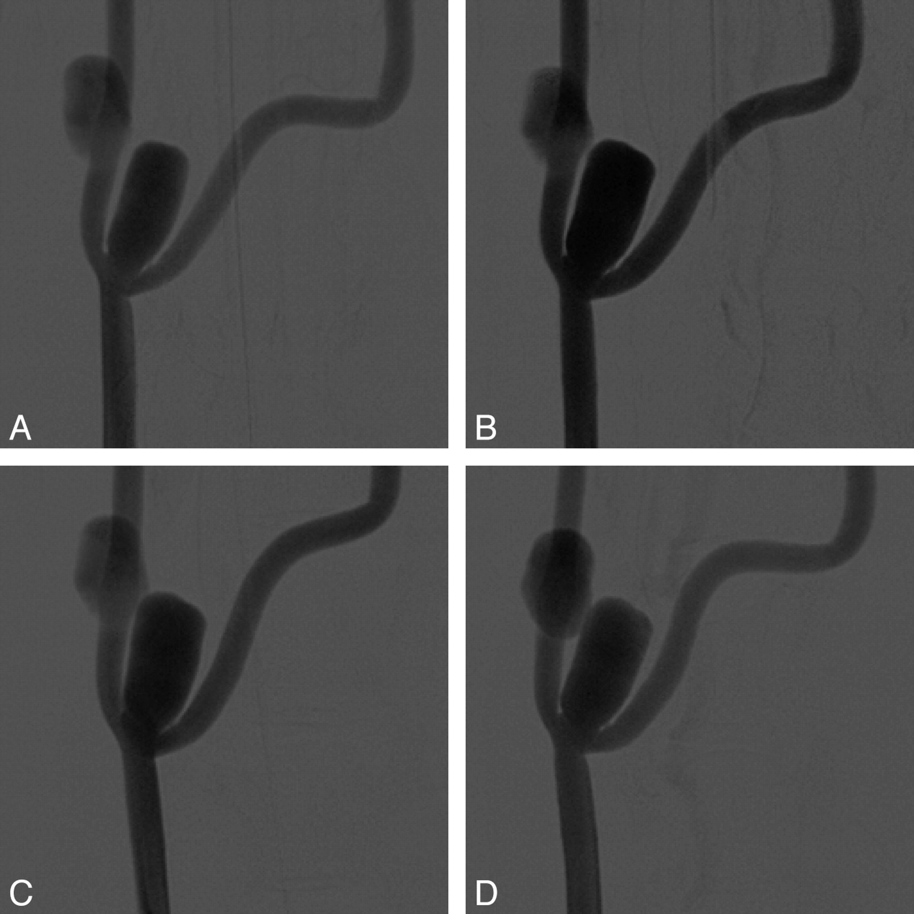
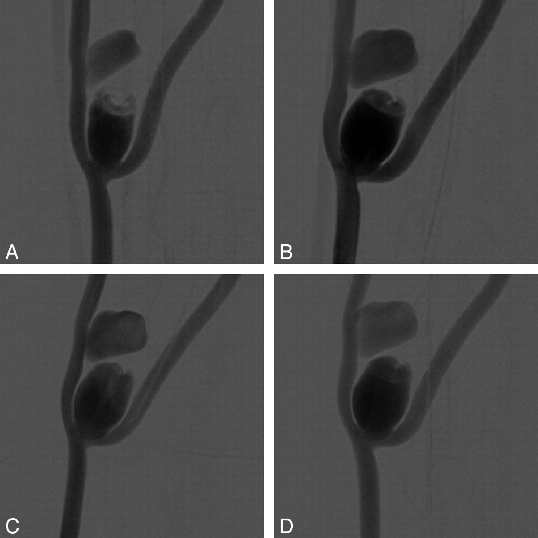
Four of 5 aneurysms were patent during this follow-up period (Fig 1). One aneurysm was regarded as a partially thrombosed aneurysm at 1 month. However, the extent of partial thrombosis was less at 10 months than at the time of aneurysm creation (Fig 2).

Representative DSA 1 (A), 4 (B), 7 (C), and 10 (D) months after creation. The venous pouch bifurcation aneurysms progressively increase in size during the first several months before appearing to stabilize.

Representative DSA 1 (A), 4 (B), 7 (C), and 10 (D) months after creation. The extent of partial thrombosis is less at 10 months than at the time of aneurysm creation.
Aneurysm Height
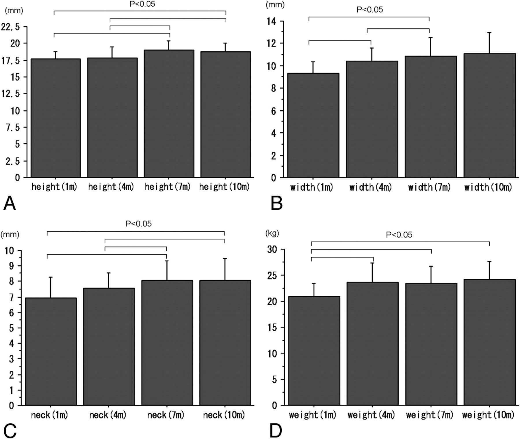
Mean aneurysm heights 1, 4, 7, and 10 months after creation were 17.8 ± 1.1, 17.9 ± 1.8, 19.3 ± 1.3, and 18.7 ± 1.3 mm, respectively. There were significant differences among these heights (P = .0057). The heights at 7 and 10 months after creation were significantly larger than those at 1 and 4 months (Table 1 and Fig 3A).

Bar graphs illustrate mean sizes of height (A), width (B), neck diameter (C), and body weight (D) in each group.
Aneurysm morphology and animal body weight during follow-up
Aneurysm Width
Mean aneurysm widths 1, 4, 7, and 10 months after creation were 9.4 ± 1.1, 10.6 ± 1.3, 11.2 ± 1.6, and 11.1 ± 1.9 mm, respectively. There were significant differences among these widths (P = .0406). The width at 4 months was significantly larger than that at 1 month, and that at 7 months was larger than those at 1 and 4 months (Table 1 and Fig 3B).
Aneurysm Neck
Mean neck diameters 1, 4, 7, and 10 months after creation were 6.9 ± 1.5, 7.5 ± 1.1, 8.2 ± 1.4, and 8.1 ± 1.4 mm, respectively. There were significant differences among these neck diameters (P = .057). The neck diameter at 7 months after creation was significantly larger than those at 1 and 4 months, and that at 10 months was significantly larger than those at 1 and 4 months (Table 1 and Fig 3C).
Body Weight
Mean animal weights 1, 4, 7, and 10 months after creation were 21.3 ± 2.6, 24.3 ± 3.6, 23.9 ± 3.5, and 24.2 ± 3.3 kg, respectively. There were significant differences among animal weights (P = .0158). The weights at 4 months were significantly more than that at 1 month, and those at 7 and 10 months were more than that at 1 month (Table 1 and Fig 3D).
Discussion
This study shows that the venous pouch bifurcation aneurysms can progressively increase in size (aneurysm height, width, and neck diameter) during the first several months over a 10-month period. Two previous reports investigated the natural history of experimental aneurysms created in dogs.7,8 Turk et al7 evaluated canine experimental bifurcation and orthogonal sidewall aneurysms. They reported that spontaneous occlusion of the sidewall canine vein patch aneurysm occurred in less than 10% and spontaneous thrombosis almost never occurred in the bifurcation aneurysm. However, their evaluation was limited, because the average follow-up period was only 74.9 days for sidewall aneurysms and 66.5 days for bifurcation aneurysms. They did not note significant changes to the aneurysm dimensions during this relatively short evaluation period. Kallmes et al8 reported the natural history of canine orthogonal side-wall aneurysms with sonography and angiography for a similar but shorter follow-up period of 7 months. They reported that orthogonal sidewall aneurysms may undergo spontaneous thrombosis in the early postoperative period, but that experimental sidewall aneurysms in dogs appear to remain patent beyond this period. With the longest follow-up period for experimental bifurcation aneurysms created in dogs, our study confirms the low rate of spontaneous thrombosis of experimental bifurcation aneurysms in dogs and is the first to document early progressive enlargement of these experimental aneurysms.
Although there are several animal aneurysm models to evaluate new devices, it is unknown which one is optimal. Sidewall aneurysms, particularly porcine, are known to spontaneously thrombose; therefore, these experimental sidewall aneurysms have significant limitations in the new device evaluation for most types of aneurysms. As an improvement, both an elastase-induced rabbit model and a canine bifurcation aneurysm model have been more recently used for device evaluation.1,5,6,13 The natural history of elastase-induced saccular aneurysm models in rabbits has been evaluated by Ding et al,9 with follow-up as long as 24 months after creation. In their study, none of the 20 elastase-induced aneurysms showed spontaneous thrombosis or significant change in dimensions (aneurysm neck, width, and height). However, the elastase-induced aneurysms are smaller and are, therefore, limited in the evaluation of large or giant aneurysms. These studies suggest that the elastase-induced aneurysm model in rabbits is better for the evaluation of smaller aneurysms (largest diameter, 4–10 mm), particularly with a small neck (neck <4 mm), whereas the canine bifurcation aneurysm model is better for the evaluation of larger aneurysms (largest diameter, 11–25 mm).
The progressive enlargement of the aneurysms may be related to animal body weight and animal maturation, because there were no significant differences in all of the dimensions between 7 and 10 months. Nonetheless, the apparent progressive enlargement of venous pouch bifurcation aneurysms in this study needs further investigation, including surgical technique and hemodynamic stresses. This model may simulate the dynamic process of some large and giant aneurysms, particularly those that are partially thrombosed and that can increase in size in humans, so that if endovascular occlusion devices achieve lasting closure of the aneurysms in this model, they may be effective for such challenging human aneurysm counterparts.
Limitations of this study include the small number of dogs. The costs of conducting such a study for a prolonged period (eg, maintenance costs) prevented more animals and even longer-term follow-up. In one of the aneurysms, partial thrombosis was observed. Although this partial thrombus was significantly less in the 10-month follow-up angiogram, this aneurysm was the first such partially thrombosed bifurcation aneurysm created and observed. Therefore, this case was considered unusual. We feel that spontaneous thrombosis can occur early in the period, related to technical difficulties associated with surgical aneurysm creation. On the other hand, we believe that spontaneous thrombosis never occurs beyond this time point, as mentioned by Turk et al7 that there was almost never spontaneous thrombosis of the canine bifurcation aneurysm at an average of 66.5 days after aneurysm creation.
Conclusions
In this follow-up study of venous pouch bifurcation aneurysms created in dogs up to a 10-month period, early progressive aneurysm enlargement was observed. Investigators should be aware of this observation if this experimental model is used to evaluate new endovascular devices for cerebral aneurysm treatment.
Acknowledgments
We are greatful to Dr. Aquilla S. Turk and his laboratory staff for their instruction to us about aneurysm creation.
References
- Received November 29, 2007.
- Accepted after revision January 10, 2008.
- Copyright © American Society of Neuroradiology
In this issue


Jump to section
Related Articles
Cited By...
- Surgical technique for venous patch aneurysms with no neck in a rabbit model
- A Large and Giant Bifurcation Aneurysm Model in Canines: Proof of Feasibility
- In Vivo Experimental Intracranial Aneurysm Models: A Systematic Review
- Symptomatic Perianeurysmal Edema Following Bare Platinum Embolization of a Small Unruptured Cerebral Aneurysm
- Dangerous Advances in Measurements from Digital Subtraction Angiography: When Is a Millimeter Not a Millimeter?