Advertisement

Main menu

  • Home
  • Content
    • Current Issue
    • Accepted Manuscripts
    • Article Preview
    • Past Issue Archive
    • AJNR Case Collection
    • Case of the Week Archive
    • Classic Case Archive
    • Case of the Month Archive
  • Special Collections
    • Spinal CSF Leak Articles (Jan 2020-June 2024)
    • 2024 AJNR Journal Awards
    • Most Impactful AJNR Articles
  • Multimedia
    • AJNR Podcast
    • AJNR Scantastics
    • Video Articles
  • For Authors
    • Submit a Manuscript
    • Author Policies
    • Fast publishing of Accepted Manuscripts
    • Graphical Abstract Preparation
    • Manuscript Submission Guidelines
    • Imaging Protocol Submission
    • Submit a Case for the Case Collection
  • About Us
    • About AJNR
    • Editorial Board
  • More
    • Become a Reviewer/Academy of Reviewers
    • Subscribers
    • Permissions
    • Alerts
    • Feedback
    • Advertisers
    • ASNR Home
  • Other Publications
    • ajnr

User menu

  • Alerts
  • Log in

Search

  • Advanced search
American Journal of Neuroradiology

American Journal of Neuroradiology

ASHNR American Society of Functional Neuroradiology ASHNR American Society of Pediatric Neuroradiology ASSR
  • Alerts
  • Log in

Advanced Search

  • Home
  • Content
    • Current Issue
    • Accepted Manuscripts
    • Article Preview
    • Past Issue Archive
    • AJNR Case Collection
    • Case of the Week Archive
    • Classic Case Archive
    • Case of the Month Archive
  • Special Collections
    • Spinal CSF Leak Articles (Jan 2020-June 2024)
    • 2024 AJNR Journal Awards
    • Most Impactful AJNR Articles
  • Multimedia
    • AJNR Podcast
    • AJNR Scantastics
    • Video Articles
  • For Authors
    • Submit a Manuscript
    • Author Policies
    • Fast publishing of Accepted Manuscripts
    • Graphical Abstract Preparation
    • Manuscript Submission Guidelines
    • Imaging Protocol Submission
    • Submit a Case for the Case Collection
  • About Us
    • About AJNR
    • Editorial Board
  • More
    • Become a Reviewer/Academy of Reviewers
    • Subscribers
    • Permissions
    • Alerts
    • Feedback
    • Advertisers
    • ASNR Home
  • Follow AJNR on Twitter
  • Visit AJNR on Facebook
  • Follow AJNR on Instagram
  • Join AJNR on LinkedIn
  • RSS Feeds

Welcome to the new AJNR, Updated Hall of Fame, and more. Read the full announcements.


AJNR is seeking candidates for the position of Associate Section Editor, AJNR Case Collection. Read the full announcement.


  • Getting new auth cookie, if you see this message a lot, tell someone!
  • Getting new auth cookie, if you see this message a lot, tell someone!
Research ArticleSPINE

Unusual Manifestations of Vertebral Osteomyelitis: Intraosseous Lesions Mimicking Metastases

C.Y. Hsu, C.W. Yu, M.Z. Wu, B.B. Chen, K.M. Huang and T.T.F. Shih
American Journal of Neuroradiology June 2008, 29 (6) 1104-1110; DOI: https://doi.org/10.3174/ajnr.A1003
C.Y. Hsu
  • Find this author on Google Scholar
  • Find this author on PubMed
  • Search for this author on this site
C.W. Yu
  • Find this author on Google Scholar
  • Find this author on PubMed
  • Search for this author on this site
M.Z. Wu
  • Find this author on Google Scholar
  • Find this author on PubMed
  • Search for this author on this site
B.B. Chen
  • Find this author on Google Scholar
  • Find this author on PubMed
  • Search for this author on this site
K.M. Huang
  • Find this author on Google Scholar
  • Find this author on PubMed
  • Search for this author on this site
T.T.F. Shih
  • Find this author on Google Scholar
  • Find this author on PubMed
  • Search for this author on this site
  • Article
  • Figures & Data
  • Info & Metrics
  • Responses
  • References
  • PDF
Loading

Article Figures & Data

Figures

  • Tables
  • Fig 1.
    • Download figure
    • Open in new tab
    • Download powerpoint
    Fig 1.

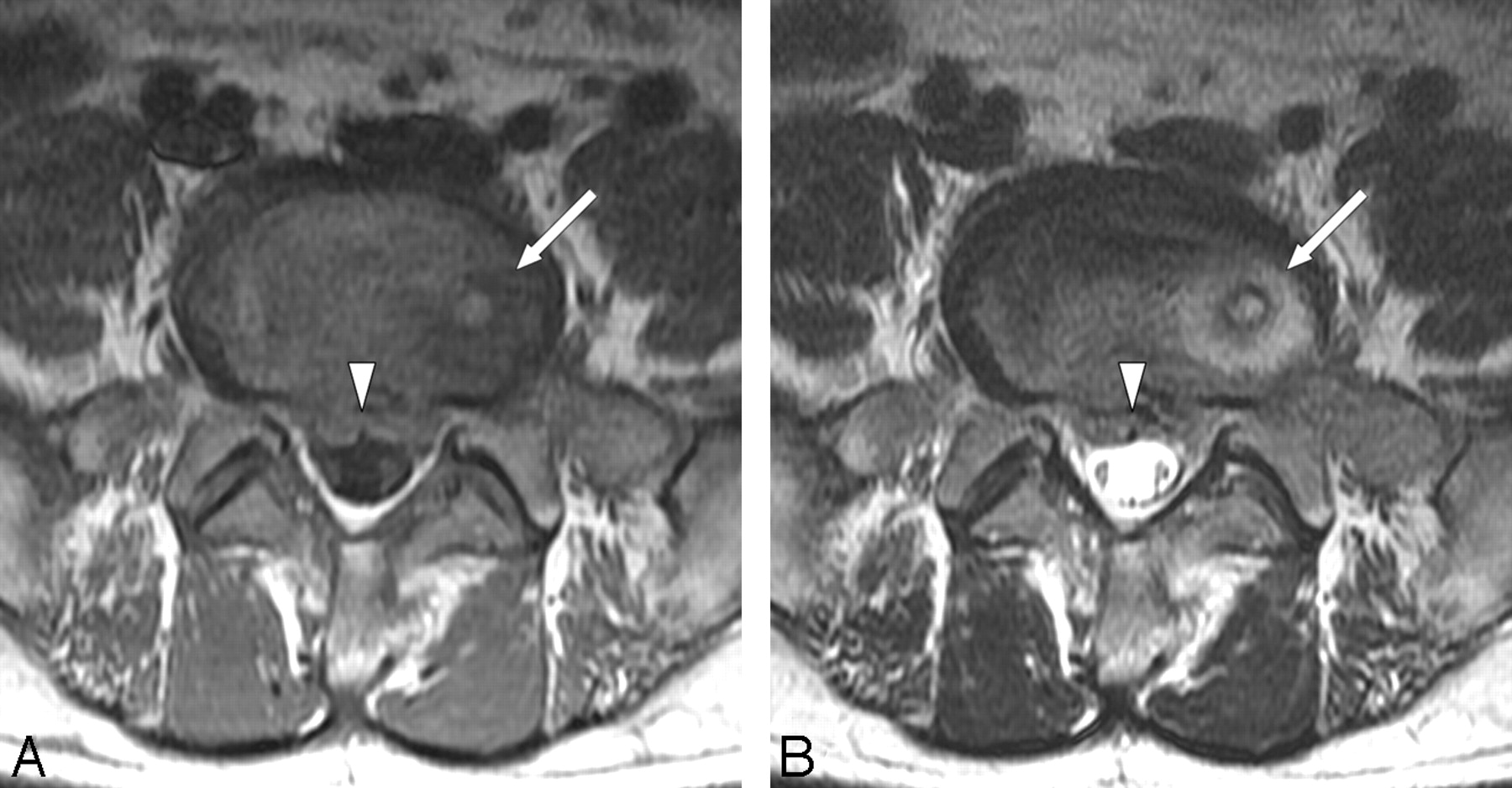
    A 42-year-old man experienced low-back pain and fever for 5 days. A and B, Axial T1-weighted (TR/TE, 600/16) (A) and axial T2-weighted (TR/TE, 4900/111) (B) MR images show an intraosseous lesion of T1-weighted low signal intensity and T2-weighted high signal intensity with an inner rim sign and an outer halo sign (arrow) near the upper endplate of S1. Note the limited amount of epidural soft-tissue inflammation (arrowhead).

  • Fig 2.
    • Download figure
    • Open in new tab
    • Download powerpoint
    Fig 2.

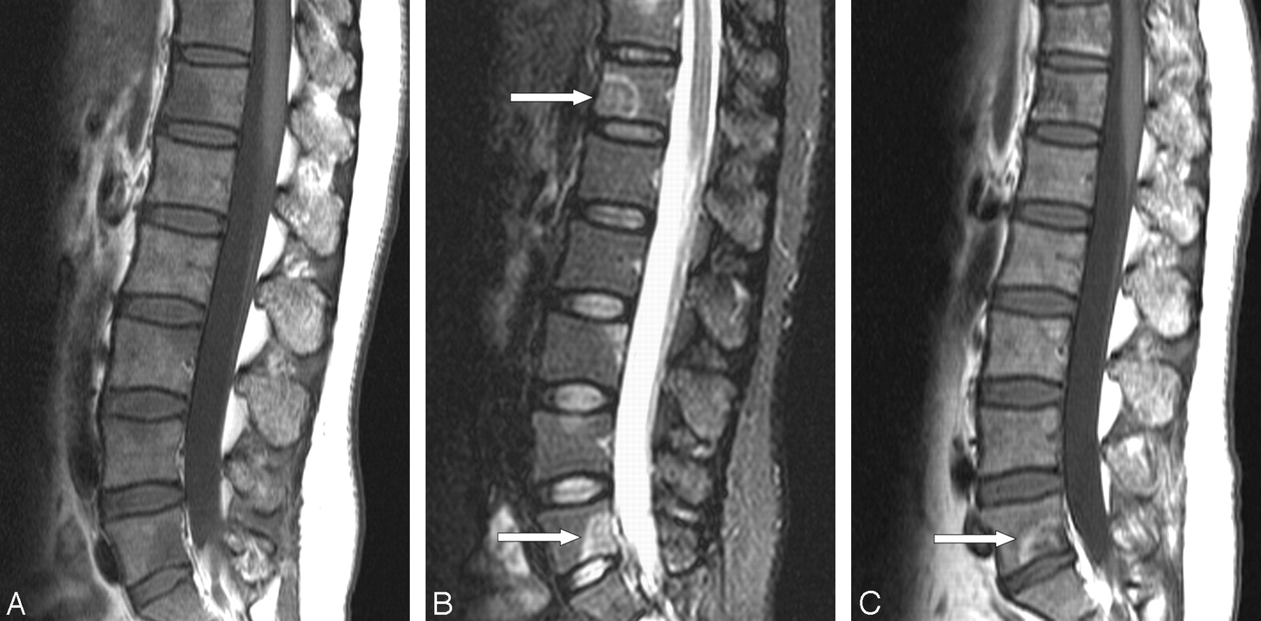
    A 46-year-old woman with fever for 2 days. A and B, Sagittal T1-weighted (TR/TE, 550/12) (A) and sagittal STIR (TR/TE/TI, 3130/65/160) (B) MR images show multiple intraosseous lesions of T1-weighted low signal intensity and STIR high signal intensity. Note some osseous lesions with a positive halo sign (arrows) and the absence of paraspinal or epidural inflammatory soft tissue. C, Sagittal T1-weighted (TR/TE, 550/12) follow-up MR image obtained 12 months later shows gradually reconstituted fatty marrow with a decrease in the number and extent of infectious foci. Note a peripheral rim of increased signal intensity (arrow) in a residual subchondral lesion of low signal intensity at the lower endplate of L5.

  • Fig 3.
    • Download figure
    • Open in new tab
    • Download powerpoint
    Fig 3.

    A 56-year-old man with fever for 1 week. A, Sagittal T1-weighted (TR/TE, 550/12) MR image shows multiple intraosseous lesions of low signal intensity involving the bodies and posterior elements of the vertebrae superimposed on the diffuse abnormal-signal-intensity marrow. Note an osseous lesion with a positive rim sign (arrow) at the T11 vertebra. B, Sagittal T1-weighted (TR/TE, 700/12) fat-suppressed contrast-enhanced MR image reveals global enhancement of intraosseous lesions (arrows). Note the absence of paraspinal or epidural inflammatory soft tissue. C, Sagittal T1-weighted (TR/TE, 700/12) fat-suppressed contrast-enhanced follow-up MR image obtained 5 months later shows an increase in extent, signal-intensity abnormality, and enhancement of T11, L1, L3, and apparently new L4 lesions (arrows) as well as a decrease in the extent of the L2 lesion (arrowhead). D, Sagittal T1-weighted (TR/TE, 700/12) follow-up MR image obtained 25 months later reveals almost complete reconstitution of fatty marrow in previously affected bone.

Tables

  • Figures
    • View popup
    Table 1:

    Demographic data, histopathologic findings, and culture results in 7 patients with unusual manifestations of vertebral osteomyelitis

    Patient No.Sex/Age (yr)Biopsy Sites and Histopathologic FindingsCulture Results of Microorganisms
    1M/53CT-guided bone biopsy, L5 vertebra: chronic osteomyelitisBone: gram-positive cocci
    Blood: S aureus
    2M/42CT-guided bone biopsy, S1 upper endplate: chronic osteomyelitisBone: (−)
    Blood: (−)
    3M/80CT-guided bone biopsy, L1 vertebra: chronic osteomyelitisBone: (−)
    Blood: (−)
    4F/46CT-guided bone biopsy, T11 vertebra: chronic osteomyelitisBone: (−)
    Blood: Salmonella organisms
    5M/56CT-guided bone biopsy, T11 vertebra: chronic osteomyelitisBone: (−)
    Blood: (−)
    6F/53CT-guided bone biopsy, L3 vertebra: chronic osteomyelitis, skin biopsy, shoulder: mycobacterial infectionBone: (−)
    Blood: (−)
    7M/66CT-guided bone biopsy, L3 vertebra: chronic fibrosis, lymph node biopsy, neck: mycobacterial infectionBone: (−)
    Blood: (−)
    • Note:—(−) indicates negative.

    • View popup
    Table 2:

    MR imaging findings in 7 patients with unusual manifestations of vertebral osteomyelitis

    Patient No.Involved Bones*Intraosseous LesionsSI of Intraosseous LesionsEnhancement of Intraosseous LesionsRim SignHalo SignPreservation of Intervertebral DisksSoft-Tissue Inflammation†
    T1WT2WSTIR
    1Spine (L5)Solitary↓−− or ↑↑GlobalYesYesYesYes
    2Spine (S1)Solitary↓↑↑GlobalYesYesYesYes
    3Spine (T10, L1, upper sacrum)Multiple↓↑↑GlobalYesYesYesYes
    4Spine (T5-L5, upper sacrum), bilateral pelvic girdlesMultiple↓−− or ↑↑Global or marginalNoYesYesYes
    5Spine (whole spine), bilateral ribsMultiple↓−− or ↑↑GlobalYesYesYesNo
    6Spine (whole spine)Multiple↓−− or ↑↑Global or marginalYesYesYesYes
    7Spine (whole spine), bilateral ribs, sternum, bilateral pectoral girdles, bilateral pelvic girdlesMultiple↓−− or ↑↑Global or marginalYesYesYesYes
    • Note:—SI indicates signal intensity; T1W, T1-weighted MR images; T2W, T2-weighted MR images; STIR, short τ inversion recovery; ↑, hyperintense; −−, isointense; ↓, hypointense.

    • * Posterior element involvement of the vertebrae is noted in 3 patients (patients 5–7).

    • † Limited amount of the paraspinal or epidural inflammatory tissue is noted in 6 patients (patients 1–4, 6, and 7).

    • View popup
    Table 3:

    Therapy, follow-up MR imaging studies, and clinical outcome in 7 patients with unusual manifestations of vertebral osteomyelitis

    Patient No.TherapyInterval between Initial and Subsequent MRI (months)Sequential Change between Initial and Last MRIClinical Outcome after Treatment
    1Antibiotic2HealingImproved
    2Surgical debridement and antibiotic5HealingImproved
    3Antibiotic4HealingImproved
    4Antibiotic3, 8, 12HealingImproved
    5Antibiotic and anti-TB5, 10.5, 25Almost healedCured
    6Antibiotic and anti-TB6.5HealingImproved
    7Antibiotic and anti-TB7HealingImproved
    • Note:—MRI indicates MR imaging; TB, tuberculosis.

PreviousNext
Back to top

In this issue

American Journal of Neuroradiology: 29 (6)
American Journal of Neuroradiology
Vol. 29, Issue 6
June 2008
  • Table of Contents
  • Index by author
Advertisement
Print
Download PDF
Email Article

Thank you for your interest in spreading the word on American Journal of Neuroradiology.

NOTE: We only request your email address so that the person you are recommending the page to knows that you wanted them to see it, and that it is not junk mail. We do not capture any email address.

Enter multiple addresses on separate lines or separate them with commas.
Unusual Manifestations of Vertebral Osteomyelitis: Intraosseous Lesions Mimicking Metastases
(Your Name) has sent you a message from American Journal of Neuroradiology
(Your Name) thought you would like to see the American Journal of Neuroradiology web site.
CAPTCHA
This question is for testing whether or not you are a human visitor and to prevent automated spam submissions.
Cite this article
C.Y. Hsu, C.W. Yu, M.Z. Wu, B.B. Chen, K.M. Huang, T.T.F. Shih
Unusual Manifestations of Vertebral Osteomyelitis: Intraosseous Lesions Mimicking Metastases
American Journal of Neuroradiology Jun 2008, 29 (6) 1104-1110; DOI: 10.3174/ajnr.A1003

Citation Manager Formats

  • BibTeX
  • Bookends
  • EasyBib
  • EndNote (tagged)
  • EndNote 8 (xml)
  • Medlars
  • Mendeley
  • Papers
  • RefWorks Tagged
  • Ref Manager
  • RIS
  • Zotero
0 Responses
Respond to this article
Share
Bookmark this article
Unusual Manifestations of Vertebral Osteomyelitis: Intraosseous Lesions Mimicking Metastases
C.Y. Hsu, C.W. Yu, M.Z. Wu, B.B. Chen, K.M. Huang, T.T.F. Shih
American Journal of Neuroradiology Jun 2008, 29 (6) 1104-1110; DOI: 10.3174/ajnr.A1003
del.icio.us logo Twitter logo Facebook logo Mendeley logo
  • Tweet Widget
  • Facebook Like
  • Google Plus One
Purchase

Jump to section

  • Article
    • Abstract
    • Methods
    • Results
    • Discussion
    • Conclusion
    • References
  • Figures & Data
  • Info & Metrics
  • Responses
  • References
  • PDF

Related Articles

  • No related articles found.
  • PubMed
  • Google Scholar

Cited By...

  • No citing articles found.
  • Crossref (15)
  • Google Scholar

This article has been cited by the following articles in journals that are participating in Crossref Cited-by Linking.

  • Antibiotic treatment in patients with chronic low back pain and vertebral bone edema (Modic type 1 changes): a double-blind randomized clinical controlled trial of efficacy
    Hanne B. Albert, Joan S. Sorensen, Berit Schiott Christensen, Claus Manniche
    European Spine Journal 2013 22 4
  • Imaging of Spine Infection
    Felix E. Diehn
    Radiologic Clinics of North America 2012 50 4
  • Percutaneous needle biopsy in diagnosis and identification of causative organisms in cases of suspected vertebral osteomyelitis
    Jennifer K. Sehn, Louis A. Gilula
    European Journal of Radiology 2012 81 5
  • Pyogenic Spinal Infections
    E. Turgut Tali, A. Yusuf Oner, A. Murat Koc
    Neuroimaging Clinics of North America 2015 25 2
  • Benign Bone Conditions That May Be FDG-avid and Mimic Malignancy
    Thomas C. Kwee, John M.H. de Klerk, Maarten Nix, Ben G.F. Heggelman, Stefan V. Dubois, Hugo J.A. Adams
    Seminars in Nuclear Medicine 2017 47 4
  • Candida Spondylitis: Comparison of MRI Findings With Bacterial and Tuberculous Causes
    Sheen-Woo Lee, Sang Hoon Lee, Hye Won Chung, Min Jee Kim, Min Jeong Seo, Myung Jin Shin
    American Journal of Roentgenology 2013 201 4
  • Diagnostic Value of Biopsy Techniques in Lumbar Spondylodiscitis: Percutaneous Needle Biopsy and Open Biopsy
    Kyoung Hyup Nam, Geun Sung Song, In Ho Han, Byung Kwan Choi, Seung Heon Cha
    Korean Journal of Spine 2011 8 4
  • Percutaneous Vertebroplasty
    Michael C. Hurley, Rami Kaakaji, Guilherme Dabus, Ali Shaibani, Mathew T. Walker, Richard G. Fessler, Bernard R. Bendok
    Neurosurgery Clinics of North America 2009 20 3
  • Imaging Findings of Solitary Spinal Bony Lesions and the Differential Diagnosis of Benign and Malignant Lesions
    Young Soo Kim, In Ho Han, In Sook Lee, Jung Sub Lee, Byung Kwan Choi
    Journal of Korean Neurosurgical Society 2012 52 2
  • Imaging of Bone Tumors and Tumor-Like Lesions
    Prudencia N. M. Tyrrell, Victor N. Cassar-Pullicino, Radhesh K. Lalam, Bernhard J. Tins
    2009

More in this TOC Section

  • Bern Score Validity for SIH
  • MP2RAGE 7T in MS Lesions of the Cervical Spine
  • Deep Learning for STIR Spine MRI Quality
Show more Spine

Similar Articles

Advertisement

Indexed Content

  • Current Issue
  • Accepted Manuscripts
  • Article Preview
  • Past Issues
  • Editorials
  • Editors Choice
  • Fellow Journal Club
  • Letters to the Editor

Cases

  • Case Collection
  • Archive - Case of the Week
  • Archive - Case of the Month
  • Archive - Classic Case

Special Collections

  • Special Collections

Resources

  • News and Updates
  • Turn around Times
  • Submit a Manuscript
  • Author Policies
  • Manuscript Submission Guidelines
  • Evidence-Based Medicine Level Guide
  • Publishing Checklists
  • Graphical Abstract Preparation
  • Imaging Protocol Submission
  • Submit a Case
  • Become a Reviewer/Academy of Reviewers
  • Get Peer Review Credit from Publons

Multimedia

  • AJNR Podcast
  • AJNR SCANtastic
  • Video Articles

About Us

  • About AJNR
  • Editorial Board
  • Not an AJNR Subscriber? Join Now
  • Alerts
  • Feedback
  • Advertise with us
  • Librarian Resources
  • Permissions
  • Terms and Conditions

American Society of Neuroradiology

  • Not an ASNR Member? Join Now

© 2025 by the American Society of Neuroradiology All rights, including for text and data mining, AI training, and similar technologies, are reserved.
Print ISSN: 0195-6108 Online ISSN: 1936-959X

Powered by HighWire