
Article Figures & Data
Figures
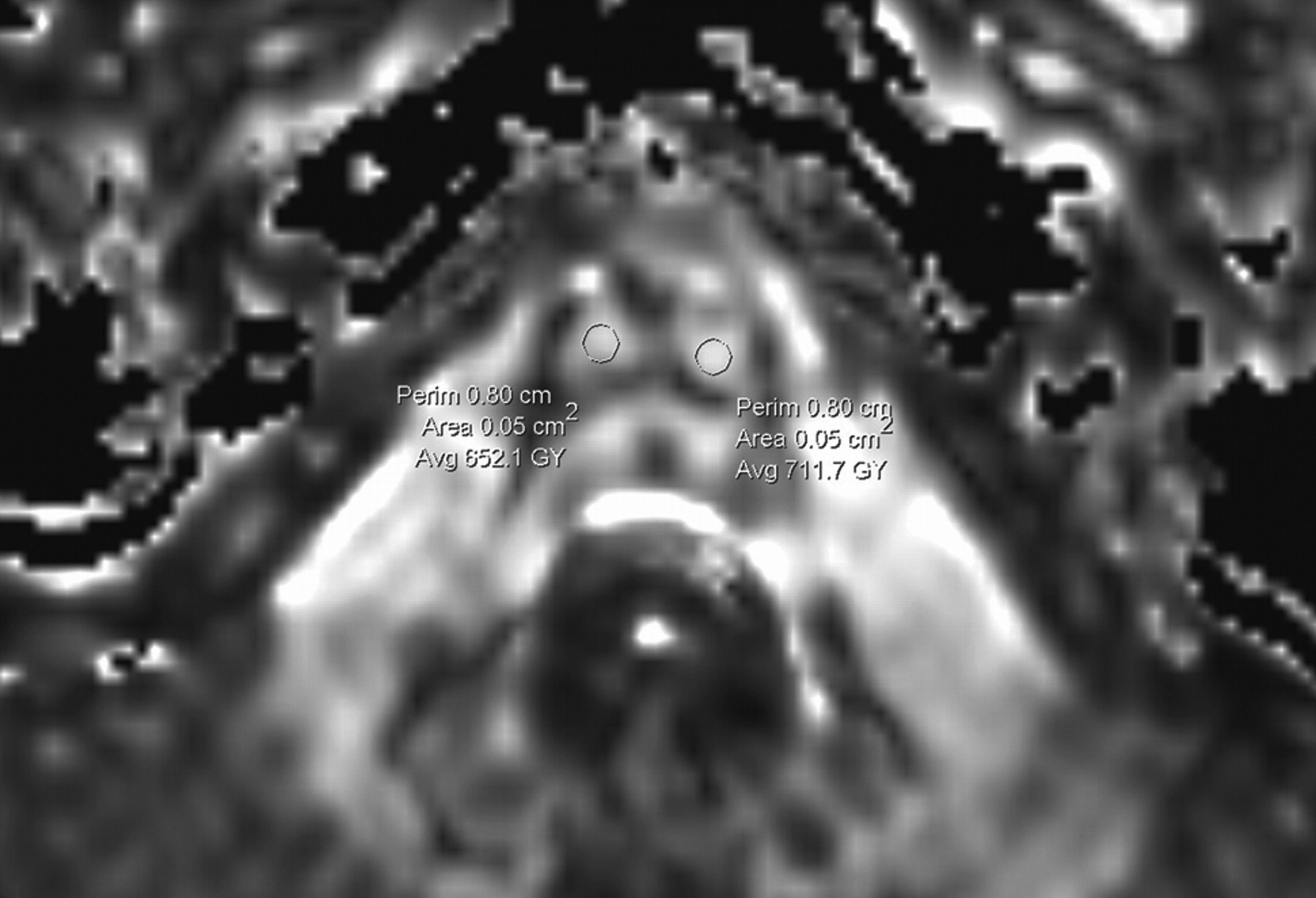
- Fig 1.
Normal subject. FA map shows highly directional white matter tracts (corticospinal tracts) in the region of brain stem. The ROIs (50 mm2) for the estimation of the FA values are placed exclusively within the central portion of the visualized white matter tracts.

Tables
- Table 1:
Descriptive statistics of FA values (×103) (SD), as well as results of the performed t test (P) in the examined anatomic structures on the basis of 2 b-values (mm2/s) and both scanning sessions.
Anatomic Structure b-Value Session 1 Session 2 Right internal capsule anterior limb 1000 691 (71) 672 (54) 700 702 (63) 718 (60) Right internal capsule genu 1000 702 (51)* 737 (48) P = .02 700 745 (67)* 760 (51) Right internal capsule posterior limb 1000 745 (33) 732 (45) 700 761 (54) 751 (37) Corpus callosum genu 1000 782 (71) 816 (64) 700 769 (74) 782 (70) Corpus callosum splenium 1000 823 (71) 836 (69) 700 829 (53) 851 (47) Left internal capsule anterior limb 1000 617 (81)* 682 (65) Left internal capsule genu 1000 729 (53) 746 (65) 700 741 (44) 752 (57) Left internal capsule posterior limb 1000 766 (51) 745 (55) 700 781 (53) 767 (65) Right corticospinal tract 1000 646 (67)† 570 (54)† P = .048 700 661 (54) 631 (60) Left corticospinal tract 1000 672 (73) 649 (61) 700 662 (91) 672 (72) Right thalamus 1000 349 (36)† 316 (24)† P = .024 700 358 (41) 340 (39) Note:—The values are based on the readings of the first rater.
* Statistically significant changes in the obtained FA between the 2 b-values are shown.
† Statistically significant changes in the obtained FA between the 2 scanning sessions are shown.
- Table 2:
Intraclass correlation coefficients for interrater agreement between 2 observers for the examined FA values in various anatomic locations
Anatomic Location b-Values Intraclass Correlation Coefficients (95% Confidence Intervals), mm2/s Right internal capsule anterior limb 1000 0.70 (0.49–0.85) 700 0.71 (0.61–0.92) Left internal capsule anterior limb 1000 0.58 (0.31–0.72) 700 0.37 (0.20–0.56) Right internal capsule posterior limb 1000 0.40 (0.28–0.60) 700 0.31 (0.10–0.52) Left internal capsule posterior limb 1000 0.46 (0.20–0.67) 700 0.53 (0.33–0.87) Right internal capsule genu 1000 0.58 (0.31–0.77) 700 0.39 (0.20–0.62) Left internal capsule genu 1000 0.38 (0.17–0.49) 700 0.28 (0.13–0.47) Corpus callosum genu 1000 0.73 (0.47–0.89) 700 0.60 (0.47–0.79) Corpus callosum splenium 1000 0.82 (0.61–0.90) 700 0.88 (0.59–0.96) Right corticospinal tract 1000 0.35 (0.18–0.58) 700 0.38 (0.16–0.53) Left corticospinal tract 1000 0.34 (0.29–0.62) 700 0.47 (0.29–0.75) Right thalamus 1000 0.64 (0.56–0.79) 700 0.65 (0.51–0.89) Note:—The coefficients are calculated for 2 b-values.
- Table 3:
Mean difference (SD) and 95% limits of agreement of the FA values (×103) for the ROI analysis between the 2 sessions, as well as measurement error and repeatability for the serial FA measurements in healthy subjects
Anatomic Structure b-Value Mean Difference (SD) 95% Limits of Agreement Within-Subject SD Within-Subject Coefficient of Variation, % Repeatability Coefficient Significant Change for a Single Subject, % Right internal capsule anterior limb 1000 −18.4 (95.71) −206 to 169.2 17 1.3 13 19 700 −27 (88.03) −145 to 199.5 40 6 11 16 Right internal capsule genu 1000 74.7 (99.31) −119.9 to 269.4 16 1.2 11 16 700 −5.7 (130.17) −260.9 to 249.4 26 3 9 10 Right internal capsule posterior limb 1000 16.9 (35.48) −52.6 to 86.5 13 0.6 7 7 700 9.5 (28.99) −47.3 to 66.3 14 2 5 5 Corpus callosum genu 1000 −22.2 (41.42) −103.4 to 59.0 12 0.4 4 4 700 12.9 (49.71) −84.5 to 110.3 22 3 6 7 Corpus callosum splenium 1000 10.6 (43.98) −103.4 to 59.0 20 2 7 6 700 −22.7 (37.51) −92.9 to 50.9 21 2 6 7 Left internal capsule anterior limb 1000 −4.1 (59.45) −199.0 to 190.8 51 6 12 18 700 −20.7 (43.58) −106.2 to 64.0 40 6 13 17 Left internal capsule genu 1000 −8.5 (66.63) −139.2 to 122.0 33 4 8 11 700 −3.4 (80.07) −160.4 to 153.5 46 5 11 15 Left internal capsule posterior limb 1000 23.0 (52.81) −80.5 to 126.5 31 3 7 19 700 28.3 (28.3) −41.0 to 97.5 22 2 6 7 Right corticospinal tract 1000 74.7 (99.31) −119.9 to 269.4 60 8 16 24 700 −6.7 (130.17) −260.9 to 249.4 62 10 17 27 Left corticospinal tract 1000 54.9 (99.27) −139.7 to 249.5 51 7 15 20 700 −11.5 (131.9) −139.7 to 247.1 63 9 18 26 Right thalamus 1000 36.5 (29.32) −20.9 to 94.0 22 6 16 17 700 10.9 (56.26) −95.4 to 117.3 26 8 7 21
In this issue


Jump to section
Related Articles
Cited By...
- Early Prediction of Delayed Ischemia and Functional Outcome in Acute Subarachnoid Hemorrhage: Role of Diffusion Tensor Imaging
- Toward Precision and Reproducibility of Diffusion Tensor Imaging: A Multicenter Diffusion Phantom and Traveling Volunteer Study
- A Validation Study of Multicenter Diffusion Tensor Imaging: Reliability of Fractional Anisotropy and Diffusivity Values
- Multicentre imaging measurements for oncology and in the brain