Advertisement

Main menu

  • Home
  • Content
    • Current Issue
    • Accepted Manuscripts
    • Article Preview
    • Past Issue Archive
    • AJNR Case Collection
    • Case of the Week Archive
    • Classic Case Archive
    • Case of the Month Archive
  • Special Collections
    • Spinal CSF Leak Articles (Jan 2020-June 2024)
    • 2024 AJNR Journal Awards
    • Most Impactful AJNR Articles
  • Multimedia
    • AJNR Podcast
    • AJNR Scantastics
    • Video Articles
  • For Authors
    • Submit a Manuscript
    • Author Policies
    • Fast publishing of Accepted Manuscripts
    • Graphical Abstract Preparation
    • Manuscript Submission Guidelines
    • Imaging Protocol Submission
    • Submit a Case for the Case Collection
  • About Us
    • About AJNR
    • Editorial Board
  • More
    • Become a Reviewer/Academy of Reviewers
    • Subscribers
    • Permissions
    • Alerts
    • Feedback
    • Advertisers
    • ASNR Home
  • Other Publications
    • ajnr

User menu

  • Alerts
  • Log in

Search

  • Advanced search
American Journal of Neuroradiology

American Journal of Neuroradiology

ASHNR American Society of Functional Neuroradiology ASHNR American Society of Pediatric Neuroradiology ASSR
  • Alerts
  • Log in

Advanced Search

  • Home
  • Content
    • Current Issue
    • Accepted Manuscripts
    • Article Preview
    • Past Issue Archive
    • AJNR Case Collection
    • Case of the Week Archive
    • Classic Case Archive
    • Case of the Month Archive
  • Special Collections
    • Spinal CSF Leak Articles (Jan 2020-June 2024)
    • 2024 AJNR Journal Awards
    • Most Impactful AJNR Articles
  • Multimedia
    • AJNR Podcast
    • AJNR Scantastics
    • Video Articles
  • For Authors
    • Submit a Manuscript
    • Author Policies
    • Fast publishing of Accepted Manuscripts
    • Graphical Abstract Preparation
    • Manuscript Submission Guidelines
    • Imaging Protocol Submission
    • Submit a Case for the Case Collection
  • About Us
    • About AJNR
    • Editorial Board
  • More
    • Become a Reviewer/Academy of Reviewers
    • Subscribers
    • Permissions
    • Alerts
    • Feedback
    • Advertisers
    • ASNR Home
  • Follow AJNR on Twitter
  • Visit AJNR on Facebook
  • Follow AJNR on Instagram
  • Join AJNR on LinkedIn
  • RSS Feeds

Welcome to the new AJNR, Updated Hall of Fame, and more. Read the full announcements.


AJNR is seeking candidates for the position of Associate Section Editor, AJNR Case Collection. Read the full announcement.


OtherBRAIN

Inclusion or Exclusion of Intratumoral Vessels in Relative Cerebral Blood Volume Characterization in Low-Grade Gliomas: Does It Make a Difference?

G. Brasil Caseiras, J.S. Thornton, T. Yousry, C. Benton, J. Rees, A.D. Waldman and H.R. Jäger
American Journal of Neuroradiology June 2008, 29 (6) 1140-1141; DOI: https://doi.org/10.3174/ajnr.A0993
G. Brasil Caseiras
  • Find this author on Google Scholar
  • Find this author on PubMed
  • Search for this author on this site
J.S. Thornton
  • Find this author on Google Scholar
  • Find this author on PubMed
  • Search for this author on this site
T. Yousry
  • Find this author on Google Scholar
  • Find this author on PubMed
  • Search for this author on this site
C. Benton
  • Find this author on Google Scholar
  • Find this author on PubMed
  • Search for this author on this site
J. Rees
  • Find this author on Google Scholar
  • Find this author on PubMed
  • Search for this author on this site
A.D. Waldman
  • Find this author on Google Scholar
  • Find this author on PubMed
  • Search for this author on this site
H.R. Jäger
  • Find this author on Google Scholar
  • Find this author on PubMed
  • Search for this author on this site
  • Article
  • Figures & Data
  • Supplemental
  • Info & Metrics
  • Responses
  • References
  • PDF
Loading

Abstract

SUMMARY: We assessed the influence of inclusion (method 1) and exclusion (method 2) of intratumoral vessels when determining maximum relative cerebral blood volume (rCBVmax) in 3 types of low-grade gliomas (LGGs): astrocytomas, oligoastrocytomas, and oligodendrogliomas. Method 1 yielded significantly higher mean rCBVmax than method 2. However, only method 2 demonstrated a significant (P = .026) association between rCBVmax and membership of a differently ranked histologic category. Exclusion of intratumoral vessels appears, therefore, preferable when determining rCBVmax in LGGs.

Dynamic susceptibility contrast-enhanced (DSC) MR perfusion imaging has become an important technique for studying brain tumors. Intratumoral relative cerebral blood volume (CBV [rCBV]) correlates with histologic and angiographic measures of tumor vascularity,1 helps predict the histologic grade of gliomas,2 and correlates with outcome in low-grade gliomas (LGGs).3 High-grade gliomas usually have a higher rCBV than low-grade tumors, but rCBVs may also be elevated in low-grade oligodendrogliomas (ODs),4 which confounds tumor grading.5

Techniques of data acquisition, postprocessing, and analysis influence rCBV measurements. Gradient-echo echo-planar imaging (GE-EPI) pulse sequences are more sensitive to larger vessels than spin-echo echo-planar imaging (SE-EPI) methods and provide better differentiation between histopathologic tumor grades.6

Wetzel et al7 found that interobserver and intraobserver reproducibility for intratumoral rCBV were best when the highest CBV from several regions of interest (ROIs) was chosen and highlighted the importance of excluding large vessels. Inclusion or exclusion of intratumoral vessels is often not explicitly stated in glioma perfusion studies, though some investigators focused on excluding peritumoral vessels.6 The significance of intratumoral vessels for rCBV measurements has, to our knowledge, not been formally examined.

Here we studied the influence of intratumoral vessels on rCBV characterization in 3 histologic categories of low-grade glial tumors: astrocytomas (ACs), ODs, and oligoastrocytomas (OAs).

Technique and Results

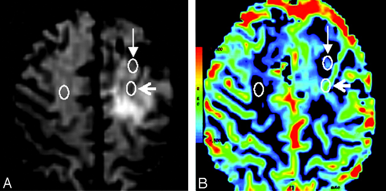
Thirty-four patients with LGGs, composed of 21 ACs, 8 ODs, and 5 OAs, had DSC GE-EPI (TR = 1200 ms; TE = 40 ms; flip angle = 20°; FOV = 26 cm; matrix = 96 × 128; section thickness = 5 mm) at 1.5T (GE Signa Horizon Echospeed LX9.1; GE Healthcare, Waukesha, Wis) with a bolus of 0.1 mmol/kg of body weight of gadoterate meglumine at 5 mL/s. Color maps of rCBV were generated with FuncTool 1.9 (GE Healthcare) and analyzed by 2 neuroradiologists (H.R.J. and G.B.C., with 15 and 7 years of experience, respectively), reaching a consensus for placement of ROIs. At least 6 intratumoral ROIs with a size of 9 pixels were placed over areas showing mostly elevated CBV on color perfusion maps. Blood vessels within the tumor were identified on unprocessed perfusion images acquired between the time points of maximum arterial and venous signal intensity drop. Sections above and below intratumoral vessels were viewed to identify potentially confounding large-vessel partial volume effects. We used 2 different methods for selecting the ROI with the maximum intratumoral CBV: method 1 included and method 2 excluded ROIs situated over intratumoral blood vessels and associated partial volume effects. The maximum rCBV (rCBVmax) was then obtained by dividing the highest intratumoral CBV by the mean CBV obtained from a contralateral normal-appearing white matter ROI (Fig 1).

Fig 1.
  • Download figure
  • Open in new tab
  • Download powerpoint
Fig 1.

T2*-weighted image during maximum arterial signal intensity drop (A) and rCBV map (B) in low-grade OD, demonstrating the position of ROIs used to calculate rCBVmax. Method 1 (open arrowhead, posterior ROI in the left cerebral hemisphere, overlying an intratumoral vessel) and method 2 (closed arrowhead, anterior ROI in the left cerebral hemisphere, lying outside intratumoral vessels). The mean CBV from contralateral normal-appearing white matter (ROI in the right cerebral hemisphere) was used to normalize the data for each method. White circles have been superimposed on the original color-coded ROIs generated by FuncTool.

Mean rCBVmaxs obtained for each group with each method are shown in the Table. Method 1 yielded higher mean values and wider ranges than method 2 in all 3 of the histologic tumor types, particularly in OA and OD. For the patient group as a whole, there was a significant difference between mean rCBVmax obtained using each method (P < .001, Wilcoxon test). Ordinal regression was used to assess the relationship of rCBVmax and tumor histology, categorized in 3 groups (0: OA; 1: AC; 2: OD). Only method 2 showed a significant association between rCBVmax and the risk of being in a histologic category with a higher ordinate. Using method 2, the odds ratio of being in a higher category was 4.25 (95% confidence interval: 1.19-15.14 for each additional unit rCBV increment; P = .026). Method 1 did not demonstrate a significant association between rCBVmax and the risk of being in a higher category (P = .638).

View this table:
  • View inline
  • View popup

CBVmax obtained using methods 1 and 2

Discussion

Several studies have demonstrated increased rCBV in high-grade gliomas compared with low-grade tumors.2,4–6,8–10 There is, however, considerable variation in the reported rCBV values for low- and high-grade tumors (On-line Table). In SE-EPI DSC imaging, transverse relaxation rates peak at a vessel diameter of 1-2 μm, whereas in GE-EPI DSC imaging they plateau at 3–4 μm and then remain independent of vessel size, explaining the lower rCBVs found with SE techniques.6 Differences may also be due to variations in the histologic types of LGG examined and inclusion or exclusion of intratumoral vessels in the analysis, often not specified.

We demonstrated that inclusion of large intratumoral vessels significantly increases rCBVmax values in all types of LGG. Their identification may be difficult on rCBV color maps alone and necessitates reviewing of unprocessed perfusion data. The diameter of intratumoral vessels clearly identifiable on GE-EPI images lies in the millimeter range (approximating the size of peripheral leptomeningeal vessels), whereas glioma neoangiogenetic vessels in animal models measure between 40 and 250 μm.6

In concordance with previous investigators, we found higher rCBVmax in tumors with oligodendral elements than in purely astrocytic tumors,5 explained by the “chicken wire” hypervascularity seen in the former. Cha et al4 chose intratumoral ROIs with an automated method targeting areas of maximum signal intensity decrease during the first pass of the gadolinium-based contrast bolus. This method probably incorporated intratumoral vessels and yielded mean rCBV for ODs of 3.68. Spampinato et al8 presented one of the few reports specifying exclusion of large intratumoral vessels for ROI analysis. Their mean rCBV measurement for a mixed group of low-grade OA and OD (1.61) lies between our group mean rCBV measurements of OA (1.53) and OD (2.21) using method 2. Our findings highlight the importance of using a consistent ROI placement technique, particularly if rCBV data are to be pooled in multicenter studies. The relationship between rCBVmax on MR perfusion imaging and histologic classification as OA versus OD has not been reported previously. We were able to demonstrate significant association between maximum intratumoral rCBV values and histopathologic classification as AC, OD, and AO, but only when using method 2. As a preferred technique we, therefore, recommend exclusion of intratumoral vessels when determining maximum intratumoral rCBV from GE-EPI DSC-derived data. Although beyond the scope of this work, future research will address the influence of intratumoral vessels on rCBV measurements in high-grade gliomas, which are naturally subject to greater variability.

Footnotes

  • indicates supplemental on-line table.

References

  1. ↵
    Sugahara T, Koroghi Y, Kochi M, et al. Correlation of MR imaging-determined cerebral blood maps with histologic and angiographic determination of vascularity of gliomas. AJR Am J Roentgenol 1998;171:1479–86
    CrossRefPubMed
  2. ↵
    Law M, Yang S, Wang H, et al. Glioma grading: sensitivity, specificity, and predictive values of perfusion MR imaging and proton MR spectroscopic imaging compared with conventional MR imaging. AJNR Am J Neuroradiol 2003;24:1989–98
    Abstract/FREE Full Text
  3. ↵
    Law M, Oh S, Babb JS, et al. Low-grade gliomas: dynamic susceptibility-weighted contrast-enhanced perfusion MR imaging–prediction of patient clinical response. Radiology 2006;238:658–67
    CrossRefPubMed
  4. ↵
    Cha S, Tihan T, Crawford F, et al. Differentiation of low-grade oligodendrogliomas from low-grade astrocytomas by using quantitative blood-volume measurements derived from dynamic susceptibility contrast-enhanced MR imaging. AJNR Am J Neuroradiol 2005;26:266–73
    Abstract/FREE Full Text
  5. ↵
    Lev MH, Ozsunar Y, Henson JW, et al. Glial tumor grading and outcome prediction using dynamic spin-echo MR susceptibility mapping compared with conventional contrast-enhanced MR: confounding effect of elevated rCBV of oligodendrogliomas [corrected]. AJNR Am J Neuroradiol 2004;25:214–21
    Abstract/FREE Full Text
  6. ↵
    Sugahara T, Korogi Y, Kochi M, et al. Perfusion-sensitive MR imaging of gliomas: comparison between gradient-echo and spin-echo echo-planar imaging techniques. AJNR Am J Neuroradiol 2001;22:1306–15
    Abstract/FREE Full Text
  7. ↵
    Wetzel SG, Cha S, Johnson G, et al. Relative cerebral blood volume measurements in intracranial mass lesions: interobserver and intraobserver reproducibility study. Radiology 2002;224:797–803
    CrossRefPubMed
  8. ↵
    Spampinato MV, Smith JK, Kwock L, et al. Cerebral blood volume measurements and proton MR spectroscopy in grading of oligodendroglial tumors. AJR Am J Roentgenol 2007;188:204–12
    CrossRefPubMed
  9. Yang D, Korogi T, Sugahara T, et al. Cerebral gliomas: prospective comparison of multivoxel 2D chemical-shift imaging proton MR spectroscopy, echoplanar perfusion and diffusion-weighted MRI. Neuroradiol 2002;44:656–66
    CrossRefPubMed
  10. ↵
    Hakyemez B, Erdogan C, Ercan I, et al. High-grade and low-grade gliomas: differentiation by using perfusion MR imaging. Clin Radiol 2005;60:493–502
    CrossRefPubMed
  • Received June 20, 2007.
  • Accepted after revision September 10, 2007.
  • Copyright © American Society of Neuroradiology
View Abstract
PreviousNext
Back to top

In this issue

American Journal of Neuroradiology: 29 (6)
American Journal of Neuroradiology
Vol. 29, Issue 6
June 2008
  • Table of Contents
  • Index by author
Advertisement
Print
Download PDF
Email Article

Thank you for your interest in spreading the word on American Journal of Neuroradiology.

NOTE: We only request your email address so that the person you are recommending the page to knows that you wanted them to see it, and that it is not junk mail. We do not capture any email address.

Enter multiple addresses on separate lines or separate them with commas.
Inclusion or Exclusion of Intratumoral Vessels in Relative Cerebral Blood Volume Characterization in Low-Grade Gliomas: Does It Make a Difference?
(Your Name) has sent you a message from American Journal of Neuroradiology
(Your Name) thought you would like to see the American Journal of Neuroradiology web site.
CAPTCHA
This question is for testing whether or not you are a human visitor and to prevent automated spam submissions.
Cite this article
G. Brasil Caseiras, J.S. Thornton, T. Yousry, C. Benton, J. Rees, A.D. Waldman, H.R. Jäger
Inclusion or Exclusion of Intratumoral Vessels in Relative Cerebral Blood Volume Characterization in Low-Grade Gliomas: Does It Make a Difference?
American Journal of Neuroradiology Jun 2008, 29 (6) 1140-1141; DOI: 10.3174/ajnr.A0993

Citation Manager Formats

  • BibTeX
  • Bookends
  • EasyBib
  • EndNote (tagged)
  • EndNote 8 (xml)
  • Medlars
  • Mendeley
  • Papers
  • RefWorks Tagged
  • Ref Manager
  • RIS
  • Zotero
0 Responses
Respond to this article
Share
Bookmark this article
Inclusion or Exclusion of Intratumoral Vessels in Relative Cerebral Blood Volume Characterization in Low-Grade Gliomas: Does It Make a Difference?
G. Brasil Caseiras, J.S. Thornton, T. Yousry, C. Benton, J. Rees, A.D. Waldman, H.R. Jäger
American Journal of Neuroradiology Jun 2008, 29 (6) 1140-1141; DOI: 10.3174/ajnr.A0993
del.icio.us logo Twitter logo Facebook logo Mendeley logo
  • Tweet Widget
  • Facebook Like
  • Google Plus One
Purchase

Jump to section

  • Article
    • Abstract
    • Technique and Results
    • Discussion
    • Footnotes
    • References
  • Figures & Data
  • Supplemental
  • Info & Metrics
  • Responses
  • References
  • PDF

Related Articles

  • PubMed
  • Google Scholar

Cited By...

  • ASFNR Recommendations for Clinical Performance of MR Dynamic Susceptibility Contrast Perfusion Imaging of the Brain
  • Semi-automated and automated glioma grading using dynamic susceptibility-weighted contrast-enhanced perfusion MRI relative cerebral blood volume measurements
  • Correlation of MR Relative Cerebral Blood Volume Measurements with Cellular Density and Proliferation in High-Grade Gliomas: An Image-Guided Biopsy Study
  • Crossref (20)
  • Google Scholar

This article has been cited by the following articles in journals that are participating in Crossref Cited-by Linking.

  • ASFNR Recommendations for Clinical Performance of MR Dynamic Susceptibility Contrast Perfusion Imaging of the Brain
    K. Welker, J. Boxerman, A. Kalnin, T. Kaufmann, M. Shiroishi, M. Wintermark
    American Journal of Neuroradiology 2015 36 6
  • Magnetic Resonance Perfusion and Permeability Imaging in Brain Tumors
    Saulo Lacerda, Meng Law
    Neuroimaging Clinics of North America 2009 19 4
  • Principles of T2*‐weighted dynamic susceptibility contrast MRI technique in brain tumor imaging
    Mark S. Shiroishi, Gloria Castellazzi, Jerrold L. Boxerman, Francesco D'Amore, Marco Essig, Thanh B. Nguyen, James M. Provenzale, David S. Enterline, Nicoletta Anzalone, Arnd Dörfler, Àlex Rovira, Max Wintermark, Meng Law
    Journal of Magnetic Resonance Imaging 2015 41 2
  • Low-Grade Gliomas: Six-month Tumor Growth Predicts Patient Outcome Better than Admission Tumor Volume, Relative Cerebral Blood Volume, and Apparent Diffusion Coefficient
    Gisele Brasil Caseiras, Olga Ciccarelli, Daniel R. Altmann, Christopher E. Benton, Daniel J. Tozer, Paul S. Tofts, Tarek A. Yousry, Jeremy Rees, Adam D. Waldman, Hans Rolf Jäger
    Radiology 2009 253 2
  • Relative cerebral blood volume measurements of low-grade gliomas predict patient outcome in a multi-institution setting
    Gisele B. Caseiras, Sophie Chheang, James Babb, Jeremy H. Rees, Nicole Pecerrelli, Daniel J. Tozer, Christopher Benton, David Zagzag, Glyn Johnson, Adam D. Waldman, H.R. Jäger, Meng Law
    European Journal of Radiology 2010 73 2
  • Correlation of MR Relative Cerebral Blood Volume Measurements with Cellular Density and Proliferation in High-Grade Gliomas: An Image-Guided Biopsy Study
    S.J. Price, H.A.L. Green, A.F. Dean, J. Joseph, P.J. Hutchinson, J.H. Gillard
    American Journal of Neuroradiology 2011 32 3
  • Machine learning in preoperative glioma MRI: Survival associations by perfusion‐based support vector machine outperforms traditional MRI
    Kyrre E. Emblem, Paulina Due‐Tonnessen, John K. Hald, Atle Bjornerud, Marco C. Pinho, David Scheie, Lothar R. Schad, Torstein R. Meling, Frank G. Zoellner
    Journal of Magnetic Resonance Imaging 2014 40 1
  • Quantification of serial changes in cerebral blood volume and metabolism in patients with recurrent glioblastoma undergoing antiangiogenic therapy
    Andreas Stadlbauer, Petra Pichler, Marianne Karl, Sebastian Brandner, Claudia Lerch, Bertold Renner, Gertraud Heinz
    European Journal of Radiology 2015 84 6
  • Perfusion and diffusion MRI combined with 11C-methionine PET in the preoperative evaluation of suspected adult low-grade gliomas
    Shala Ghaderi Berntsson, Anna Falk, Irina Savitcheva, Andrea Godau, Maria Zetterling, Göran Hesselager, Irina Alafuzoff, Elna-Marie Larsson, Anja Smits
    Journal of Neuro-Oncology 2013 114 2
  • Signal intensity in T2’ magnetic resonance imaging is related to brain glioma grade
    Laura Saitta, Oliver Heese, Ann-Freya Förster, Jakob Matschke, Susanne Siemonsen, Lucio Castellan, Manfred Westphal, Jens Fiehler, Einar Goebell
    European Radiology 2011 21 5

More in this TOC Section

  • Predictors of Reperfusion in Patients with Acute Ischemic Stroke
  • Qualitative and Quantitative Analysis of MR Imaging Findings in Patients with Middle Cerebral Artery Stroke Implanted with Mesenchymal Stem Cells
  • Multimodal CT Provides Improved Performance for Lacunar Infarct Detection
Show more Brain

Similar Articles

Advertisement

Indexed Content

  • Current Issue
  • Accepted Manuscripts
  • Article Preview
  • Past Issues
  • Editorials
  • Editors Choice
  • Fellow Journal Club
  • Letters to the Editor

Cases

  • Case Collection
  • Archive - Case of the Week
  • Archive - Case of the Month
  • Archive - Classic Case

Special Collections

  • Special Collections

Resources

  • News and Updates
  • Turn around Times
  • Submit a Manuscript
  • Author Policies
  • Manuscript Submission Guidelines
  • Evidence-Based Medicine Level Guide
  • Publishing Checklists
  • Graphical Abstract Preparation
  • Imaging Protocol Submission
  • Submit a Case
  • Become a Reviewer/Academy of Reviewers
  • Get Peer Review Credit from Publons

Multimedia

  • AJNR Podcast
  • AJNR SCANtastic
  • Video Articles

About Us

  • About AJNR
  • Editorial Board
  • Not an AJNR Subscriber? Join Now
  • Alerts
  • Feedback
  • Advertise with us
  • Librarian Resources
  • Permissions
  • Terms and Conditions

American Society of Neuroradiology

  • Not an ASNR Member? Join Now

© 2025 by the American Society of Neuroradiology All rights, including for text and data mining, AI training, and similar technologies, are reserved.
Print ISSN: 0195-6108 Online ISSN: 1936-959X

Powered by HighWire