Advertisement

Main menu

  • Home
  • Content
    • Current Issue
    • Accepted Manuscripts
    • Article Preview
    • Past Issue Archive
    • AJNR Case Collection
    • Case of the Week Archive
    • Classic Case Archive
    • Case of the Month Archive
  • Special Collections
    • Spinal CSF Leak Articles (Jan 2020-June 2024)
    • 2024 AJNR Journal Awards
    • Most Impactful AJNR Articles
  • Multimedia
    • AJNR Podcast
    • AJNR Scantastics
    • Video Articles
  • For Authors
    • Submit a Manuscript
    • Author Policies
    • Fast publishing of Accepted Manuscripts
    • Graphical Abstract Preparation
    • Manuscript Submission Guidelines
    • Imaging Protocol Submission
    • Submit a Case for the Case Collection
  • About Us
    • About AJNR
    • Editorial Board
  • More
    • Become a Reviewer/Academy of Reviewers
    • Subscribers
    • Permissions
    • Alerts
    • Feedback
    • Advertisers
    • ASNR Home
  • Other Publications
    • ajnr

User menu

  • Alerts
  • Log in

Search

  • Advanced search
American Journal of Neuroradiology

American Journal of Neuroradiology

ASHNR American Society of Functional Neuroradiology ASHNR American Society of Pediatric Neuroradiology ASSR
  • Alerts
  • Log in

Advanced Search

  • Home
  • Content
    • Current Issue
    • Accepted Manuscripts
    • Article Preview
    • Past Issue Archive
    • AJNR Case Collection
    • Case of the Week Archive
    • Classic Case Archive
    • Case of the Month Archive
  • Special Collections
    • Spinal CSF Leak Articles (Jan 2020-June 2024)
    • 2024 AJNR Journal Awards
    • Most Impactful AJNR Articles
  • Multimedia
    • AJNR Podcast
    • AJNR Scantastics
    • Video Articles
  • For Authors
    • Submit a Manuscript
    • Author Policies
    • Fast publishing of Accepted Manuscripts
    • Graphical Abstract Preparation
    • Manuscript Submission Guidelines
    • Imaging Protocol Submission
    • Submit a Case for the Case Collection
  • About Us
    • About AJNR
    • Editorial Board
  • More
    • Become a Reviewer/Academy of Reviewers
    • Subscribers
    • Permissions
    • Alerts
    • Feedback
    • Advertisers
    • ASNR Home
  • Follow AJNR on Twitter
  • Visit AJNR on Facebook
  • Follow AJNR on Instagram
  • Join AJNR on LinkedIn
  • RSS Feeds

Welcome to the new AJNR, Updated Hall of Fame, and more. Read the full announcements.


AJNR is seeking candidates for the position of Associate Section Editor, AJNR Case Collection. Read the full announcement.


Research ArticleBRAIN

Metabolic Assessment of Gliomas Using 11C-Methionine, [18F] Fluorodeoxyglucose, and 11C-Choline Positron-Emission Tomography

T. Kato, J. Shinoda, N. Nakayama, K. Miwa, A. Okumura, H. Yano, S. Yoshimura, T. Maruyama, Y. Muragaki and T. Iwama
American Journal of Neuroradiology June 2008, 29 (6) 1176-1182; DOI: https://doi.org/10.3174/ajnr.A1008
T. Kato
  • Find this author on Google Scholar
  • Find this author on PubMed
  • Search for this author on this site
J. Shinoda
  • Find this author on Google Scholar
  • Find this author on PubMed
  • Search for this author on this site
N. Nakayama
  • Find this author on Google Scholar
  • Find this author on PubMed
  • Search for this author on this site
K. Miwa
  • Find this author on Google Scholar
  • Find this author on PubMed
  • Search for this author on this site
A. Okumura
  • Find this author on Google Scholar
  • Find this author on PubMed
  • Search for this author on this site
H. Yano
  • Find this author on Google Scholar
  • Find this author on PubMed
  • Search for this author on this site
S. Yoshimura
  • Find this author on Google Scholar
  • Find this author on PubMed
  • Search for this author on this site
T. Maruyama
  • Find this author on Google Scholar
  • Find this author on PubMed
  • Search for this author on this site
Y. Muragaki
  • Find this author on Google Scholar
  • Find this author on PubMed
  • Search for this author on this site
T. Iwama
  • Find this author on Google Scholar
  • Find this author on PubMed
  • Search for this author on this site
  • Article
  • Figures & Data
  • Info & Metrics
  • Responses
  • References
  • PDF
Loading

Article Figures & Data

Figures

  • Tables
  • Fig 1.
    • Download figure
    • Open in new tab
    • Download powerpoint
    Fig 1.

    Graph showing the correlation between tracer uptake and tumor grade. *P < .05, †P < .01, ‡P < .001.
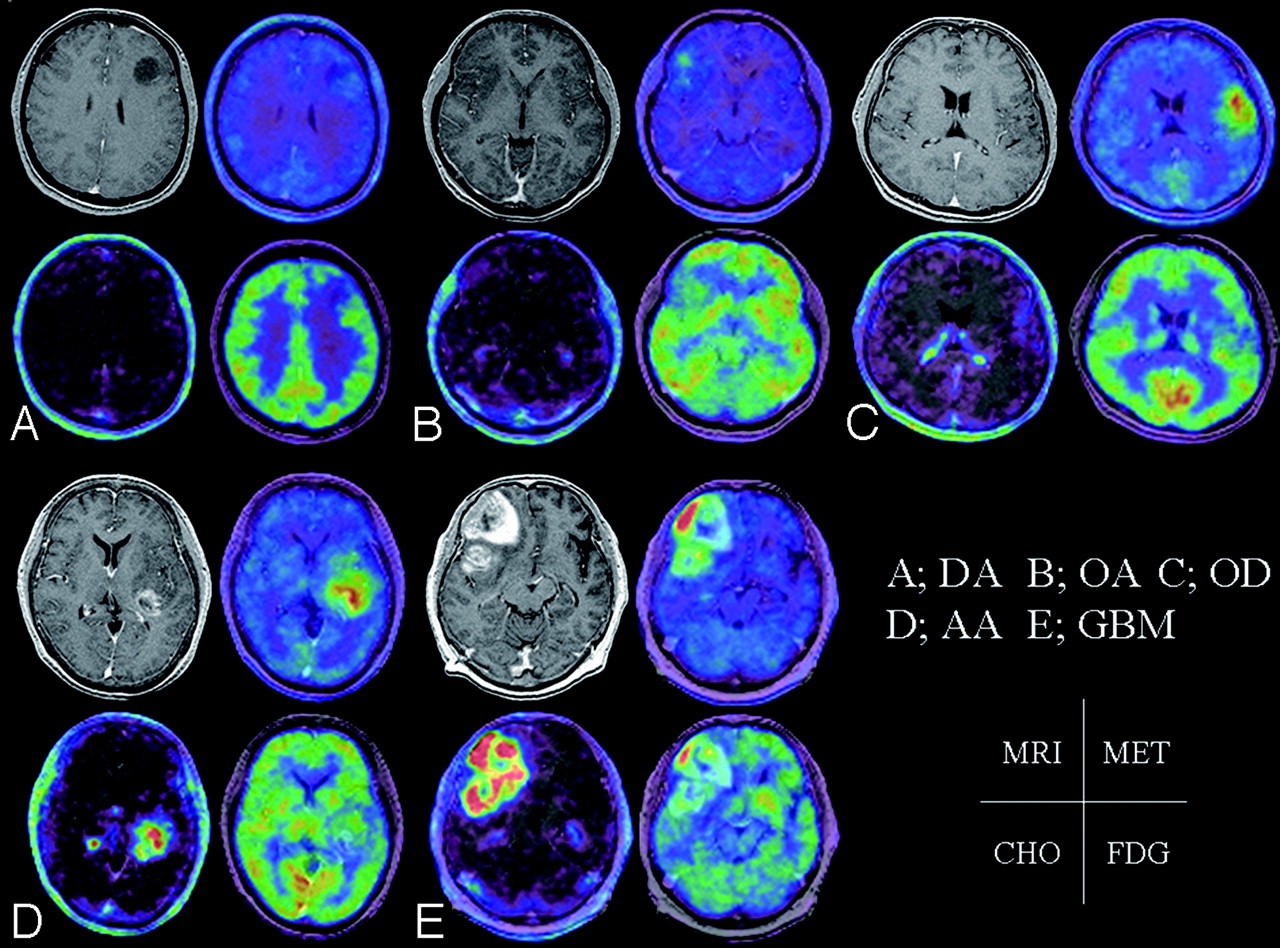
  • Fig 2.
    • Download figure
    • Open in new tab
    • Download powerpoint
    Fig 2.

    Left top, Contrast-enhanced, T1-weighted image. Right top, MET PET is superimposed on MR imaging. Left bottom, CHO PET is superimposed on MR imaging. Right bottom, FDG PET is superimposed on MR imaging. A, A 32-year-old woman presented with diffuse astrocytoma. MET T/N ratio = 1.72, CHO T/N ratio = 1.38, and FDG T/N ratio = 0.66. B, A 23-year-old woman presented with oligoastrocytoma. MET T/N ratio = 2.76, CHO T/N ratio = 1.82, and FDG T/N ratio = 0.92. C, A 44-year-old man presented with oligodendroglioma. MET T/N ratio = 3.71, CHO T/N ratio = 2.74, and FDG T/N ratio = 1.07. D, A 62-year-old woman presented with anaplastic astrocytoma. MET T/N ratio = 4.26, CHO T/N ratio = 10.17, and FDG T/N ratio = 1.24. E, A 68-year-old man presented with glioblastoma multiforme. MET T/N ratio = 6.85, CHO T/N ratio = 33.38, and FDG T/N ratio = 2.55.
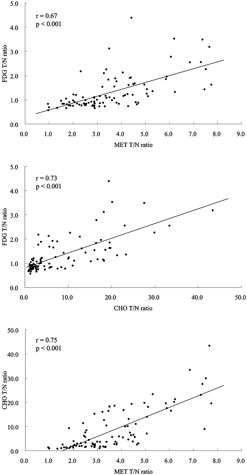
  • Fig 3.
    • Download figure
    • Open in new tab
    • Download powerpoint
    Fig 3.

    Graph showing the correlation between MET T/N ratio and FDG T/N ratio (r = 0.67; P < .001), CHO T/N ratio and FDG T/N ratio (r = 0.73; P < .001), and MET T/N ratio and CHO T/N ratio (r = 0.75; P < .001) in all of the gliomas.
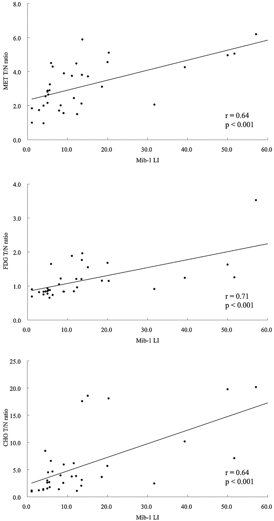
  • Fig 4.
    • Download figure
    • Open in new tab
    • Download powerpoint
    Fig 4.

    Graph showing the correlation between Mib-1 LI and MET T/N ratio (r = 0.64; P < .001), FDG T/N ratio (r = 0.71; P < .001), and CHO T/N ratio (r = 0.64; P < .001) in ATs.

Tables

  • Figures
    • View popup
    Table 1:

    Histopathologic results according to WHO classification and MRI findings

    WHO ClassificationNo. of PatientsAge, Mean ± SD, yTumor Size, Mean ± SD, cm3MRI Enhancement*
    NoneWeakStrong
    Grade II
    Diffuse astrocytoma1437.9 ± 13.451.1 ± 44.41112
    Oligodendroglioma936.8 ± 12.465.7 ± 56.7630
    Oligoastrocytoma1438.6 ± 14.148.3 ± 41.01211
    Grade III
    Anaplastic astrocytoma1943.2 ± 13.648.7 ± 23.3586
    Anaplastic oligodendroglioma1350.2 ± 14.840.4 ± 27.6427
    Anaplastic oligoastrocytoma544.2 ± 18.835.2 ± 10.2320
    Grade IV
    Glioblastoma multiforme2160.0 ± 10.943.7 ± 32.70318
    Total9545.9 ± 15.647.6 ± 35.6412034
    • Note:—MRI indicates MR imaging; WHO, World Health Organization.

    • * None indicates no enhancement; weak, partial or slight enhancement; strong, obvious enhancement.

    • View popup
    Table 2:

    T/N ratio in gliomas

    TumorGrade II, Mean ± SDGrade III, Mean ± SDGrade IV, Mean ± SD
    Astrocytic tumor, n141921
        MET2.24 ± 0.903.03 ± 1.025.03 ± 1.65
        FDG0.79 ± 0.081.27 ± 0.461.88 ± 0.78
        CHO2.69 ± 2.044.76 ± 3.0418.35 ± 6.73
    Oligodendroglial tumor913
        MET3.95 ± 1.604.46 ± 1.55
        FDG1.03 ± 0.401.71 ± 1.09
        CHO3.46 ± 2.5212.71 ± 12.21
    Oligoastrocytic tumor145
        MET2.60 ± 0.912.83 ± 0.99
        FDG1.00 ± 0.450.85 ± 0.15
        CHO3.78 ± 3.363.02 ± 1.74
    • Note:—T/N ratio indicates tumor/normal brain uptake ratio; MET, 11C-methionine; FDG, [18F] fluorodeoxyglucose; CHO, 11C-choline.

    • View popup
    Table 3:

    Summary statistics of ANCOVAs

    VariableMET, PFDG, PCHO, P
    Size.07.39.19
    Grade<.005<.005<.001
    Type<.05.33.12
    Gd-DTPA enhancement<.05.30<.01
    • Note:—MET indicates 11C-methionine; FDG, [18F] fluorodeoxyglucose; CHO, 11C-choline; Gd-DTPA, gadopentetate dimeglumine.

    • View popup
    Table 4:

    Percentage of T/N ratio more than 2.0 in gliomas

    VariableGrade II (37), n (%)Grade III (37), n (%)Grade IV (21), n (%)Overall (95), n (%)*
    MET28 (75.7)34 (91.9)21 (100.0)83 (87.4)
    FDG1 (2.7)6 (16.2)6 (28.6)13 (13.7)
    CHO18 (48.6)29 (78.4)21 (100.0)68 (71.6)
    • Note:—T/N ratio indicates tumor/normal brain uptake ratio; MET, 11C-methionine; FDG, [18F] fluorodeoxyglucose; CHO, 11C-choline.

    • * There were significant differences in the percentage of T/N ratio more than 2.0 among 3 tracers by using X2 test with Bonferroni correction. (MET/FDG and CHO/FDG: P < .001; MET/CHO: P < .01).

    • View popup
    Table 5:

    Correlation between tracer uptake and proliferation index

    VariableTumorMib-1 LI
    Pr
    METAstrocytic tumor<.0010.64
    Oligodendroglial tumor.63−0.13
    Oligoastrocytic tumor.840.05
    All tumor<.010.31
    FDGAstrocytic tumor<.0010.71
    Oligodendroglial tumor.270.29
    Oligoastrocytic tumor.78−0.07
    All tumor<.0010.42
    CHOAstrocytic tumor<.0010.64
    Oligodendroglial tumor.670.11
    Oligoastrocytic tumor.440.19
    All tumor<.0010.42
    • Note:—Proliferation index was measured by Mib-1 labeling index; MET indicates 11C-methionine; FDG, [18F] fluorodeoxyglucose; CHO, 11C-choline; LI, labeling index. P and r values were calculated by using Spearman correlation coefficients.

PreviousNext
Back to top

In this issue

American Journal of Neuroradiology: 29 (6)
American Journal of Neuroradiology
Vol. 29, Issue 6
June 2008
  • Table of Contents
  • Index by author
Advertisement
Print
Download PDF
Email Article

Thank you for your interest in spreading the word on American Journal of Neuroradiology.

NOTE: We only request your email address so that the person you are recommending the page to knows that you wanted them to see it, and that it is not junk mail. We do not capture any email address.

Enter multiple addresses on separate lines or separate them with commas.
Metabolic Assessment of Gliomas Using 11C-Methionine, [18F] Fluorodeoxyglucose, and 11C-Choline Positron-Emission Tomography
(Your Name) has sent you a message from American Journal of Neuroradiology
(Your Name) thought you would like to see the American Journal of Neuroradiology web site.
CAPTCHA
This question is for testing whether or not you are a human visitor and to prevent automated spam submissions.
Cite this article
T. Kato, J. Shinoda, N. Nakayama, K. Miwa, A. Okumura, H. Yano, S. Yoshimura, T. Maruyama, Y. Muragaki, T. Iwama
Metabolic Assessment of Gliomas Using 11C-Methionine, [18F] Fluorodeoxyglucose, and 11C-Choline Positron-Emission Tomography
American Journal of Neuroradiology Jun 2008, 29 (6) 1176-1182; DOI: 10.3174/ajnr.A1008

Citation Manager Formats

  • BibTeX
  • Bookends
  • EasyBib
  • EndNote (tagged)
  • EndNote 8 (xml)
  • Medlars
  • Mendeley
  • Papers
  • RefWorks Tagged
  • Ref Manager
  • RIS
  • Zotero
0 Responses
Respond to this article
Share
Bookmark this article
Metabolic Assessment of Gliomas Using 11C-Methionine, [18F] Fluorodeoxyglucose, and 11C-Choline Positron-Emission Tomography
T. Kato, J. Shinoda, N. Nakayama, K. Miwa, A. Okumura, H. Yano, S. Yoshimura, T. Maruyama, Y. Muragaki, T. Iwama
American Journal of Neuroradiology Jun 2008, 29 (6) 1176-1182; DOI: 10.3174/ajnr.A1008
del.icio.us logo Twitter logo Facebook logo Mendeley logo
  • Tweet Widget
  • Facebook Like
  • Google Plus One
Purchase

Jump to section

  • Article
    • Abstract
    • Materials and Methods
    • Results
    • Discussion
    • Conclusions
    • Acknowledgments
    • References
  • Figures & Data
  • Info & Metrics
  • Responses
  • References
  • PDF

Related Articles

  • No related articles found.
  • PubMed
  • Google Scholar

Cited By...

  • The Role of Metabolic Plasticity in Blood and Brain Stem Cell Pathophysiology
  • Molecular imaging of 1p/19q deletion in oligodendroglial tumours with 11C-methionine positron emission tomography
  • Diagnostic and Prognostic Value of 11C-Methionine PET for Nonenhancing Gliomas
  • Utility of Diffusion Tensor Imaging in Evaluation of the Peritumoral Region in Patients with Primary and Metastatic Brain Tumors
  • 62Cu-Diacetyl-Bis (N4-Methylthiosemicarbazone) PET in Human Gliomas: Comparative Study with [18F]Fluorodeoxyglucose and L-Methyl-[11C]Methionine PET
  • Multimodal Elucidation of Choline Metabolism in a Murine Glioma Model Using Magnetic Resonance Spectroscopy and 11C-Choline Positron Emission Tomography
  • Application of 62Cu-Diacetyl-Bis (N4-Methylthiosemicarbazone) PET Imaging to Predict Highly Malignant Tumor Grades and Hypoxia-Inducible Factor-1{alpha} Expression in Patients with Glioma
  • Voxel-Based Analysis of Dual-Time-Point 18F-FDG PET Images for Brain Tumor Identification and Delineation
  • Preliminary Study of 11C-Choline PET/CT for T Staging of Locally Advanced Nasopharyngeal Carcinoma: Comparison with 18F-FDG PET/CT
  • Examination of 11C-Methionine Metabolism by the Standardized Uptake Value in the Normal Brain of Children
  • Switching on the Lights for Real-Time Multimodality Tumor Neuroimaging: The Integrated Positron-Emission Tomography/MR Imaging System
  • Crossref (175)
  • Google Scholar

This article has been cited by the following articles in journals that are participating in Crossref Cited-by Linking.

  • Value of 11C-methionine PET in imaging brain tumours and metastases
    Andor W. J. M. Glaudemans, Roelien H. Enting, Mart A. A. M. Heesters, Rudi A. J. O. Dierckx, Ronald W. J. van Rheenen, Annemiek M. E. Walenkamp, Riemer H. J. A. Slart
    European Journal of Nuclear Medicine and Molecular Imaging 2013 40 4
  • Molecular imaging of gliomas with PET: Opportunities and limitations
    C. la Fougere, B. Suchorska, P. Bartenstein, F.-W. Kreth, J.-C. Tonn
    Neuro-Oncology 2011 13 8
  • Detecting response of rat C6 glioma tumors to radiotherapy using hyperpolarized [1‐13C]pyruvate and 13C magnetic resonance spectroscopic imaging
    Sam E. Day, Mikko I. Kettunen, Murali Krishna Cherukuri, James B. Mitchell, Martin J. Lizak, H. Douglas Morris, Shingo Matsumoto, Alan P. Koretsky, Kevin M. Brindle
    Magnetic Resonance in Medicine 2011 65 2
  • Differentiating radiation necrosis from tumor recurrence in high-grade gliomas: Assessing the efficacy of 18F-FDG PET, 11C-methionine PET and perfusion MRI
    Yong Hwy Kim, So Won Oh, You Jung Lim, Chul-Kee Park, Se-Hoon Lee, Keon Wook Kang, Hee-Won Jung, Kee Hyun Chang
    Clinical Neurology and Neurosurgery 2010 112 9
  • Brain Tumors
    Karl Herholz, Karl-Josef Langen, Christiaan Schiepers, James M. Mountz
    Seminars in Nuclear Medicine 2012 42 6
  • Comparison of &lt;sup&gt;11&lt;/sup&gt;C-Methionine, &lt;sup&gt;11&lt;/sup&gt;C-Choline, and &lt;sup&gt;18&lt;/sup&gt;F-Fluorodeoxyglucose-PET for Distinguishing Glioma Recurrence from Radiation Necrosis
    Shunsuke TAKENAKA, Yoshitaka ASANO, Jun SHINODA, Yuichi NOMURA, Shingo YONEZAWA, Kazuhiro MIWA, Hirohito YANO, Toru IWAMA
    Neurologia medico-chirurgica 2014 54 4
  • The role of positron emission tomography using carbon-11 and fluorine-18 choline in tumors other than prostate cancer: a systematic review
    Giorgio Treglia, Elisabetta Giovannini, Davide Di Franco, Maria Lucia Calcagni, Vittoria Rufini, Maria Picchio, Alessandro Giordano
    Annals of Nuclear Medicine 2012 26 6
  • Application of62Cu-Diacetyl-Bis (N4-Methylthiosemicarbazone) PET Imaging to Predict Highly Malignant Tumor Grades and Hypoxia-Inducible Factor-1α Expression in Patients with Glioma
    K. Tateishi, U. Tateishi, M. Sato, S. Yamanaka, H. Kanno, H. Murata, T. Inoue, N. Kawahara
    American Journal of Neuroradiology 2013 34 1
  • Differential reorganization of three syntax-related networks induced by a left frontal glioma
    Ryuta Kinno, Shinri Ohta, Yoshihiro Muragaki, Takashi Maruyama, Kuniyoshi L. Sakai
    Brain 2014 137 4
  • Correlation between the Uptake of 18F-Fluorodeoxyglucose (18F-FDG) and the Expression of Proliferation-Associated Antigen Ki-67 in Cancer Patients: A Meta-Analysis
    Sheng-ming Deng, Wei Zhang, Bin Zhang, Yin-yin Chen, Ji-hui Li, Yi-wei Wu, Elda Tagliabue
    PLOS ONE 2015 10 6

More in this TOC Section

  • Progression of Microstructural Damage in Spinocerebellar Ataxia Type 2: A Longitudinal DTI Study
  • Usefulness of Quantitative Susceptibility Mapping for the Diagnosis of Parkinson Disease
  • Evaluating the Effects of White Matter Multiple Sclerosis Lesions on the Volume Estimation of 6 Brain Tissue Segmentation Methods
Show more Brain

Similar Articles

Advertisement

Indexed Content

  • Current Issue
  • Accepted Manuscripts
  • Article Preview
  • Past Issues
  • Editorials
  • Editors Choice
  • Fellow Journal Club
  • Letters to the Editor

Cases

  • Case Collection
  • Archive - Case of the Week
  • Archive - Case of the Month
  • Archive - Classic Case

Special Collections

  • Special Collections

Resources

  • News and Updates
  • Turn around Times
  • Submit a Manuscript
  • Author Policies
  • Manuscript Submission Guidelines
  • Evidence-Based Medicine Level Guide
  • Publishing Checklists
  • Graphical Abstract Preparation
  • Imaging Protocol Submission
  • Submit a Case
  • Become a Reviewer/Academy of Reviewers
  • Get Peer Review Credit from Publons

Multimedia

  • AJNR Podcast
  • AJNR SCANtastic
  • Video Articles

About Us

  • About AJNR
  • Editorial Board
  • Not an AJNR Subscriber? Join Now
  • Alerts
  • Feedback
  • Advertise with us
  • Librarian Resources
  • Permissions
  • Terms and Conditions

American Society of Neuroradiology

  • Not an ASNR Member? Join Now

© 2025 by the American Society of Neuroradiology All rights, including for text and data mining, AI training, and similar technologies, are reserved.
Print ISSN: 0195-6108 Online ISSN: 1936-959X

Powered by HighWire