
Article Figures & Data
Figures
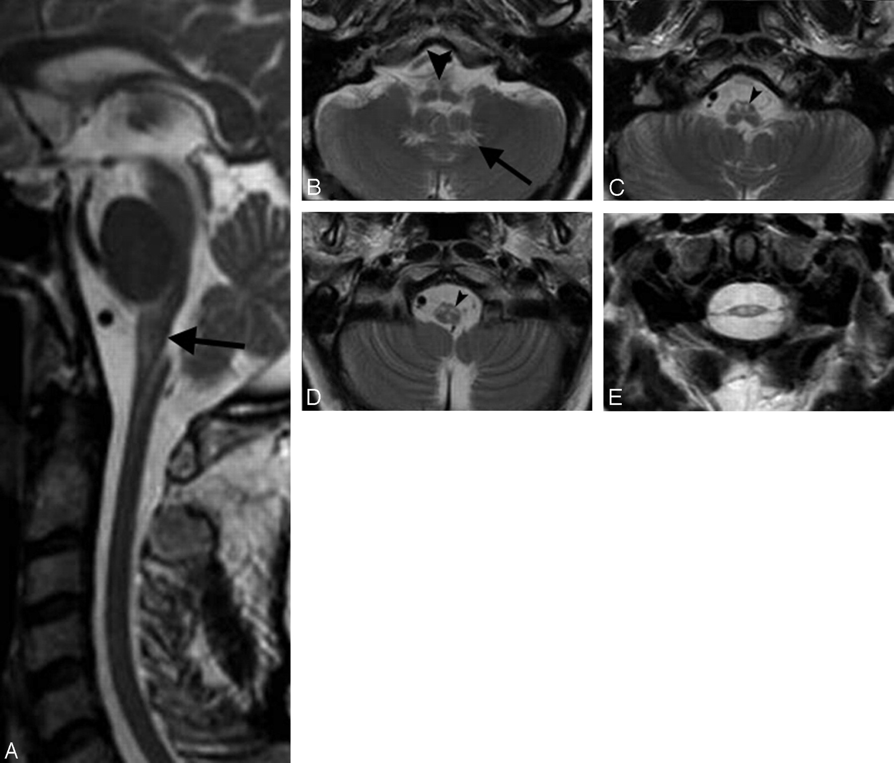
- Fig 1.
A, T2-weighted sagittal section in patient 9 shows thinning of the medulla oblongata and spinal cord with hyperintensity in the medulla; arrow indicates its posterior concave profile. B–D, Axial T2-weighted sections (thickness 3 mm) in the same patient demonstrate increased signal intensity involving the hilum of the dentate nuclei (arrow on the left side), the medial lemniscus (arrowhead, B), and the corticospinal tracts (arrowheads on the left side) in the medulla (C and D). In patient 11 (E), axial spinal cord T2-weighted section at the C1-C2 level shows severe atrophy with mild changes in signal intensity.
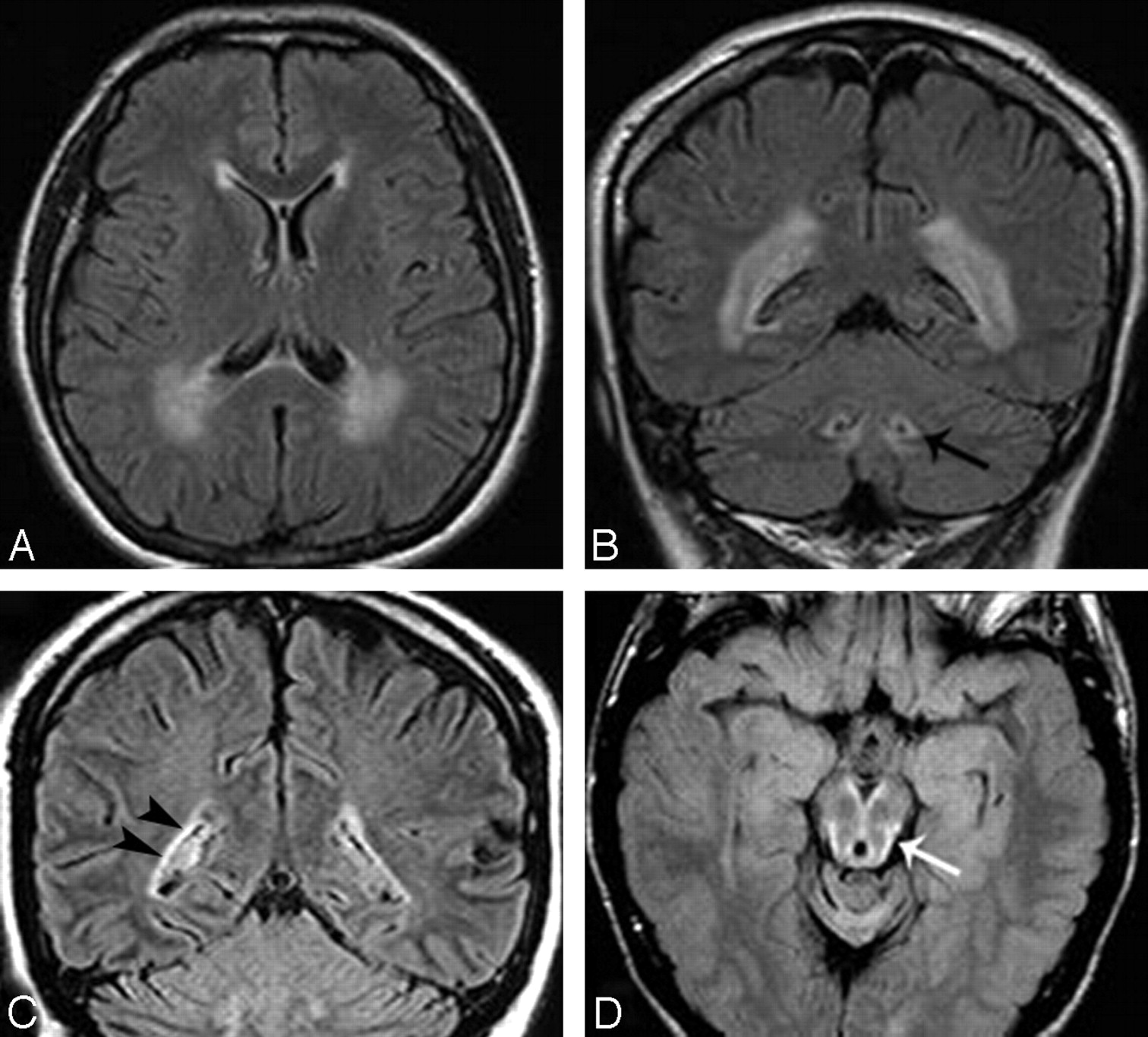
- Fig 2.
Axial and coronal FLAIR images in patient 6 show signal intensity changes prevalent in the cerebral posterior periventricular regions (A and B) and involvement of the hilum of the dentate nuclei (arrow on the left side, B). In patient 11, coronal FLAIR image (C) shows a thin bilateral band of periventricular hyperintensity (arrowheads on the right) not recognizable on T2-weighted images (not shown). In patient 3, midbrain peripheral rim of hyperintensity (arrow) is seen only on FLAIR section (D).
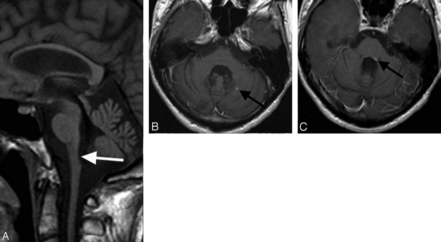
- Fig 3.
In patient 3, sagittal T1-weighted section (A) shows severe atrophy of the medulla oblongata (arrow) and spinal cord. Moderate cerebellar and cerebral atrophy is also present. Postcontrast axial sections (B and C) demonstrate 2 small areas of abnormal enhancement (arrows).


Tables
Pt/Age/Sex Disease Duration (y) Spastic Paraparesis Ataxia Dysphagia Dysarthria Dysphonia Palatal Myoclonus Others 1/26/M 7 + ± + + − Ny, diplopia, sphincter d. 2/36/F 2 + − + + − sphincter d. (f) 3/26/M 13 − + + + + Ny, ptosis, sphincter d., dysautonomia, scoliosis 4/39/F 5 − ± + + + Sphincter d., diplopia 5/30/M 0 − − − − − 6/43/F 3 months − + − + + Ny, ptosis 7/61/M 4 ± ± + + − Ny, shoulder girdle wasting & weakness (f) 8/58/M 4 + + − − − Ny, sphincter d. 9/52/F 0 − − − − − (f) 10/64/M 2 + − − − − 11/45/M 2 ± ± + + + Ny, ptosis, sphincter d. (f) Note:—AOAD indicates adult-onset Alexander disease; Ny, nystagmus; sphincter d., sphincter disturbances; +, moderate to severe; ±, mild; (f), familial case based on clinical, MR imaging, or genetic findings in relatives.
Patient/ Age/Sex Supratentorial Periventricular White Matter Abnormalities Brain stem: M.O. Atrophy/Signal Changes Cerebellum: Dentate Hilum Signal Changes Spinal Cord: C1-C2 Atrophy and Signal Changes Postcontrast Enhancement 1/26/M Thin band, also visible on T2-weighted images (garl) Severe atrophy/severe abnormalities Absent Present Present, mult 2/36/F Thin band, also visible on T2-weighted images Severe atrophy/severe abnormalities Present Present Not performed 3/26/M Large band of signal changes (garl) Severe atrophy/mild to moderate Present Present Present, mult 4/39/F Thin band, also visible on T2-weighted images Mild to moderate atrophy/severe abnormalities Present Present Present 5/30/M Very thin band recognizable on FLAIR, not on T2-weighted images Severe atrophy/mild to moderate Present Present Present, mult 6/43/F Large band of signal changes Mild to moderate atrophy/severe abnormalities Present Present Absent 7/61/M Absent Severe atrophy/severe abnormalities Present Present Absent 8/58/M Absent Mild to moderate atrophy/severe abnormalities Absent Present Absent 9/52/F Thin band, also visible on T2-weighted images Severe atrophy/severe abnormalities Present Present Present 10/64/M Absent Mild to moderate atrophy/severe abnormalities Absent Present Absent 11/45/M Very thin band recognizable on FLAIR, not on T2-weighted images Severe atrophy/severe abnormalities Present Present Absent Note:—AOAD indicates adult-onset Alexander disease; garl, ventricular garlands; M.O., medulla oblongata; mult, multiple areas of enhancement; FLAIR, fluid-attenuated inversion recovery.
Pt/Age/Sex Missense Mutations Exon Nucleotide Change Amino Acid Substitution 1/26/M 6 c.1076T>C p.L359P* 2/36/F 8 c.1178G>T p.S393I* 3/26/M 8 c.1246C>T‡ p.R416W 4/39/F 1 c.209G>A p.R70Q* 5/30/M 3 c.613G>A p.E205K† 6/43/F 1 c.208C>T‡ p.R70W 7/61/M 6 c.994G>A p.E332K 8/58/M 3 c.613G>A p.E205K† 9/52/F 8 c.1193C>A p.S398Y† 10/64/M 1 c.382G>A p.D128N† 11/45/M§ − − − Note:—GFAP indicates glial fibrillary acidic protein; AOAD, adult-onset Alexander disease.
* Mutations p.L359P,17 p.S393I,18 and p.R70Q19 have been found in these patients for the first time.
† These mutations have not been reported previously.
‡ Molecular data of patients 3 and 6 have been reported previously.19
§ Patient 11 carried no causative mutations but harbored the p.D157N rare polymorphism.
In this issue


Jump to section
Related Articles
Cited By...
- Teaching NeuroImages: Neuroimaging in Adult-Onset Alexander Disease
- Practical approach to the diagnosis of adult-onset leukodystrophies: an updated guide in the genomic era
- Spinal cord involvement in adult-onset metabolic and genetic diseases
- CSF and Blood Levels of GFAP in Alexander Disease
- Adult-onset Alexander's disease mimicking degenerative disease
- Characteristic abnormal signals in medulla oblongata--"eye spot" sign: Four cases of elderly-onset Alexander disease
- A practical approach to diagnosing adult onset leukodystrophies
- Neuroimaging and clinical features in type II (late-onset) Alexander disease
- Reviewing the genetic causes of spastic-ataxias
- GFAP mutations, age at onset, and clinical subtypes in Alexander disease
- A case of sporadic adult Alexander disease presenting with acute onset, remission and relapse
- MR Imaging Characteristics and Neuropathology of the Spinal Cord in Adult-Onset Autosomal Dominant Leukodystrophy with Autonomic Symptoms