
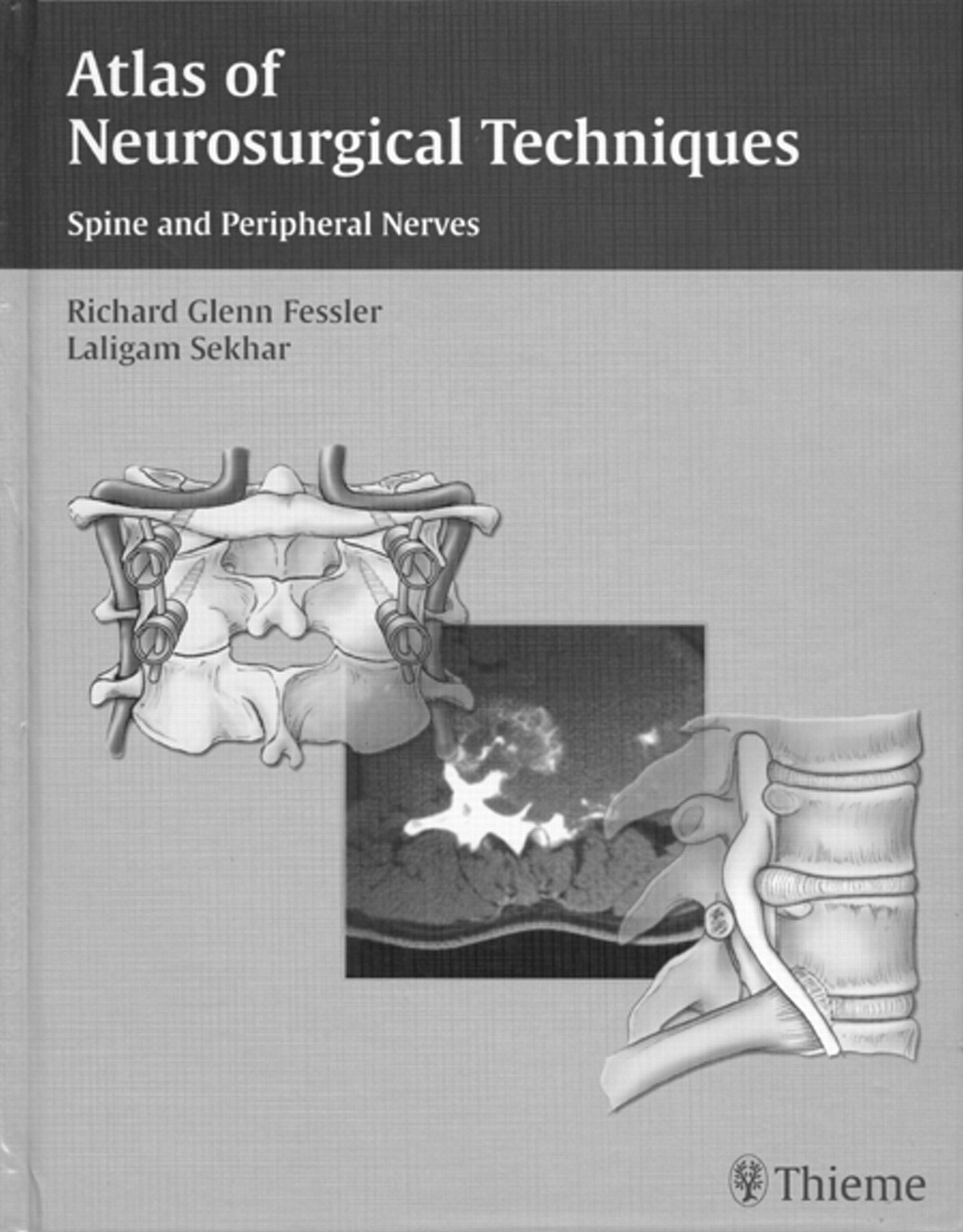
R.G. Fessler and L.N. Sekhar, eds. New York: Thieme Medical Publishers; 2006, 2048 pages, 2614 illustrations, $599.95.
This 2-volume set, Volume 1: Brain and Volume 2: Spine, lays out in artist's color drawings, line diagrams, intraoperative photographs, radiographs, and in an easily digestible written format the breadth and scope of virtually every neurosurgical procedure. This enormous work covers 2048 pages and is edited by 2 leading neurosurgeons, Drs. Sekhar and Fessler, with contributions from 156 authors (5 of whom are interventional neuroradiologists: Drs. Cloft, Debrun, Dion, Russell, and Valavanis). In an extensive work like this, it is virtually impossible to critique all areas, so this review will quickly summarize the organization of the book and then point out why, in this reviewer's eyes, the atlas should be available in every neuroradiology sectional library.
As a first observation, the set is far more than an “atlas of neurosurgical techniques.” If one were to presume by glancing at the title that these 2 books contained only a description of the surgical approaches and techniques in dealing with various brain and spine abnormalities, they would be totally wrong. These books are far more than that, and, hopefully, by a few selected examples, this review will emphasize that fact.
Volume 1: Brain
This volume is divided into 14 sections: “Introduction,” “Aneurysms,” “Arteriovenous Malformations,” “Vascular Disease,” “Brain Tumors,” “Intraventricular Lesions,” “Pineal Region Lesions,” “Cranial Base Lesions,” “Epilepsy and Functional Pain Disorders,” “Craniocerebral Trauma,” “Hydrocephalus,” “Central Nervous System Infections,” “Stereotactic Radiation Therapy,” and “Minimally Invasive Surgery.” Within each of these sections there are multiple chapters (91 chapters in all) dealing with specific topics. Each chapter is structured differently but follows a general pattern of discussing surgical indications, imaging, surgical techniques, and complications. The illustrations (artist's drawings) are highly educational and allow nonsurgeons to understand the approaches to various lesions and the potential pitfalls one may encounter in the operating room. There is, however, an unevenness of the material in the book in terms of illustrated material and descriptions. One needs only to compare the excellent detailed anatomy shown in the chapter on “Surgical Approaches to Lesions Located in the Lateral, Third, and Fourth Ventricles” to the more sparsely illustrated anatomy in other chapters to recognize this difference in approach from chapter to chapter.
Imaging is abundant in virtually all of the chapters but serves only to show the entity under consideration not to describe full imaging characteristics; that certainly is a reasonable approach. It is, however, disappointing that there is no incorporation of advanced imaging techniques into the text or illustrations. For example, one would have expected to see more than one example of functional MR (cortical activation) imaging when mapping out the eloquent cortex in patients undergoing brain tumor resection. The one example is shown in the metastatic brain tumor section but there is no in-depth discussion of exactly how that information was used in tumor resection, if in fact that patient was even operated on. Why more is not present in a neurosurgical text published in 2006 is bothersome, particularly when many neurosurgical departments use that information in their preoperative planning and when abundant articles using advanced imaging techniques appear in the neurosurgical literature. Even the chapter on “Tumors in Eloquent Areas” shows no fMRI; that is a serious omission in this reviewer's mind. Similarly, there is a striking absence of single voxel MR spectroscopy, multivoxel MR spectroscopy, and chemical shift imaging. These days, when describing the surgery to remove or debulk an intraparenchymal tumor, there should be some recognition of the value of MR spectroscopy to define the tumor extent beyond the margins shown by routine imaging. Equally disappointing is the failure of authors to incorporate diffusion tensor imaging in their material. Knowing whether white matter tracts are displaced or invaded by a tumor has been recognized as potentially important in surgical planning. Why this is missing from a text devoted to the approach to brain tumors is mystifying. One would hope that future editions of this text take in account what modern MR techniques can add to surgical planning.
Volume 2: Spine and Peripheral Nerves
This volume, edited by Dr. Fessler, follows, in general, the same format as Volume 1. It is virtually identical in size (1027 pages), with 7 sections, 137 chapters, and 173 authors. The first 5 sections are divided as follows: “Occipitocervical Junction,” “Midcervical Spine,” “Cervicothoracic Junction,” “Thoracic and Thoracolumbar Spine,” and “Lumbar and Lumbosacral Spine.” In these sections, the layout is identical, with discussions on the pathology of the areas under consideration and the various surgical approaches used. The remaining 2 sections are concerned with “Minimally Invasive Spine Procedures” (cervical, thoracic, and lumbar) and “Peripheral Nerves” (brachial plexus, lumbosacral plexus, and other specific nerves).
The value of this book to the neuroradiologist is to show the possible approaches to different pathologic conditions of the spine. This is accomplished with a host of excellent artist's drawings, most of which are in color. The imaging presented in each chapter is relatively straightforward and, to the neuroradiologist, the imaging, per se, would not be of vital interest. A few words are necessary, however, concerning the description of the images, because closer editing and greater input from a neuroradiologist would have been helpful. For example, a sagittal MR of a C1 assimilation is labeled as a T2-weighted image (but it is clearly a T1-weighted image), and even more to the point, a possible syrinx associated with this base of the skull abnormality is left undescribed. If the authors felt this was an artifact, this needed to be noted. Also, many of the images are dated, even with examples of common diseases, such as cervical canal stenosis. In that case, for example, the images are significantly underdescribed. Compressive changes are causing an alteration of signal intensity of the spinal cord (high signal intensity on a conventional spin-echo), but this is not mentioned, nor is a T1-weighted image included to judge whether there may be necrotic/cystic alterations in the cord or whether the T2 signal intensity is potentially irreversible with spine decompression. Then, to make matters worse (at least from an imaging standpoint), the postoperative sagittal MR image is a poor gradient-echo image. So one never knows if the surgery altered the high signal intensity or not. Furthermore, a separate chapter on intraoperative MR imaging would have been appropriate; whether or not this becomes widely used, there is enough interest to devote space to this subject.
Despite these misgivings, both volumes can help fill a need for practicing neuroradiology and in training programs in neuroradiology. Few venture into the neurosurgical suite to witness these procedures. At least with this book as a reference, an understanding of the surgical management of intracranial and intraspinal abnormalities can be appreciated. This reviewer believes that this text borders on being mandatory in any neuroradiology library, particularly where fellows and residents are being trained. Questions often arise concerning approaches, complications, and indications, which, on occasion, cannot be answered by the attending neuroradiologist. For these reasons, the Atlas of Neurosurgical Techniques is highly recommended as an addition to a department's collection of important clinical textbooks.

- Copyright © American Society of Neuroradiology
In this issue


Jump to section
Related Articles
Cited By...
- No citing articles found.