
Article Figures & Data
Figures
- Fig 1.
FA images across the spinal cord for a representative subject.
- Fig 2.
Eigenvalue and anisotropy distributions for the entire spinal cord. A, Primary eigenvalue (λ1) histogram for control group. Bin frequency is shown as gray-scale level across the entire spinal cord (bin size = 1 × 10⁻⁵ mm2/s). Superimposed on the gray-scale histogram is the group mean and SD (solid black line and error bars). B, Secondary eigenvalue (λ2) histogram for control group. Bin frequency is shown as a gray-scale level across the entire spinal cord (bin size = 1 × 10⁻⁵ mm2/s). Superimposed on the gray-scale histogram is the group mean and SD (solid black line and error bars). C, Tertiary eigenvalue (λ3) histogram for the control group. Bin frequency is shown as a gray-scale level across the spinal cord (bin size = 1 × 10⁻⁵ mm2/s). Superimposed on the gray-scale histogram is the group mean and SD (solid black line and error bars). D, Fractional anisotropy histogram for control group. Bin frequency is shown as a gray-scale level across the spinal cord (bin size = 0.01). Superimposed on the gray-scale histogram is the group mean and SD (solid black line and error bars).
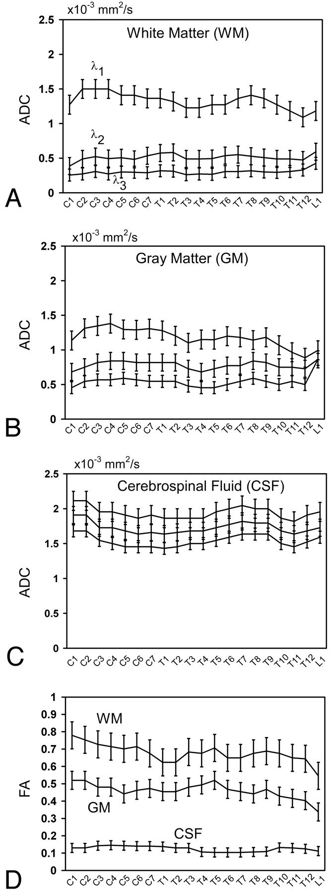
- Fig 3.
Primary eigenvalue, λ1; secondary eigenvalue, λ2; and tertiary eigenvalue, λ3, across the spinal cord for WM regions (A), ventral GM (B), and CSF (C). FA across the spinal cord (D) for individual WM regions, ventral GM, and CSF. Mean values are shown as solid black lines. Error bars indicate SD across subjects.
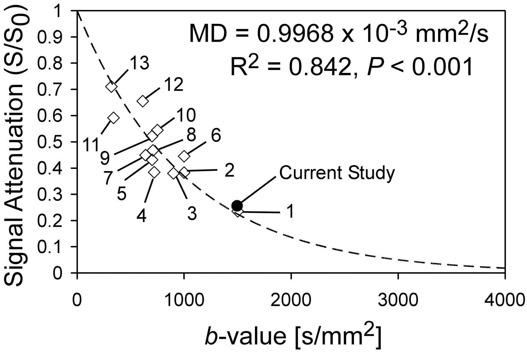
- Fig 4.
Regression analysis of mean apparent diffusion coefficients from DTI studies of the human spinal cord with use of a variety of pulse sequences. Numbers represent identification numbers and are located in the accompanying table. Results of logarithmic regression are also shown. Solid circle indicates average signal intensity attenuation for this study.


Tables
Identification numbers (ID), study name/date, and pulse sequence used for studies included in regression analysis (Fig. 4)
ID Study Pulse Sequence 1 Ellingson et al, 20067 SS-EPI + Fuzzy logic 2 Robertson et al, 200026 LSDI 3 Cercignani et al, 200314 SENSE-EPI 4 Clark et al, 19999 Nav. echo - SE 5 Murphy et al, 200127 LSDI 6 Mamata et al, 20053 LSDI 7 Holder et al, 200010 Multishot SE-EPI 8 Bammer et al, 200012 Phase nav., IEPI 9 Wheeler-Kingshott et al, 200223 ZOOM-EPI 10 Maier and Mamata, 200528 LSDI 11 Ries et al, 20008 Multishot SE-EPI 12 Bammer et al, 200229 FSE & IEPI 13 Clark et al, 20002 Nav. echo - SE Note:—SS-EPI indicates single-shot, echo-planar imaging; LSDI, line-scan diffusion imaging; IEPI, interleaved echo-planar imaging; SENSE, sensitivity encoding; SE, spin-echo; FSE, fast spin-echo; ZOOM-EPI, zonally magnified oblique multisection echo-planar imaging; nav., navigator.
In this issue


Jump to section
Related Articles
Cited By...
- Cervical Spinal Cord DTI Is Improved by Reduced FOV with Specific Balance between the Number of Diffusion Gradient Directions and Averages
- Pulse-Triggered DTI Sequence with Reduced FOV and Coronal Acquisition at 3T for the Assessment of the Cervical Spinal Cord in Patients with Myelitis
- Diffusion Tensor Imaging of the Normal Pediatric Spinal Cord Using an Inner Field of View Echo-Planar Imaging Sequence
- Diffusion Tensor Imaging of the Pediatric Spinal Cord at 1.5T: Preliminary Results
- Quantification of Diffusivities of the Human Cervical Spinal Cord Using a 2D Single-Shot Interleaved Multisection Inner Volume Diffusion-Weighted Echo-Planar Imaging Technique