Advertisement

Main menu

  • Home
  • Content
    • Current Issue
    • Accepted Manuscripts
    • Article Preview
    • Past Issue Archive
    • Video Articles
    • AJNR Case Collection
    • Case of the Week Archive
    • Case of the Month Archive
    • Classic Case Archive
  • Special Collections
    • AJNR Awards
    • Low-Field MRI
    • Alzheimer Disease
    • ASNR Foundation Special Collection
    • Photon-Counting CT
    • View All
  • Multimedia
    • AJNR Podcasts
    • AJNR SCANtastic
    • Trainee Corner
    • MRI Safety Corner
    • Imaging Protocols
  • For Authors
    • Submit a Manuscript
    • Submit a Video Article
    • Submit an eLetter to the Editor/Response
    • Manuscript Submission Guidelines
    • Statistical Tips
    • Fast Publishing of Accepted Manuscripts
    • Graphical Abstract Preparation
    • Imaging Protocol Submission
    • Author Policies
  • About Us
    • About AJNR
    • Editorial Board
    • Editorial Board Alumni
  • More
    • Become a Reviewer/Academy of Reviewers
    • Subscribers
    • Permissions
    • Alerts
    • Feedback
    • Advertisers
    • ASNR Home

User menu

  • Alerts
  • Log in

Search

  • Advanced search
American Journal of Neuroradiology

American Journal of Neuroradiology

ASHNR American Society of Functional Neuroradiology ASHNR American Society of Pediatric Neuroradiology ASSR
  • Alerts
  • Log in

Advanced Search

  • Home
  • Content
    • Current Issue
    • Accepted Manuscripts
    • Article Preview
    • Past Issue Archive
    • Video Articles
    • AJNR Case Collection
    • Case of the Week Archive
    • Case of the Month Archive
    • Classic Case Archive
  • Special Collections
    • AJNR Awards
    • Low-Field MRI
    • Alzheimer Disease
    • ASNR Foundation Special Collection
    • Photon-Counting CT
    • View All
  • Multimedia
    • AJNR Podcasts
    • AJNR SCANtastic
    • Trainee Corner
    • MRI Safety Corner
    • Imaging Protocols
  • For Authors
    • Submit a Manuscript
    • Submit a Video Article
    • Submit an eLetter to the Editor/Response
    • Manuscript Submission Guidelines
    • Statistical Tips
    • Fast Publishing of Accepted Manuscripts
    • Graphical Abstract Preparation
    • Imaging Protocol Submission
    • Author Policies
  • About Us
    • About AJNR
    • Editorial Board
    • Editorial Board Alumni
  • More
    • Become a Reviewer/Academy of Reviewers
    • Subscribers
    • Permissions
    • Alerts
    • Feedback
    • Advertisers
    • ASNR Home
  • Follow AJNR on Twitter
  • Visit AJNR on Facebook
  • Follow AJNR on Instagram
  • Join AJNR on LinkedIn
  • RSS Feeds

AJNR Awards, New Junior Editors, and more. Read the latest AJNR updates

Research ArticleBRAIN

Atrophy Measurement of the Anterior Commissure and Substantia Innominata with 3T High-Resolution MR Imaging: Does the Measurement Differ for Patients with Frontotemporal Lobar Degeneration and Alzheimer Disease and for Healthy Subjects?

W.-J. Moon, H.-J. Kim, H.G. Roh and S.-H. Han
American Journal of Neuroradiology August 2008, 29 (7) 1308-1313; DOI: https://doi.org/10.3174/ajnr.A1103
W.-J. Moon
  • Find this author on Google Scholar
  • Find this author on PubMed
  • Search for this author on this site
H.-J. Kim
  • Find this author on Google Scholar
  • Find this author on PubMed
  • Search for this author on this site
H.G. Roh
  • Find this author on Google Scholar
  • Find this author on PubMed
  • Search for this author on this site
S.-H. Han
  • Find this author on Google Scholar
  • Find this author on PubMed
  • Search for this author on this site
  • Article
  • Figures & Data
  • Info & Metrics
  • Responses
  • References
  • PDF
Loading

Article Figures & Data

Figures

  • Tables
  • Fig 1.
    • Download figure
    • Open in new tab
    • Download powerpoint
    Fig 1.

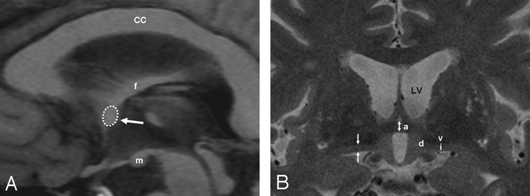
    Schema of the MR imaging measurements. A, A sagittal T1-weighted inversion-recovery image shows the AC area (dotted circle and arrow) in a control subject. The corpus callosum (CC), fornix (f), and mamillary body (m) are identified. B, A coronal thin-sectional T2-weighted image shows the AC (a), the SI (i), the diagonal gyrus (d), and the ventral pallidum (v). AC (bidirectional arrow) and the SI thicknesses (arrows) are depicted. LV indicates left ventricle.

  • Fig 2.
    • Download figure
    • Open in new tab
    • Download powerpoint
    Fig 2.

    Measurement of the AC and SI thicknesses. Coronal T2-weighted fast spin-echo (TR/TE, 3000/125.4 ms; section thickness, 2 mm) MR images show the AC thickness (bidirectional arrow) in a control subject (A; AC thickness, 4.30 mm), a patient with AD (B; AC thickness, 3.01 mm), and a patient with FTLD (C; AC thickness, 2.15 mm). The arrows indicate the measured part of the thickness of the SI. Thinning of the SI is noted in the patient with AD and to a lesser degree in the patient with FTLD.

  • Fig 3.
    • Download figure
    • Open in new tab
    • Download powerpoint
    Fig 3.

    Boxplot of the AC area in patients with FTLD and AD and control subjects. The line across the box represents the median value. The box ends represent the first and third quartiles. The end points of each graph represent the smallest and largest values. The median AC area is the lowest in the FTLD group of patients compared with the patients with AD and control subjects. None of the AC cross-sectional area measurements in the FTLD group of patients reached the lowest value obtained for the control subjects.

  • Fig 4.
    • Download figure
    • Open in new tab
    • Download powerpoint
    Fig 4.

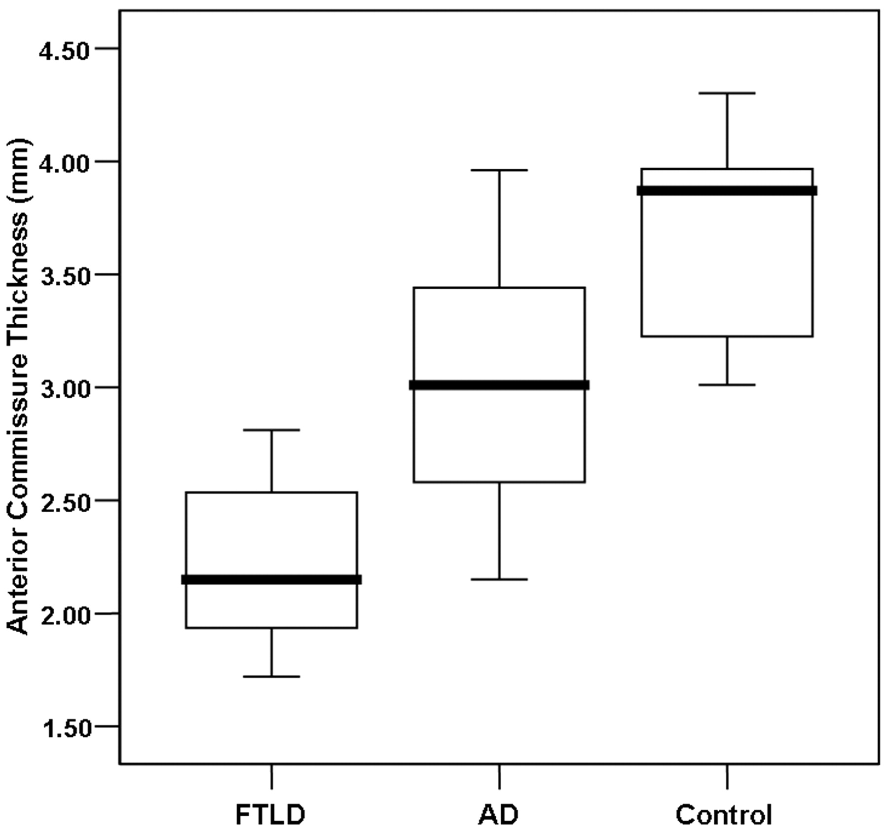
    Boxplot of the AC thickness in patients with FTLD and AD and control subjects. The line across the box represents the median value. The box ends represent the first and third quartiles. The end points of each graph represent the smallest and largest values. The median AC thickness is the lowest in the FTLD group of patients compared with the patients with AD and control subjects. Although considerable overlap of the values between the FTLD and AD groups of patients exists, none of the AC measurements in the FTLD group of patients reached the lowest value obtained for the control subjects.

  • Fig 5.
    • Download figure
    • Open in new tab
    • Download powerpoint
    Fig 5.

    Boxplot of the SI thickness in patients with FTLD and AD and control subjects. The line across the box represents the median value. The box ends represent the first and third quartiles. The end points of each graph represent the smallest and largest values. The median SI thickness tends to be lower in the AD group of patients compared with the FTLD group of patients and control subjects. Note the considerable overlap of the values between the patients with FTLD and control subjects.

Tables

  • Figures
    • View popup
    Table 1:

    Demographic data*

    FTLD (n = 7)AD (n = 20)Control (n = 16)
    Men/women4:34:165:11
    Age (years)69.3 ± 6.470.4 ± 4.368.0 ± 5.3
    MMSE18.1 ± 4.418.5 ± 4.029.1 ± 0.7
    CDR score0.71 ± 0.270.90 ± 0.450.00 ± 0.00
    • Note:—FTLD indicates frontotemporal lobar degeneration; AD, Alzheimer disease; MMSE, Mini-Mental State Examination; CDR, Clinical Dementia Rating.

    • * Values are mean ± SD.

    • View popup
    Table 2:

    MR Measurement of the AC and SI thicknesses*

    FTLD (n = 7)AD (n = 20)Control (n = 16)P
    AC area (mm2), AC thickness† (mm)5.45 ± 1.80, 2.23 ± 0.428.97 ± 1.84, 2.97 ± 0.5012.47 ± 1.42, 3.67 ± 0.48<.001†, <.001
    Average SI thickness (mm)2.31 ± 0.172.17 ± 0.502.45 ± 0.33.147
    Right SI thickness (mm)2.35 ± 0.192.11 ± 0.532.50 ± 0.36.041‡
    Left SI thickness (mm)2.28 ± 0.192.25 ± 0.562.45 ± 0.37.419
    • Note:—AC indicates anterior commissure; SI, substantia innominata; FTLD, frontotemporal lobe degeneration; AD, Alzheimer disease.

    • * Values are mean ± SD.

    • † P < .01 between FTLD versus AD, FTLD versus control, and AD versus control.

    • ‡ P < .05 between AD versus control.

PreviousNext
Back to top

In this issue

American Journal of Neuroradiology: 29 (7)
American Journal of Neuroradiology
Vol. 29, Issue 7
August 2008
  • Table of Contents
  • Index by author
Advertisement
Print
Download PDF
Email Article

Thank you for your interest in spreading the word on American Journal of Neuroradiology.

NOTE: We only request your email address so that the person you are recommending the page to knows that you wanted them to see it, and that it is not junk mail. We do not capture any email address.

Enter multiple addresses on separate lines or separate them with commas.
Atrophy Measurement of the Anterior Commissure and Substantia Innominata with 3T High-Resolution MR Imaging: Does the Measurement Differ for Patients with Frontotemporal Lobar Degeneration and Alzheimer Disease and for Healthy Subjects?
(Your Name) has sent you a message from American Journal of Neuroradiology
(Your Name) thought you would like to see the American Journal of Neuroradiology web site.
CAPTCHA
This question is for testing whether or not you are a human visitor and to prevent automated spam submissions.
Cite this article
W.-J. Moon, H.-J. Kim, H.G. Roh, S.-H. Han
Atrophy Measurement of the Anterior Commissure and Substantia Innominata with 3T High-Resolution MR Imaging: Does the Measurement Differ for Patients with Frontotemporal Lobar Degeneration and Alzheimer Disease and for Healthy Subjects?
American Journal of Neuroradiology Aug 2008, 29 (7) 1308-1313; DOI: 10.3174/ajnr.A1103

Citation Manager Formats

  • BibTeX
  • Bookends
  • EasyBib
  • EndNote (tagged)
  • EndNote 8 (xml)
  • Medlars
  • Mendeley
  • Papers
  • RefWorks Tagged
  • Ref Manager
  • RIS
  • Zotero
0 Responses
Respond to this article
Share
Bookmark this article
Atrophy Measurement of the Anterior Commissure and Substantia Innominata with 3T High-Resolution MR Imaging: Does the Measurement Differ for Patients with Frontotemporal Lobar Degeneration and Alzheimer Disease and for Healthy Subjects?
W.-J. Moon, H.-J. Kim, H.G. Roh, S.-H. Han
American Journal of Neuroradiology Aug 2008, 29 (7) 1308-1313; DOI: 10.3174/ajnr.A1103
del.icio.us logo Twitter logo Facebook logo Mendeley logo
  • Tweet Widget
  • Facebook Like
  • Google Plus One
Purchase

Jump to section

  • Article
    • Abstract
    • Methods
    • Results
    • Discussion
    • Footnotes
    • References
  • Figures & Data
  • Info & Metrics
  • Responses
  • References
  • PDF

Related Articles

  • No related articles found.
  • PubMed
  • Google Scholar

Cited By...

  • Evaluation of Common Structural Brain Changes in Aging and Alzheimer Disease with the Use of an MRI-Based Brain Atrophy and Lesion Index: A Comparison Between T1WI and T2WI at 1.5T and 3T
  • Crossref
  • Google Scholar

This article has not yet been cited by articles in journals that are participating in Crossref Cited-by Linking.

More in this TOC Section

  • White Matter Alterations in the Brains of Patients with Active, Remitted, and Cured Cushing Syndrome: A DTI Study
  • Qualitative and Quantitative Analysis of MR Imaging Findings in Patients with Middle Cerebral Artery Stroke Implanted with Mesenchymal Stem Cells
  • Fast Contrast-Enhanced 4D MRA and 4D Flow MRI Using Constrained Reconstruction (HYPRFlow): Potential Applications for Brain Arteriovenous Malformations
Show more Brain

Similar Articles

Advertisement

Indexed Content

  • Current Issue
  • Accepted Manuscripts
  • Article Preview
  • Past Issues
  • Editorials
  • Editor's Choice
  • Fellows' Journal Club
  • Letters to the Editor
  • Video Articles

Cases

  • Case Collection
  • Archive - Case of the Week
  • Archive - Case of the Month
  • Archive - Classic Case

More from AJNR

  • Trainee Corner
  • Imaging Protocols
  • MRI Safety Corner
  • Book Reviews

Multimedia

  • AJNR Podcasts
  • AJNR Scantastics

Resources

  • Turnaround Time
  • Submit a Manuscript
  • Submit a Video Article
  • Submit an eLetter to the Editor/Response
  • Manuscript Submission Guidelines
  • Statistical Tips
  • Fast Publishing of Accepted Manuscripts
  • Graphical Abstract Preparation
  • Imaging Protocol Submission
  • Evidence-Based Medicine Level Guide
  • Publishing Checklists
  • Author Policies
  • Become a Reviewer/Academy of Reviewers
  • News and Updates

About Us

  • About AJNR
  • Editorial Board
  • Editorial Board Alumni
  • Alerts
  • Permissions
  • Not an AJNR Subscriber? Join Now
  • Advertise with Us
  • Librarian Resources
  • Feedback
  • Terms and Conditions
  • AJNR Editorial Board Alumni

American Society of Neuroradiology

  • Not an ASNR Member? Join Now

© 2025 by the American Society of Neuroradiology All rights, including for text and data mining, AI training, and similar technologies, are reserved.
Print ISSN: 0195-6108 Online ISSN: 1936-959X

Powered by HighWire