Advertisement

Main menu

  • Home
  • Content
    • Current Issue
    • Accepted Manuscripts
    • Article Preview
    • Past Issue Archive
    • AJNR Case Collection
    • Case of the Week Archive
    • Classic Case Archive
    • Case of the Month Archive
  • Special Collections
    • Spinal CSF Leak Articles (Jan 2020-June 2024)
    • 2024 AJNR Journal Awards
    • Most Impactful AJNR Articles
  • Multimedia
    • AJNR Podcast
    • AJNR Scantastics
    • Video Articles
  • For Authors
    • Submit a Manuscript
    • Author Policies
    • Fast publishing of Accepted Manuscripts
    • Graphical Abstract Preparation
    • Manuscript Submission Guidelines
    • Imaging Protocol Submission
    • Submit a Case for the Case Collection
  • About Us
    • About AJNR
    • Editorial Board
  • More
    • Become a Reviewer/Academy of Reviewers
    • Subscribers
    • Permissions
    • Alerts
    • Feedback
    • Advertisers
    • ASNR Home
  • Other Publications
    • ajnr

User menu

  • Alerts
  • Log in

Search

  • Advanced search
American Journal of Neuroradiology

American Journal of Neuroradiology

ASHNR American Society of Functional Neuroradiology ASHNR American Society of Pediatric Neuroradiology ASSR
  • Alerts
  • Log in

Advanced Search

  • Home
  • Content
    • Current Issue
    • Accepted Manuscripts
    • Article Preview
    • Past Issue Archive
    • AJNR Case Collection
    • Case of the Week Archive
    • Classic Case Archive
    • Case of the Month Archive
  • Special Collections
    • Spinal CSF Leak Articles (Jan 2020-June 2024)
    • 2024 AJNR Journal Awards
    • Most Impactful AJNR Articles
  • Multimedia
    • AJNR Podcast
    • AJNR Scantastics
    • Video Articles
  • For Authors
    • Submit a Manuscript
    • Author Policies
    • Fast publishing of Accepted Manuscripts
    • Graphical Abstract Preparation
    • Manuscript Submission Guidelines
    • Imaging Protocol Submission
    • Submit a Case for the Case Collection
  • About Us
    • About AJNR
    • Editorial Board
  • More
    • Become a Reviewer/Academy of Reviewers
    • Subscribers
    • Permissions
    • Alerts
    • Feedback
    • Advertisers
    • ASNR Home
  • Follow AJNR on Twitter
  • Visit AJNR on Facebook
  • Follow AJNR on Instagram
  • Join AJNR on LinkedIn
  • RSS Feeds

Welcome to the new AJNR, Updated Hall of Fame, and more. Read the full announcements.


AJNR is seeking candidates for the position of Associate Section Editor, AJNR Case Collection. Read the full announcement.


Research ArticleHEAD & NECK

Interfrontal Sinus Septal Cell: A Cause of Obstructing Inflammation and Mucoceles

P.M. Som and W. Lawson
American Journal of Neuroradiology August 2008, 29 (7) 1369-1371; DOI: https://doi.org/10.3174/ajnr.A1107
P.M. Som
  • Find this author on Google Scholar
  • Find this author on PubMed
  • Search for this author on this site
W. Lawson
  • Find this author on Google Scholar
  • Find this author on PubMed
  • Search for this author on this site
  • Article
  • Figures & Data
  • Info & Metrics
  • Responses
  • References
  • PDF
Loading

Abstract

BACKGROUND AND PURPOSE: Recent interest in the origin and complications associated with frontal intersinus septal cells prompted us to review our material looking for cases of the uncommon occurrence of a mucocele developing within such a cell. The purpose of this article was to present the imaging findings on 4 cases of mucoceles arising within surgically proven frontal intersinus septal cells.

MATERIALS AND METHODS: A retrospective review of the case material in our department of otolaryngology since 2000 was made looking for cases of patients with surgically proven frontal intersinus septal cell mucoceles. Institutional review board approval for the review was obtained. Four cases were identified that also had CT and/or MR imaging studies. Examples of 4 additional classic frontal sinus mucoceles and 3 anterior ethmoid mucoceles were also identified for comparison.

RESULTS: All 4 of the cases of frontal intersinus septal cell mucoceles had an expanded midline frontal sinus cell, which thinned the posterior and/or anterior frontal sinus tables. The classic frontal sinus mucoceles were either to the left or right side, and any table thinning was off midline. The ethmoid mucoceles were clearly centered below the frontal sinuses.

CONCLUSIONS: The rare occurrence of a frontal intersinus septal mucocele can be diagnosed on CT and MR imaging studies, because its appearance in the midline is clearly distinct from the more common classic frontal sinus mucoceles that develop within the left or right frontal sinus proper. Distinction is also routinely made from large anterior ethmoid mucoceles.

There has been some recent interest in the origin of, and complications associated with, frontal intersinus septal cells.1–6 The complications are the result of disease within these septal cells obstructing the adjacent frontal sinuses. This is especially true in the uncommon occurrence of a mucocele developing within such a cell. The purpose of this article was to present the imaging findings on 4 cases of mucoceles arising within surgically proven frontal intersinus septal cells.

Materials and Methods

A retrospective review was made of the paranasal sinus cases operated on at our institution since 2000, which involved disease within a frontal intersinus septal cell. From this material, we found 4 cases of surgically proven mucoceles arising within a frontal intersinus sinus septal cell. These cases were imaged either with CT scans or MR images on a variety of scanners both within and outside our institution. The study was performed with approval of the internal review board No. 06-254. The patients ranged in age from 37 to 68 years, and there were 3 women and 1 man. The images were reviewed by a head and neck radiologist and the otolaryngologist who operated on the cases.

Results

All 4 of the cases showed an expanded central cell in the frontal sinus obstructing the more laterally positioned frontal sinus cavities (Fig 1). There was thinning of the midline anterior and/or posterior frontal sinus tables in all of the cases. In one case, a Pott puffy tumor developed from an expanding central frontal cell (Fig 4). For purposes of comparison, 4 cases of classic frontal sinus mucoceles were reviewed, and representative images are shown in Fig 5. Three cases of ethmoid mucoceles were also reviewed, and a representative image is shown in Fig 6. The classic frontal sinus mucoceles clearly arose in 1 side of the frontal sinuses, and if the frontal sinus tables were thinned, it was off of the midline. The large anterior ethmoid mucoceles all had their epicenter in the ethmoid complex, caudal to the frontal sinuses, and only impinged off of the midline on the lower frontal sinus. Thus, the frontal intersinus septal cell mucoceles were routinely distinguished from these other mucoceles.

Fig 1.
  • Download figure
  • Open in new tab
  • Download powerpoint
Fig 1.

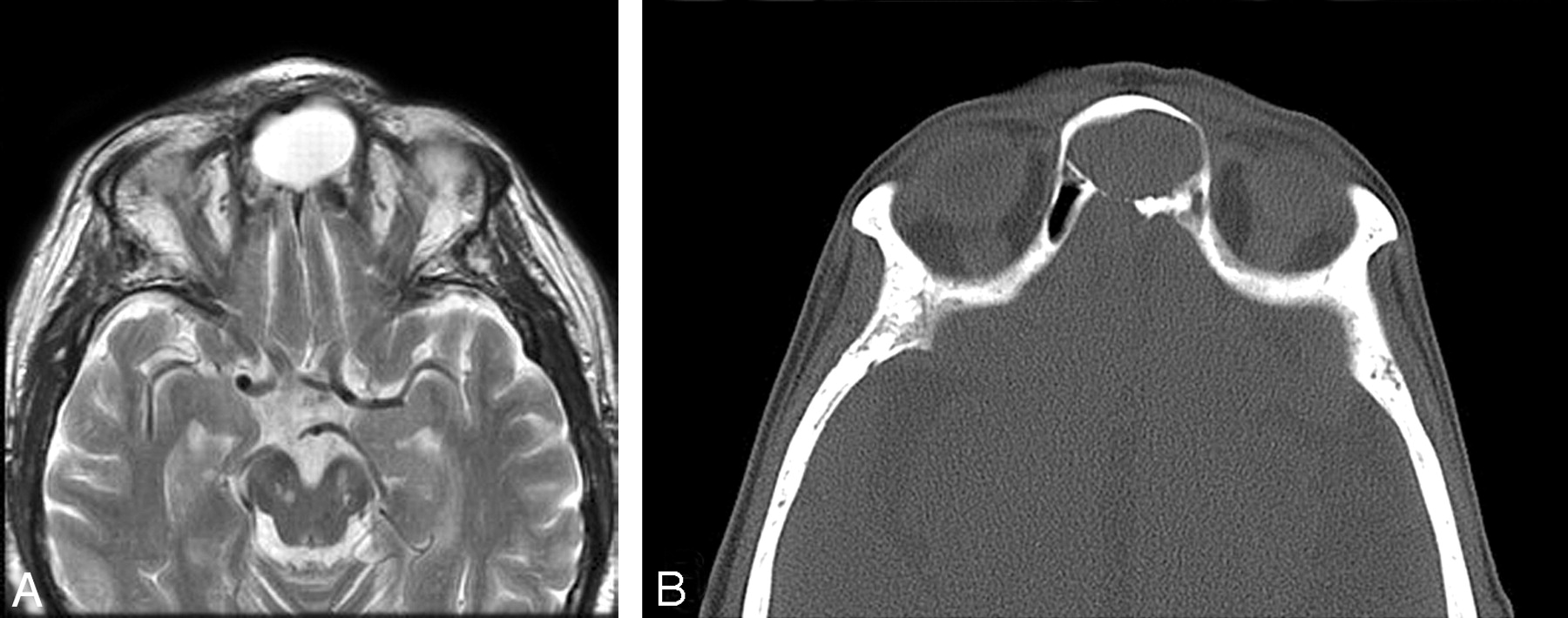
A 37-year-old woman presented with a feeling of pressure in her forehead. A, An axial T2-weighted MR image shows an expanded interfrontal sinus septal cell with high signal intensity. B, A CT scan shows the mucocele thinning the midline posterior frontal sinus table.

Fig 2.
  • Download figure
  • Open in new tab
  • Download powerpoint
Fig 2.

A 43-year-old man presented with a headache. An axial T1-weighted MR image shows an expanded interfrontal sinus septal cell with high (proteinaceous) signal intensity. There was thinning of the central posterior sinus table.

Fig 3.
  • Download figure
  • Open in new tab
  • Download powerpoint
Fig 3.

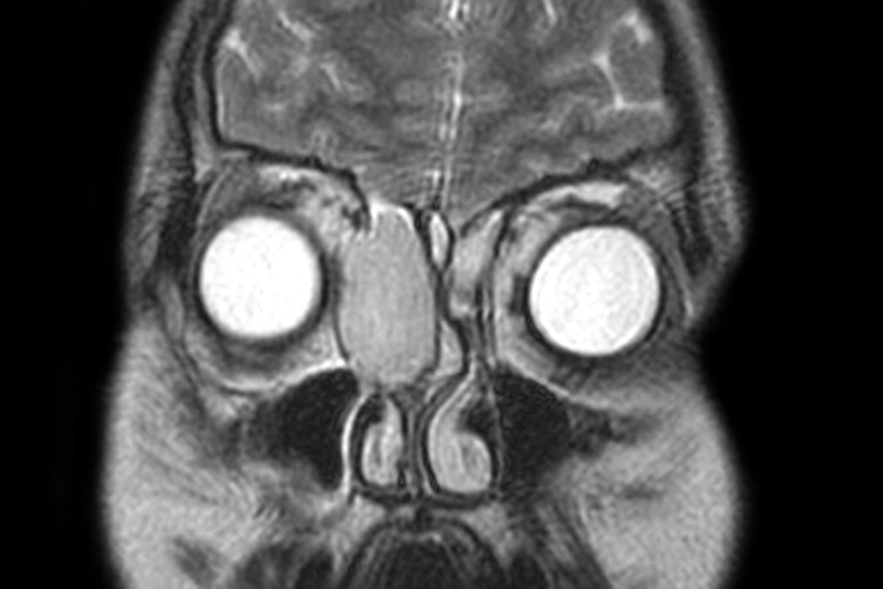
A 57-year-old woman presented with a slowly growing mass in her lower forehead. A coronal T2-weighted MR image shows an expanded interfrontal sinus septal cell with high signal intensity. This mucocele has obstructed the left frontal sinus proper. At surgery, the right frontal sinus drainage was patent and just behind the mucocele.

Fig 4.
  • Download figure
  • Open in new tab
  • Download powerpoint
Fig 4.

A 68-year-old woman presented with a painful forehead mass. An axial T2-weighted MR image shows a minimally expanded interfrontal sinus septal cell with high signal intensity. This mucocele extended through the midline anterior table and caused a Pott puffy tumor in the overlying scalp (arrow).

Fig 5.
  • Download figure
  • Open in new tab
  • Download powerpoint
Fig 5.

A 53-year-old man presented with a mass in his upper medial right orbit. A coronal T2-weighted MR image shows a right frontal sinus mucocele that has expanded the sinus and depressed the superomedial orbital rim. Inflammatory mucosal disease is present in the left frontal sinus, both ethmoid sinuses, and the right maxillary sinus. B, A 46-year-old woman presented with a headache. An axial T1-weighted MR image shows a mucocele expanding the right frontal sinus and thinning the posterior sinus table to the right side, off of the midline.

Fig 6.
  • Download figure
  • Open in new tab
  • Download powerpoint
Fig 6.

A 32-year-old man presented with a bulge in his medial right orbit and difficulty breathing through the right side of his nose. A coronal T2-weighted MR image shows a large right anterior ethmoid mucocele. It is clearly centered within the ethmoid complex, caudal to the frontal sinuses. Inflammatory mucosal disease is also present in the left ethmoid cells and the right antrum.

Discussion

There are 3 principal groups of cells that impact the frontal sinuses. Of these, the supraorbital cells have received the greatest attention with regard to their role in the pathogenesis of frontal sinus disease and its management.7 These cells pneumatize the orbital plate of the frontal bone posterior to the frontal recess.8–10 They generally arise from anterior ethmoid cells, but they can also develop from a posterior cell, and they may be multiple. They drain into the frontal recess posterior to the frontal sinus ostium. Their incidence has been estimated at 5%–15%11 in dissection studies, but imaging studies have detected a much greater prevalence (62%).3

The intrafrontal (intrabullar, type III and IV) cells have the lowest incidence estimated at 1%–2%. On review of approximately 600 CT scans, Bent et al12 detected them in 9 skulls (1.5%). In a later study of 50 patients examined with multiplanar CT, Lee et al3 found type III cells in 8% of cases.

Finally, there is the intersinus septum cell (ISSC), which is common but has received the least attention in the literature. These cells are defined as a pneumatization within the midline or paramedian bony lamella (septum) between the frontal sinuses. Van Alyea11 noted them to be the most frequent cell, present in 28 (11.6%) of his 242 dissection specimens. In a coronal CT scan study of 300 heads (200 patients with chronic sinusitis and 100 cadavers) by Merrit et al,13 an ISSC was identified in 34%, 10 having multiple cells. The cells extended various distances along the height of the septum. Paradoxically, a multiplanar 3D-CT scan study of 50 heads found a prevalence of 14%.3 In a prospective CT scan study performed in the coronal and axial planes of 200 consecutive nonsinusitis patients, Som and Lawson4 found ISSC in 30.5% of the cases, also having variable extension along the septum. However, this study revealed communications between most of these cells (85.5%) and the frontal sinus, suggesting that most of these cells may arise as diverticular extensions from the frontal sinuses rather than by the evagination of anterior ethmoid air cells from the frontal recess.

The literature contains few references regarding pathology of the ISSC. Van Alya11 noted that the ISSC may pneumatize sufficiently to obstruct the frontal ostium. Merrit et al13 reported 5 patients in their series of 200 patients with chronic sinusitis to have disease in an ISSC, of which 3 cases failed treatment. Chiu and Vaughan1 reported enlarging a narrow frontal recess by removing an ISSC. Lawson also stated that, when a deviated intersinus septum is found to impinge on the frontal sinus ostium, an air cell in the lower part of the septum should be suspected.

The present study documents that an ISSC may become a mucocele sufficiently large to unilaterally block the frontal sinus outflow, or it may enlarge massively to form a giant mucocele involving both frontal sinuses. Such a mucocele usually erodes the anterior and/or posterior sinus tables.

Current management of frontal and frontoethmoidal mucoceles is principally by endoscopic marsupialization into the nasal cavity, with external procedures reserved for inaccessible and recurrent lesions. Intersinus septal mucoceles present an algorithm shift. Although many small mucoceles may be successfully treated endonasally, large ones expand bilaterally, often sequestering the lateral recesses of the frontal sinus, and require external surgery (osteoplastic flap) primarily for management.

Conclusions

When a frontal sinus mucocele is seen to arise in the midline rather than primarily to one side of the frontal sinus, an uncommon mucocele arising within an interfrontal sinus septal cell should be considered. This may impact what the surgeon must do to correct the problem.

References

  1. ↵
    Chiu AG, Vaughan WC. Using the frontal intersinus septal cell to widen the narrow frontal recess. Laryngoscope 2004;114:1315–17
    CrossRefPubMed
  2. Goldsztein H, Pletcher SD, Reh DD, et al. The frontal wishbone: anatomic and clinical implications. Am J Rhinol 2007;21:725–28
    CrossRefPubMed
  3. ↵
    Lee WT, Kuhn FA, Citardi MJ. 3D computed tomographic analysis of frontal recess anatomy in patients without frontal sinusitis. Otolaryngol Head Neck Surg 2004;3:164–73
  4. ↵
    Som PM, Lawson W. The frontal intersinus septal air cell—a new hypothesis of its origin. AJNR Am J Neuroradiol 2008;29:1215–17. Epub 2008 Apr 3
    Abstract/FREE Full Text
  5. Chandra RK, Palmer JN, Tangsujarittham T, et al. Factors associated with failure of frontal sinusotomy in the early follow-up period. Otolaryngol Head Neck Surg 2004;131:514–18
    Abstract/FREE Full Text
  6. ↵
    Kountakis SE, Senior B, Draf W, eds. The Frontal Sinus. Berlin, Germany: Springer-Verlag;2005 :21–31
  7. ↵
    McLaughlin RB Jr, Rehl RM, Lanza DC. Clinically relevant frontal sinus anatomy and physiology. Otolaryngol Clin North Am 2001;34:1–22
    CrossRefPubMed
  8. ↵
    Owen RG Jr, Kuhn FA. Supraorbital ethmoid cell. Otolaryngol Head Neck Surg 1997;116:254–61
    FREE Full Text
  9. Cryer MH. Some variation of the frontal sinus. JAMA 1907;48:284–89
  10. ↵
    Kasper KA. Nasofrontal connections: a study based on one hundred consecutive dissections. Arch Otolaryngol 1936;23:322–43
    CrossRef
  11. ↵
    Van Alyea OE. Frontal cells: an anatomic study of these cells with consideration of their clinical significance. Arch Otol 1941;34:11–23
  12. ↵
    Bent JP, Cuilty-Siller C, Kuhn FA. The frontal cell as a cause of frontal sinus obstruction. Am J Rhinol 1994;8:185–91
    CrossRef
  13. ↵
    Merrit R, Bent JP, Kuhn FA. The intersinus septal cell. Am J Rhinol 1996;299–302
  • Received February 26, 2008.
  • Accepted after revision March 2, 2008.
  • Copyright © American Society of Neuroradiology
View Abstract
PreviousNext
Back to top

In this issue

American Journal of Neuroradiology: 29 (7)
American Journal of Neuroradiology
Vol. 29, Issue 7
August 2008
  • Table of Contents
  • Index by author
Advertisement
Print
Download PDF
Email Article

Thank you for your interest in spreading the word on American Journal of Neuroradiology.

NOTE: We only request your email address so that the person you are recommending the page to knows that you wanted them to see it, and that it is not junk mail. We do not capture any email address.

Enter multiple addresses on separate lines or separate them with commas.
Interfrontal Sinus Septal Cell: A Cause of Obstructing Inflammation and Mucoceles
(Your Name) has sent you a message from American Journal of Neuroradiology
(Your Name) thought you would like to see the American Journal of Neuroradiology web site.
CAPTCHA
This question is for testing whether or not you are a human visitor and to prevent automated spam submissions.
Cite this article
P.M. Som, W. Lawson
Interfrontal Sinus Septal Cell: A Cause of Obstructing Inflammation and Mucoceles
American Journal of Neuroradiology Aug 2008, 29 (7) 1369-1371; DOI: 10.3174/ajnr.A1107

Citation Manager Formats

  • BibTeX
  • Bookends
  • EasyBib
  • EndNote (tagged)
  • EndNote 8 (xml)
  • Medlars
  • Mendeley
  • Papers
  • RefWorks Tagged
  • Ref Manager
  • RIS
  • Zotero
0 Responses
Respond to this article
Share
Bookmark this article
Interfrontal Sinus Septal Cell: A Cause of Obstructing Inflammation and Mucoceles
P.M. Som, W. Lawson
American Journal of Neuroradiology Aug 2008, 29 (7) 1369-1371; DOI: 10.3174/ajnr.A1107
del.icio.us logo Twitter logo Facebook logo Mendeley logo
  • Tweet Widget
  • Facebook Like
  • Google Plus One
Purchase

Jump to section

  • Article
    • Abstract
    • Materials and Methods
    • Results
    • Discussion
    • Conclusions
    • References
  • Figures & Data
  • Info & Metrics
  • Responses
  • References
  • PDF

Related Articles

  • No related articles found.
  • PubMed
  • Google Scholar

Cited By...

  • No citing articles found.
  • Crossref (8)
  • Google Scholar

This article has been cited by the following articles in journals that are participating in Crossref Cited-by Linking.

  • Typical and atypical presentations of paranasal sinus mucocele at computed tomography
    Bruna Vilaça de Carvalho, Izabella de Campos Carvalho Lopes, James de Brito Corrêa, Laura Filgueiras Mourão Ramos, Emília Guerra Pinto Coelho Motta, Renata Lopes Furletti Caldeira Diniz
    Radiologia Brasileira 2013 46 6
  • Correlation of frontal sinus recess anatomy with ethnicity, gender, and pathology
    Laura K. House, Scott P. Stringer, Samantha Seals
    American Journal of Otolaryngology 2017 38 4
  • Inflammation of the frontal intersinus septal air cell as a cause of headaches
    Jarosław Miłoński, Piotr Pietkiewicz, Joanna Urbaniak, Krzysztof Kuśmierczyk, Jurek Olszewski
    International Journal of Surgery Case Reports 2014 5 12
  • Two Cases of Isolated Frontal Septal Cell Inflammation Treated with the Draf IIb Procedure
    Yeon-Hee Joo
    Journal of Clinical Otolaryngology Head and Neck Surgery 2021 32 1
  • Diagnostic Imaging: Head and Neck
    2017
  • Imaging in Otolaryngology
    2018
  • Interfrontal Sinus Septal Cell Mucocele: A Rare Clinical Entity
    Hye-Jin Park, Ji-Hwan Park, Sung-Dong Kim, Kyu-Sup Cho
    Journal of Clinical Otolaryngology Head and Neck Surgery 2025 36 1
  • Two cases with an interseptal sinus cell mucocele: The different mechanisms of the development varying in the time of the onset
    Fumihiko Kuwata, Shogo Shinohara, Hiroyuki Harada, Ippei Kishimoto, Atsushi Suehiro, Keizo Fujiwara, Yasushi Naito
    Acta Oto-Laryngologica Case Reports 2017 2 1

More in this TOC Section

  • Peritumoral Signal in Vestibular Schwannomas
  • Chondrosarcoma vs Synovial Chondromatosis: Imaging
  • NI-RADS for HEAD&NECK Cancer Recurrence
Show more HEAD & NECK

Similar Articles

Advertisement

Indexed Content

  • Current Issue
  • Accepted Manuscripts
  • Article Preview
  • Past Issues
  • Editorials
  • Editors Choice
  • Fellow Journal Club
  • Letters to the Editor

Cases

  • Case Collection
  • Archive - Case of the Week
  • Archive - Case of the Month
  • Archive - Classic Case

Special Collections

  • Special Collections

Resources

  • News and Updates
  • Turn around Times
  • Submit a Manuscript
  • Author Policies
  • Manuscript Submission Guidelines
  • Evidence-Based Medicine Level Guide
  • Publishing Checklists
  • Graphical Abstract Preparation
  • Imaging Protocol Submission
  • Submit a Case
  • Become a Reviewer/Academy of Reviewers
  • Get Peer Review Credit from Publons

Multimedia

  • AJNR Podcast
  • AJNR SCANtastic
  • Video Articles

About Us

  • About AJNR
  • Editorial Board
  • Not an AJNR Subscriber? Join Now
  • Alerts
  • Feedback
  • Advertise with us
  • Librarian Resources
  • Permissions
  • Terms and Conditions

American Society of Neuroradiology

  • Not an ASNR Member? Join Now

© 2025 by the American Society of Neuroradiology All rights, including for text and data mining, AI training, and similar technologies, are reserved.
Print ISSN: 0195-6108 Online ISSN: 1936-959X

Powered by HighWire