Advertisement

Main menu

  • Home
  • Content
    • Current Issue
    • Accepted Manuscripts
    • Article Preview
    • Past Issue Archive
    • AJNR Case Collection
    • Case of the Week Archive
    • Classic Case Archive
    • Case of the Month Archive
  • Special Collections
    • Spinal CSF Leak Articles (Jan 2020-June 2024)
    • 2024 AJNR Journal Awards
    • Most Impactful AJNR Articles
  • Multimedia
    • AJNR Podcast
    • AJNR Scantastics
    • Video Articles
  • For Authors
    • Submit a Manuscript
    • Author Policies
    • Fast publishing of Accepted Manuscripts
    • Graphical Abstract Preparation
    • Manuscript Submission Guidelines
    • Imaging Protocol Submission
    • Submit a Case for the Case Collection
  • About Us
    • About AJNR
    • Editorial Board
  • More
    • Become a Reviewer/Academy of Reviewers
    • Subscribers
    • Permissions
    • Alerts
    • Feedback
    • Advertisers
    • ASNR Home
  • Other Publications
    • ajnr

User menu

  • Alerts
  • Log in

Search

  • Advanced search
American Journal of Neuroradiology

American Journal of Neuroradiology

ASHNR American Society of Functional Neuroradiology ASHNR American Society of Pediatric Neuroradiology ASSR
  • Alerts
  • Log in

Advanced Search

  • Home
  • Content
    • Current Issue
    • Accepted Manuscripts
    • Article Preview
    • Past Issue Archive
    • AJNR Case Collection
    • Case of the Week Archive
    • Classic Case Archive
    • Case of the Month Archive
  • Special Collections
    • Spinal CSF Leak Articles (Jan 2020-June 2024)
    • 2024 AJNR Journal Awards
    • Most Impactful AJNR Articles
  • Multimedia
    • AJNR Podcast
    • AJNR Scantastics
    • Video Articles
  • For Authors
    • Submit a Manuscript
    • Author Policies
    • Fast publishing of Accepted Manuscripts
    • Graphical Abstract Preparation
    • Manuscript Submission Guidelines
    • Imaging Protocol Submission
    • Submit a Case for the Case Collection
  • About Us
    • About AJNR
    • Editorial Board
  • More
    • Become a Reviewer/Academy of Reviewers
    • Subscribers
    • Permissions
    • Alerts
    • Feedback
    • Advertisers
    • ASNR Home
  • Follow AJNR on Twitter
  • Visit AJNR on Facebook
  • Follow AJNR on Instagram
  • Join AJNR on LinkedIn
  • RSS Feeds

Welcome to the new AJNR, Updated Hall of Fame, and more. Read the full announcements.


AJNR is seeking candidates for the position of Associate Section Editor, AJNR Case Collection. Read the full announcement.


Research ArticleSPINE

Postdiskogram CT Features of Lidocaine-Sensitive and Lidocaine-Insensitive Severely Painful Disks at Provocation Lumbar Diskography

W.S. Bartynski, W.E. Rothfus and M. Kurs-Lasky
American Journal of Neuroradiology September 2008, 29 (8) 1455-1460; DOI: https://doi.org/10.3174/ajnr.A1151
W.S. Bartynski
  • Find this author on Google Scholar
  • Find this author on PubMed
  • Search for this author on this site
W.E. Rothfus
  • Find this author on Google Scholar
  • Find this author on PubMed
  • Search for this author on this site
M. Kurs-Lasky
  • Find this author on Google Scholar
  • Find this author on PubMed
  • Search for this author on this site
  • Article
  • Figures & Data
  • Supplemental
  • Info & Metrics
  • Responses
  • References
  • PDF
Loading

Article Figures & Data

Figures

  • Fig 1.
    • Download figure
    • Open in new tab
    • Download powerpoint
    Fig 1.

    Postdiskogram CT features commonly identified in painful degenerative lumbar disks studied at diskography. A, Simple radial annular tear (column 1); radial annular tear with a peripheral annular pocket (column 2); annular gap or wide radial annular tear (column 3). B, Central radial tear into a peripheral annular tear (column 1); combined central and lateral radial tears into peripheral annular tear (column 2); lateral radial tear into a peripheral annular tear (column 3). C, Central radial tear with lamellar annular tears (column 1); combined central and lateral radial tears with lamellar annular tears (column 2); lateral radial tear with lamellar annular tears (column 3). D, Central lamellar annular tears (column 1); combined central and lateral lamellar annular tears (column 2); lateral lamellar annular tears (column 3). E, Central peripheral annular tear (column 1); combined central and lateral peripheral annular tear (column 2); lateral peripheral annular tear (column 3). F, Extensive circumferential lamellar annular tears (column 1); attached annular fragments, loose annular fragments, and bucket-handle annular tear (column 2); extensive free annular fragments with severe macerative annular tears and debris (column 3).

  • Fig 2.
    • Download figure
    • Open in new tab
    • Download powerpoint
    Fig 2.

    Development of annular gaps. Complex radial annular tears extending into peripheral circumferential annular tears may (A–C) lead to detachment of fragments of the annulus that become free within the central nuclear region (G). Alternatively, peripheral circumferential annular tear (D and E) may become sufficiently detached from the outer annulus with development of a “bucket-handle tear” (F) that subsequently fragments and detaches, coming to lie free within the central nuclear region.
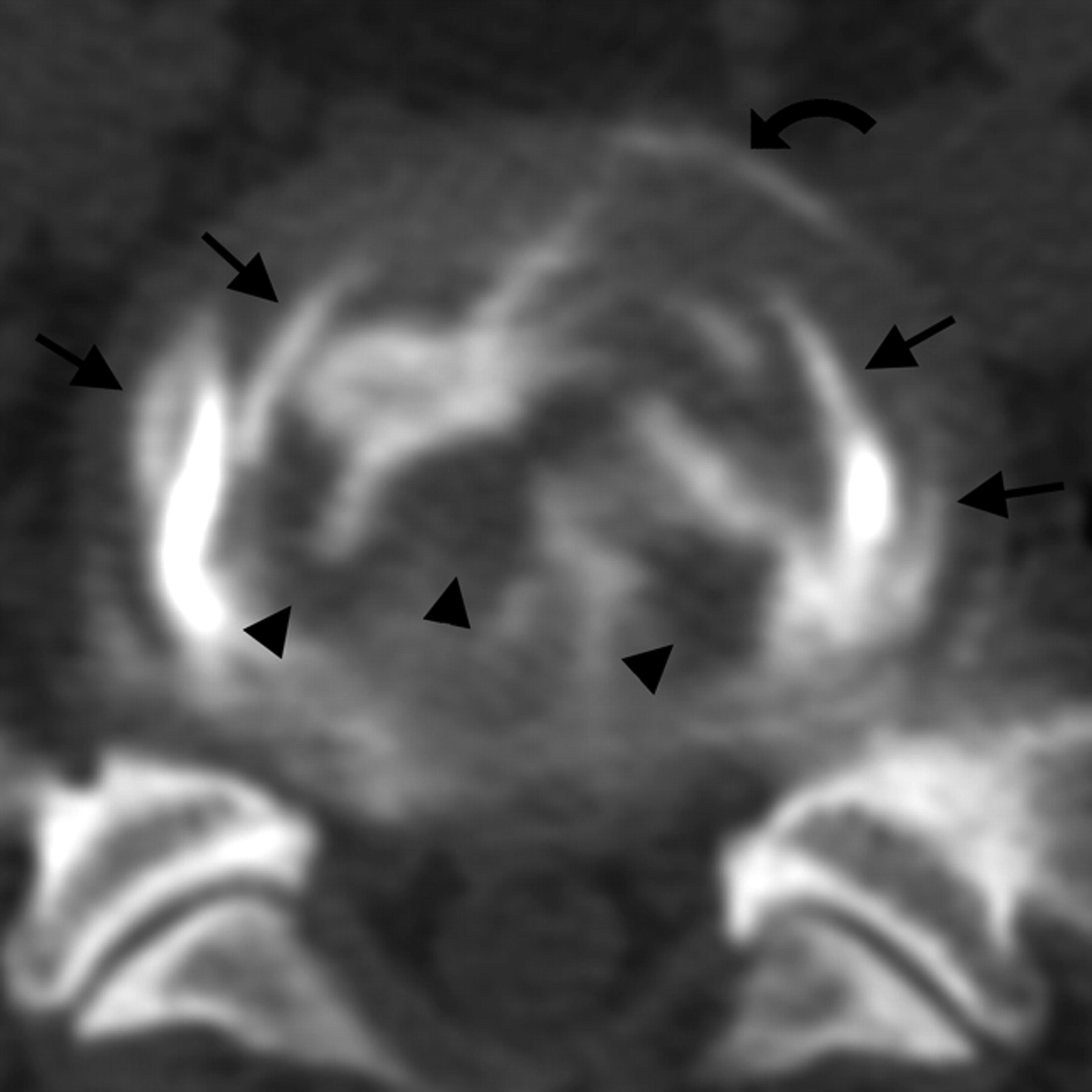
  • Fig 3.
    • Download figure
    • Open in new tab
    • Download powerpoint
    Fig 3.

    A and B, Patient is a 30-year-old man with a 4-year history of increasing severe LBP with some left leg numbness, which he relates to heavy labor at work but without clear injury incident. MR imaging demonstrated degenerative changes with asymmetric disk bulge at L4–5. Diskographic contrast injection at L4–5 (3 mL) provoked severe and concordant back pain (VAS 7/10). Lidocaine injection into this disk (1.5 mL) resulted in near-complete elimination of the provoked pain. Axial postdiskogram CT demonstrates a wide annular gap (curved arrows) along with a peripheral annular tear (arrows). Also noted are free annular fragments present in the annular gap (arrowheads). Diskographic contrast leakage was noted at the margin of the annular gap (not shown).
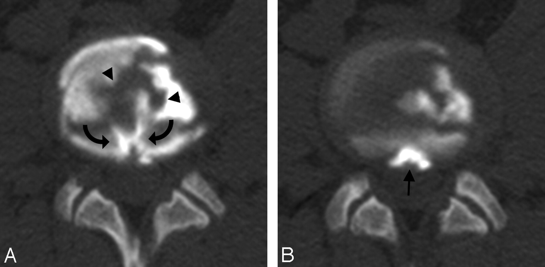
  • Fig 4.
    • Download figure
    • Open in new tab
    • Download powerpoint
    Fig 4.

    Patient is a 33-year-old man with a long-standing history of severe LBP that has worsened over the past 3 years. Outside MR imaging demonstrates disk degenerative changes, in particular at L5–S1. Diskographic contrast injection at L5–S1 (2.5 mL) provoked severe and concordant LBP (VAS 10/10). Intradiskal lidocaine injection (1.5 mL) resulted in no improvement in the provoked pain. Axial postdiskogram CT imaging demonstrates circumferential lamellar annular tears (arrows), multiple free annular fragments (arrowheads), and a peripheral annular tear (curved arrow) without diskographic contrast leakage.

  • Fig 5.
    • Download figure
    • Open in new tab
    • Download powerpoint
    Fig 5.

    A and B, Patient is a 38-year-old man who sustained a service-related injury 16 years ago with progressively increasing severe LBP with some left leg radiation. Outside MR imaging demonstrated significant degenerative disk changes at L3–4. Diskographic contrast injection (3 mL) provoked severe concordant pain (VAS 9/10) at L3–4. Lidocaine injection into the disk (1.5 mL) resulted in no improvement in the provoked pain. Postdiskogram CT imaging demonstrates a central radial tear (curved arrows) and a peripheral annular pocket (arrow), along with loose annular fragments (arrowheads) without diskographic contrast leakage.

PreviousNext
Back to top

In this issue

American Journal of Neuroradiology: 29 (8)
American Journal of Neuroradiology
Vol. 29, Issue 8
September 2008
  • Table of Contents
  • Index by author
Advertisement
Print
Download PDF
Email Article

Thank you for your interest in spreading the word on American Journal of Neuroradiology.

NOTE: We only request your email address so that the person you are recommending the page to knows that you wanted them to see it, and that it is not junk mail. We do not capture any email address.

Enter multiple addresses on separate lines or separate them with commas.
Postdiskogram CT Features of Lidocaine-Sensitive and Lidocaine-Insensitive Severely Painful Disks at Provocation Lumbar Diskography
(Your Name) has sent you a message from American Journal of Neuroradiology
(Your Name) thought you would like to see the American Journal of Neuroradiology web site.
CAPTCHA
This question is for testing whether or not you are a human visitor and to prevent automated spam submissions.
Cite this article
W.S. Bartynski, W.E. Rothfus, M. Kurs-Lasky
Postdiskogram CT Features of Lidocaine-Sensitive and Lidocaine-Insensitive Severely Painful Disks at Provocation Lumbar Diskography
American Journal of Neuroradiology Sep 2008, 29 (8) 1455-1460; DOI: 10.3174/ajnr.A1151

Citation Manager Formats

  • BibTeX
  • Bookends
  • EasyBib
  • EndNote (tagged)
  • EndNote 8 (xml)
  • Medlars
  • Mendeley
  • Papers
  • RefWorks Tagged
  • Ref Manager
  • RIS
  • Zotero
0 Responses
Respond to this article
Share
Bookmark this article
Postdiskogram CT Features of Lidocaine-Sensitive and Lidocaine-Insensitive Severely Painful Disks at Provocation Lumbar Diskography
W.S. Bartynski, W.E. Rothfus, M. Kurs-Lasky
American Journal of Neuroradiology Sep 2008, 29 (8) 1455-1460; DOI: 10.3174/ajnr.A1151
del.icio.us logo Twitter logo Facebook logo Mendeley logo
  • Tweet Widget
  • Facebook Like
  • Google Plus One
Purchase

Jump to section

  • Article
    • Abstract
    • Materials and Methods
    • Results
    • Discussion
    • Conclusions
    • Footnotes
    • References
  • Figures & Data
  • Supplemental
  • Info & Metrics
  • Responses
  • References
  • PDF

Related Articles

  • PubMed
  • Google Scholar

Cited By...

  • Immediate Pain Response to Interlaminar Lumbar Epidural Steroid Administration: Response Characteristics and Effects of Anesthetic Concentration
  • Crossref (13)
  • Google Scholar

This article has been cited by the following articles in journals that are participating in Crossref Cited-by Linking.

  • Lumbar disc nomenclature: version 2.0
    David F. Fardon, Alan L. Williams, Edward J. Dohring, F. Reed Murtagh, Stephen L. Gabriel Rothman, Gordon K. Sze
    The Spine Journal 2014 14 11
  • Lumbar Disc Nomenclature
    David F. Fardon, Alan L. Williams, Edward J. Dohring, F. Reed Murtagh, Stephen L. Gabriel Rothman, Gordon K. Sze
    Spine 2014 39 24
  • Inadvertent Intradiscal Contrast Flow During Lumbar Transforaminal Epidural Steroid Injections: A Case Series Examining the Prevalence of Intradiscal Injection as well as Potential Associated Factors and Adverse Events
    Christopher T. Plastaras, Ellen Casey, Bradley S. Goodman, Larry Chou, Daniel Roth, Joshua Rittenberg
    Pain Medicine 2010 11 12
  • Immediate Pain Response to Interlaminar Lumbar Epidural Steroid Administration: Response Characteristics and Effects of Anesthetic Concentration
    W.S. Bartynski, R.B. Jennings, W.E. Rothfus, V. Agarwal
    American Journal of Neuroradiology 2013 34 1
  • ‘Progressive-Onset’ versus Injury-Associated Discogenic Low Back Pain: Features of Disc Internal Derangement in Patients Studied with Provocation Lumbar Discography
    W.S. Bartynski, L.M. Dejohn, W.E. Rothfus, P.C. Gerszten
    Interventional Neuroradiology 2013 19 1
  • Peripheral Disc Margin Shape and Internal Disc Derangement: Imaging Correlation in Significantly Painful Discs Identified at Provocation Lumbar Discography
    W.S. Bartynski, W.E. Rothfus
    Interventional Neuroradiology 2012 18 2
  • Enhancing Annular Fissures and High-Intensity Zones: Pain, Internal Derangement, and Anesthetic Response at Provocation Lumbar Discography
    W.S. Bartynski, V. Agarwal, H. Trang, A.I. Bandos, W.E. Rothfus, J. Tsay, W.T. Delfyett, B. Nastasi
    American Journal of Neuroradiology 2023 44 1
  • Interventional Assessment of the Lumbar Disk: Provocation Lumbar Diskography and Functional Anesthetic Diskography
    Walter S. Bartynski, A. Orlando Ortiz
    Techniques in Vascular and Interventional Radiology 2009 12 1
  • MR imaging of thoracic annular fissures
    Shadi Asadollahi, Faranak Rafiee, Rodrigo Luna, David M. Yousem, Gary Gong
    Clinical Imaging 2023 101
  • Diagnostic Imaging: Spine
    2015

More in this TOC Section

  • Bern Score Validity for SIH
  • MP2RAGE 7T in MS Lesions of the Cervical Spine
  • Deep Learning for STIR Spine MRI Quality
Show more Spine

Similar Articles

Advertisement

Indexed Content

  • Current Issue
  • Accepted Manuscripts
  • Article Preview
  • Past Issues
  • Editorials
  • Editors Choice
  • Fellow Journal Club
  • Letters to the Editor

Cases

  • Case Collection
  • Archive - Case of the Week
  • Archive - Case of the Month
  • Archive - Classic Case

Special Collections

  • Special Collections

Resources

  • News and Updates
  • Turn around Times
  • Submit a Manuscript
  • Author Policies
  • Manuscript Submission Guidelines
  • Evidence-Based Medicine Level Guide
  • Publishing Checklists
  • Graphical Abstract Preparation
  • Imaging Protocol Submission
  • Submit a Case
  • Become a Reviewer/Academy of Reviewers
  • Get Peer Review Credit from Publons

Multimedia

  • AJNR Podcast
  • AJNR SCANtastic
  • Video Articles

About Us

  • About AJNR
  • Editorial Board
  • Not an AJNR Subscriber? Join Now
  • Alerts
  • Feedback
  • Advertise with us
  • Librarian Resources
  • Permissions
  • Terms and Conditions

American Society of Neuroradiology

  • Not an ASNR Member? Join Now

© 2025 by the American Society of Neuroradiology All rights, including for text and data mining, AI training, and similar technologies, are reserved.
Print ISSN: 0195-6108 Online ISSN: 1936-959X

Powered by HighWire