Advertisement

Main menu

  • Home
  • Content
    • Current Issue
    • Accepted Manuscripts
    • Article Preview
    • Past Issue Archive
    • Video Articles
    • AJNR Case Collection
    • Case of the Week Archive
    • Case of the Month Archive
    • Classic Case Archive
  • Special Collections
    • AJNR Awards
    • Low-Field MRI
    • Alzheimer Disease
    • ASNR Foundation Special Collection
    • Photon-Counting CT
    • View All
  • Multimedia
    • AJNR Podcasts
    • AJNR SCANtastic
    • Trainee Corner
    • MRI Safety Corner
    • Imaging Protocols
  • For Authors
    • Submit a Manuscript
    • Submit a Video Article
    • Submit an eLetter to the Editor/Response
    • Manuscript Submission Guidelines
    • Statistical Tips
    • Fast Publishing of Accepted Manuscripts
    • Graphical Abstract Preparation
    • Imaging Protocol Submission
    • Author Policies
  • About Us
    • About AJNR
    • Editorial Board
    • Editorial Board Alumni
  • More
    • Become a Reviewer/Academy of Reviewers
    • Subscribers
    • Permissions
    • Alerts
    • Feedback
    • Advertisers
    • ASNR Home

User menu

  • Alerts
  • Log in

Search

  • Advanced search
American Journal of Neuroradiology

American Journal of Neuroradiology

ASHNR American Society of Functional Neuroradiology ASHNR American Society of Pediatric Neuroradiology ASSR
  • Alerts
  • Log in

Advanced Search

  • Home
  • Content
    • Current Issue
    • Accepted Manuscripts
    • Article Preview
    • Past Issue Archive
    • Video Articles
    • AJNR Case Collection
    • Case of the Week Archive
    • Case of the Month Archive
    • Classic Case Archive
  • Special Collections
    • AJNR Awards
    • Low-Field MRI
    • Alzheimer Disease
    • ASNR Foundation Special Collection
    • Photon-Counting CT
    • View All
  • Multimedia
    • AJNR Podcasts
    • AJNR SCANtastic
    • Trainee Corner
    • MRI Safety Corner
    • Imaging Protocols
  • For Authors
    • Submit a Manuscript
    • Submit a Video Article
    • Submit an eLetter to the Editor/Response
    • Manuscript Submission Guidelines
    • Statistical Tips
    • Fast Publishing of Accepted Manuscripts
    • Graphical Abstract Preparation
    • Imaging Protocol Submission
    • Author Policies
  • About Us
    • About AJNR
    • Editorial Board
    • Editorial Board Alumni
  • More
    • Become a Reviewer/Academy of Reviewers
    • Subscribers
    • Permissions
    • Alerts
    • Feedback
    • Advertisers
    • ASNR Home
  • Follow AJNR on Twitter
  • Visit AJNR on Facebook
  • Follow AJNR on Instagram
  • Join AJNR on LinkedIn
  • RSS Feeds

AJNR Awards, New Junior Editors, and more. Read the latest AJNR updates

Research ArticleBRAIN

Quantitative Cerebrovascular Reserve Measured by Acetazolamide-Challenged Dynamic CT Perfusion in Ischemic Adult Moyamoya Disease: Initial Experience with Angiographic Correlation

K.H. Kang, H.S. Kim and S.Y. Kim
American Journal of Neuroradiology September 2008, 29 (8) 1487-1493; DOI: https://doi.org/10.3174/ajnr.A1129
K.H. Kang
  • Find this author on Google Scholar
  • Find this author on PubMed
  • Search for this author on this site
H.S. Kim
  • Find this author on Google Scholar
  • Find this author on PubMed
  • Search for this author on this site
S.Y. Kim
  • Find this author on Google Scholar
  • Find this author on PubMed
  • Search for this author on this site
  • Article
  • Figures & Data
  • Info & Metrics
  • Responses
  • References
  • PDF
Loading

Article Figures & Data

Figures

  • Tables
  • Fig 1.
    • Download figure
    • Open in new tab
    • Download powerpoint
    Fig 1.

    ROIs drawn in a reference CT image (A) and vessel-removed MTT map (B). The ROIs were placed on cortical regions in the MCA territory, EBZ, ACA territory, and putamen in the section of BG level around the foramen of Monro.

  • Fig 2.
    • Download figure
    • Open in new tab
    • Download powerpoint
    Fig 2.

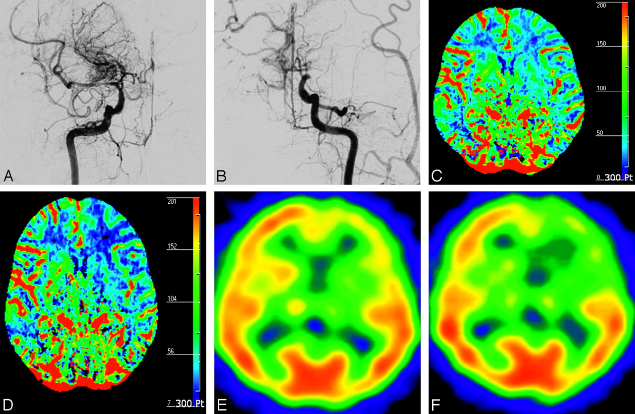
    A 48-year-old woman with transient ischemic attack (case 16). A, Anteroposterior view of right internal carotid arteriogram shows severe stenosis in the proximal M1 portion of MCA with developed BMVs (modified Suzuki stage II). B, Anteroposterior view of left internal carotid arteriogram shows occlusion of distal ICA without antegrade flow (modified Suzuki stage IV). CBF maps before (C) and after (D) ACZ administration show decreased CVR in the left anterior EBZ (white arrow) and MCA territory (black arrow) ipsilateral to the hemisphere with higher modified Suzuki stage compared with contralateral hemisphere with lower modified Suzuki stage. Corresponding SPECT before (E) and after (F) ACZ administration show decreased CVR in the same areas.

  • Fig 3.
    • Download figure
    • Open in new tab
    • Download powerpoint
    Fig 3.

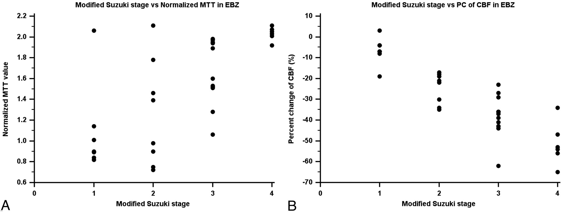
    Scatterplot between baseline MTT and PCs of CBF and angiographic stages in the anterior EBZ. A, Angiographic stage versus MTT in the EBZ. B, Angiographic stage versus PC of CBF in the EBZ.

  • Fig 4.
    • Download figure
    • Open in new tab
    • Download powerpoint
    Fig 4.

    Clustered box-and-whisker graphs show the differences in the mean values of hemodynamic parameters according to the presence of BMVs in each vascular territory. A, BMV versus normalized CBV. B, BMV versus normalized CBF. C, BMV versus normalized MTT. D, BMV versus PC of CBF.

Tables

  • Figures
    • View popup
    Table 1:

    Clinical data of 16 adult patients with ischemic Moyamoya diseases

    Patient No.Age/SexClinical SymptomsDuration of TIABypass Surgery
    139/FTIA12 monthsEIAB
    244/MHeadache, TIA7 monthsNegative*
    333/FHeadache, TIA8 monthsNegative*
    464/FHeadache, TIA4 monthsNegative*
    566/FHeadache, TIA2 monthsEIAB
    632/MHeadache, TIA24 monthsNegative*
    736/MTIA24 monthsNegative*
    845/FTIA1 monthNegative*
    942/FTIA7 monthsNegative*
    1039/FTIA36 monthsNegative*
    1156/FHeadache, TIA1 monthNegative*
    1231/FTIA24 monthsEIAB
    1335/FHeadache, TIA12 monthsNegative*
    1437/MTIA8 monthsEIAB
    1542/MTIA11 monthsNegative*
    1648/FTIA1 monthNegative*
    • Note:—TIA indicates transient ischemic attack; EIAB, external internal arterial bypass; F, female; M, male.

    • * Negative indicates a patient who did not have vascular bypass surgery.

    • View popup
    Table 2:

    Modified Suzuki angiographic stages

    StagesAngiographic Findings
    IMild-to-moderate stenosis around carotid bifurcation with absent or slightly developed ICA Moyamoya:* almost all of both ACA and MCA branches are opacified in antegrade fashion
    IISevere stenosis around carotid bifurcation or occlusion of either of proximal ACA or MCA with well-developed ICA Moyamoya:* either ACA or MCA branches or both are clearly defective, but at least several of ACA or MCA branches remain opacified in antegrade fashion
    IIIOcclusion of both proximal ACA and MCA with well-developed ICA Moyamoya:* only a few of either ACA or MCA branches or both are faintly opacified in antegrade fashion through the meshwork of ICA Moyamoya
    IVComplete occlusion of both proximal ACA and MCA with absent or small amount of ICA Moyamoya:* without opacification of either ACA or MCA branches in antegrade fashion
    • Note:—Data were modified from the staging system proposed by Suzuki and Takaku.1 When the proximal ACA was hypoplastic, stage was determined by evaluating the proximal MCA involvement, opacification of MCA branches, and degree of development of ICA Moyamoya. ICA indicates internal carotid artery; ACA, anterior cerebral artery; MCA, middle cerebral artery.

    • * ICA Moyamoya indicates Moyamoya vessels at or around the terminal part of the ICA.

    • View popup
    Table 3:

    The stages of basal Moyamoya vessels

    StagesAngiographic Findings
    INo Moyamoya vessels were seen
    IIMoyamoya vessels were localized in the area around the ICA bifurcation, and each vessel was fine and had little contrast
    IIIMoyamoya vessels had intermediate extension and thickness
    IVMoyamoya vessels extended a great deal, and each one was thick and strongly opacified
    • Note:—ICA indicates internal carotid artery.

    • View popup
    Table 4:

    Correlation coefficient between normalized baseline- and ACZ-challenged CT perfusion parameters and angiographic stages in each vascular territory

    VariableBG*ACA*MCA*EBZ*
    Observer 1
        CBV vs AS0.39†0.290.290.69†
        CBF vs AS−0.16−0.24−0.50†−0.33
        MTT vs AS0.47†0.39†0.52†0.68†
        PC of CBV vs AS−0.360.11−0.29−0.19
        PC of CBF vs AS−0.65†−0.40†−0.81†−0.82†
        PC of MTT vs AS0.250.320.330.48†
    Observer 2
        CBV vs AS0.370.260.310.67†
        CBF vs AS−0.14−0.27−0.47†−0.31
        MTT vs AS0.46†0.38†0.53†0.64†
        PC of CBV vs AS−0.330.14−0.29−0.19
        PC of CBF vs AS−0.69†−0.45†−0.83†−0.79†
        PC of MTT vs AS0.280.340.310.49†
    • Note:—CBV indicates normalized cerebral blood volume; CBF, normalized cerebral blood flow; MTT, normalized mean transit time; PC, percent change; AS, angiographic stage; BG, basal ganglia; ACA, anterior cerebral artery; MCA, middle cerebral artery; EBZ, anterior external border zone.

    • * Values are Pearson correlation coefficient (r).

    • † Values are statistically significant (P < .05).

    • View popup
    Table 5:

    Differences of the mean values of normalized hemodynamic parameters according to the presence of BMV

    VariableBGACAMCAEBZ
    BMV(+)BMV(−)BMV(+)BMV(−)BMV(+)BMV(−)BMV(+)BMV(−)
    Observer 1
        CBV1.341.221.211.141.060.981.24*1.07*
        CBF1.271.210.850.970.750.890.78*0.99*
        MTT1.101.041.45*1.21*1.461.201.76*1.20*
        PC of CBV−2%5%−5%−6%−3%4%−11%−5%
        PC of CBF−29%−15%−24%−17%−33%*−18%*−39%*−21%*
        PC of MTT25%20%31%22%30%22%37%26%
    Observer 2
        CBV1.331.201.221.171.091.011.27*1.10*
        CBF1.291.190.920.950.770.890.82*0.96*
        MTT1.111.011.45*1.24*1.421.221.71*1.22*
        PC of CBV−4%5%−7%−9%−6%1%−9%−4%
        PC of CBF−32%−16%−21%−13%−32%*−19%*−33%*−19%*
        PC of MTT27%24%35%27%27%19%34%27%
    • Note:—CBV indicates normalized cerebral blood volume; CBF, normalized cerebral blood flow; MTT, normalized mean transit time; PC, percent change; BG, basal ganglia; ACA, anterior cerebral artery; MCA, middle cerebral artery; EBZ, anterior external border zone; BMV, basal Moyamoya vessel.

    • * Values are statistically significant (P < .05) by using analysis of variance and the multiple comparison test.

PreviousNext
Back to top

In this issue

American Journal of Neuroradiology: 29 (8)
American Journal of Neuroradiology
Vol. 29, Issue 8
September 2008
  • Table of Contents
  • Index by author
Advertisement
Print
Download PDF
Email Article

Thank you for your interest in spreading the word on American Journal of Neuroradiology.

NOTE: We only request your email address so that the person you are recommending the page to knows that you wanted them to see it, and that it is not junk mail. We do not capture any email address.

Enter multiple addresses on separate lines or separate them with commas.
Quantitative Cerebrovascular Reserve Measured by Acetazolamide-Challenged Dynamic CT Perfusion in Ischemic Adult Moyamoya Disease: Initial Experience with Angiographic Correlation
(Your Name) has sent you a message from American Journal of Neuroradiology
(Your Name) thought you would like to see the American Journal of Neuroradiology web site.
CAPTCHA
This question is for testing whether or not you are a human visitor and to prevent automated spam submissions.
Cite this article
K.H. Kang, H.S. Kim, S.Y. Kim
Quantitative Cerebrovascular Reserve Measured by Acetazolamide-Challenged Dynamic CT Perfusion in Ischemic Adult Moyamoya Disease: Initial Experience with Angiographic Correlation
American Journal of Neuroradiology Sep 2008, 29 (8) 1487-1493; DOI: 10.3174/ajnr.A1129

Citation Manager Formats

  • BibTeX
  • Bookends
  • EasyBib
  • EndNote (tagged)
  • EndNote 8 (xml)
  • Medlars
  • Mendeley
  • Papers
  • RefWorks Tagged
  • Ref Manager
  • RIS
  • Zotero
0 Responses
Respond to this article
Share
Bookmark this article
Quantitative Cerebrovascular Reserve Measured by Acetazolamide-Challenged Dynamic CT Perfusion in Ischemic Adult Moyamoya Disease: Initial Experience with Angiographic Correlation
K.H. Kang, H.S. Kim, S.Y. Kim
American Journal of Neuroradiology Sep 2008, 29 (8) 1487-1493; DOI: 10.3174/ajnr.A1129
del.icio.us logo Twitter logo Facebook logo Mendeley logo
  • Tweet Widget
  • Facebook Like
  • Google Plus One
Purchase

Jump to section

  • Article
    • Abstract
    • Methods
    • Results
    • Discussion
    • Conclusion
    • Acknowledgments
    • Footnotes
    • References
  • Figures & Data
  • Info & Metrics
  • Responses
  • References
  • PDF

Related Articles

  • No related articles found.
  • PubMed
  • Google Scholar

Cited By...

  • Standardized MR Perfusion Scoring System for Evaluation of Sequential Perfusion Changes and Surgical Outcome of Moyamoya Disease
  • Clinical Significance of the Champagne Bottle Neck Sign in the Extracranial Carotid Arteries of Patients with Moyamoya Disease
  • Evaluation of CT Perfusion in the Setting of Cerebral Ischemia: Patterns and Pitfalls
  • Quantification of Cerebrovascular Reactivity by Blood Oxygen Level-Dependent MR Imaging and Correlation with Conventional Angiography in Patients with Moyamoya Disease
  • The Acetazolamide Challenge: Techniques and Applications in the Evaluation of Chronic Cerebral Ischemia
  • Crossref
  • Google Scholar

This article has not yet been cited by articles in journals that are participating in Crossref Cited-by Linking.

More in this TOC Section

  • Evaluating the Effects of White Matter Multiple Sclerosis Lesions on the Volume Estimation of 6 Brain Tissue Segmentation Methods
  • Quiet PROPELLER MRI Techniques Match the Quality of Conventional PROPELLER Brain Imaging Techniques
  • Predictors of Reperfusion in Patients with Acute Ischemic Stroke
Show more Brain

Similar Articles

Advertisement

Indexed Content

  • Current Issue
  • Accepted Manuscripts
  • Article Preview
  • Past Issues
  • Editorials
  • Editor's Choice
  • Fellows' Journal Club
  • Letters to the Editor
  • Video Articles

Cases

  • Case Collection
  • Archive - Case of the Week
  • Archive - Case of the Month
  • Archive - Classic Case

More from AJNR

  • Trainee Corner
  • Imaging Protocols
  • MRI Safety Corner
  • Book Reviews

Multimedia

  • AJNR Podcasts
  • AJNR Scantastics

Resources

  • Turnaround Time
  • Submit a Manuscript
  • Submit a Video Article
  • Submit an eLetter to the Editor/Response
  • Manuscript Submission Guidelines
  • Statistical Tips
  • Fast Publishing of Accepted Manuscripts
  • Graphical Abstract Preparation
  • Imaging Protocol Submission
  • Evidence-Based Medicine Level Guide
  • Publishing Checklists
  • Author Policies
  • Become a Reviewer/Academy of Reviewers
  • News and Updates

About Us

  • About AJNR
  • Editorial Board
  • Editorial Board Alumni
  • Alerts
  • Permissions
  • Not an AJNR Subscriber? Join Now
  • Advertise with Us
  • Librarian Resources
  • Feedback
  • Terms and Conditions
  • AJNR Editorial Board Alumni

American Society of Neuroradiology

  • Not an ASNR Member? Join Now

© 2025 by the American Society of Neuroradiology All rights, including for text and data mining, AI training, and similar technologies, are reserved.
Print ISSN: 0195-6108 Online ISSN: 1936-959X

Powered by HighWire