
Article Figures & Data
Figures
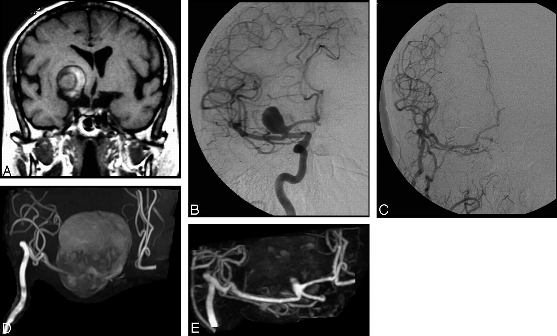
- Fig 1.
A, A large extra-axial lesion and hemorrhagic component indicative of a giant thrombosed aneurysm is apparent in the basal ganglia on the coronal T1-weighted MR image. B, Selective DSA of the right internal carotid artery before treatment reveals a patent portion of the giant thrombosed aneurysm. C, Selective DSA after embolization reveals complete occlusion of the aneurysm and a patent internal carotid artery–external carotid artery bypass. D, Follow-up 3D TOF MRA with MIP reconstruction performed at 1 year after treatment is inconclusive because of artifact related to the methemoglobin component of the thrombosed aneurysm. Additionally, flow-related signal intensity loss in the A1 segment of the anterior cerebral artery is apparent. E, Conversely, follow-up CE-MRA with MIP reconstruction demonstrates both the patent internal carotid artery–external carotid artery bypass and the presence of a small (type I) remnant at the neck of the treated aneurysm.
- Fig 2.
A, Selective DSA of the right internal carotid artery before treatment reveals a giant carotid-ophthalmic aneurysm. B, Selective DSA after embolization demonstrates complete occlusion of the aneurysm but with slightly reduced coil compaction near the neck. C and D, Follow-up 3D TOF MRA source image (C) and MIP reconstruction (D) obtained at 8 months after treatment reveal residual patency, but clear definition of the (type 2) remnant is compromised by saturation and methemoglobin artifact. E and F, Clearer depiction of the residual aneurysm is achieved on CE-MRA source images (E) and MIP reconstruction (F).
- Fig 3.
A, Selective DSA of the left internal carotid artery before treatment reveals a small aneurysm (arrow) of the anterior communicating artery. B, This aneurysm (arrow) appears completely occluded at follow-up DSA performed after embolization. C and D, Follow-up 3D TOF MRA source image (C) and MIP reconstruction (D) obtained at 3 months after treatment reveal high-signal-intensity inhomogeneities (arrow) in the region of the aneurysm neck but do not clearly demonstrate an aneurysm remnant. E and F, CE-MRA source image (E) and MIP reconstruction (F) clearly reveal the presence of a small (type 2) remnant.
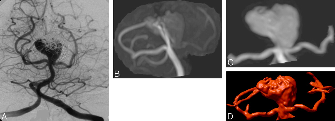
- Fig 4.
A, Follow-up DSA at 8 months after embolization of a large basilar artery aneurysm reveals only partial occlusion. B, 3D TOF MRA with MIP reconstruction (B) fails to show the (type 2) remnant entirely, due to flow effects that also compromise visualization of the left posterior cerebral artery. C and D, CE-MRA with MIP (C) and VR (D) reconstruction show both the artery and the residual aneurysm sac.


Tables
Comparison of TOF MRA with CE-MRA for visualization of residual aneurysm patency by the size of the treated aneurysm
Aneurysm Size TOF MRA Better Sequences Equal CE-MRA Better Small (n = 43) 0 37 6 Large (n = 10) 0 6 4 Giant (n = 1) 0 0 1 Total (n = 54) 0 43 11 Note:—TOF MRA indicates time-of-flight MR angiography; CE-MRA, contrast enhanced MR angiography.
In this issue


Jump to section
Related Articles
Cited By...
- Differential Subsampling with Cartesian Ordering-MRA for Classifying Residual Treated Aneurysms
- Long-Term Results and Follow-Up Examinations after Endovascular Embolization for Unruptured Cerebral Aneurysms
- Surveillance of Unruptured Intracranial Saccular Aneurysms Using Noncontrast 3D-Black-Blood MRI: Comparison of 3D-TOF and Contrast-Enhanced MRA with 3D-DSA
- Guidelines for the Management of Patients With Unruptured Intracranial Aneurysms: A Guideline for Healthcare Professionals From the American Heart Association/American Stroke Association
- Optimized Angiographic CT Using Intravenous Contrast Injection: A Noninvasive Imaging Option for the Follow-Up of Coiled Aneurysms?
- Follow-Up of Coiled Intracranial Aneurysms: Comparison of 3D Time-of-Flight MR Angiography at 3T and 1.5T in a Large Prospective Series
- Long-Term Prospective Follow-Up of Intracranial Aneurysms Treated with Endovascular Coiling Using Contrast-Enhanced MR Angiography
- A Prospective Trial of 3T and 1.5T Time-of-Flight and Contrast-Enhanced MR Angiography in the Follow-Up of Coiled Intracranial Aneurysms
- Evaluation of the Occlusion Status of Coiled Intracranial Aneurysms with MR Angiography at 3T: Is Contrast Enhancement Necessary?
- MR Angiographic Follow-Up of Intracranial Aneurysms Treated with Detachable Coils: Evaluation of a Blood-Pool Contrast Medium
- MR Angiography Follow-Up of Aneurysms Treated with Coils: Is There a Need for the Use of Gadolinium?