
Article Figures & Data
Figures
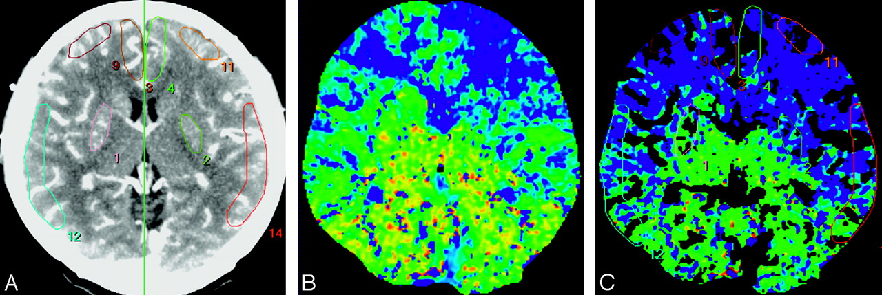
- Fig 1.
ROIs drawn in a reference CT image (A), MTT map (B), and vessel-removed MTT map (C). The ROIs were placed on cortical regions in the MCA territory, EBZ, ACA territory, and BG.
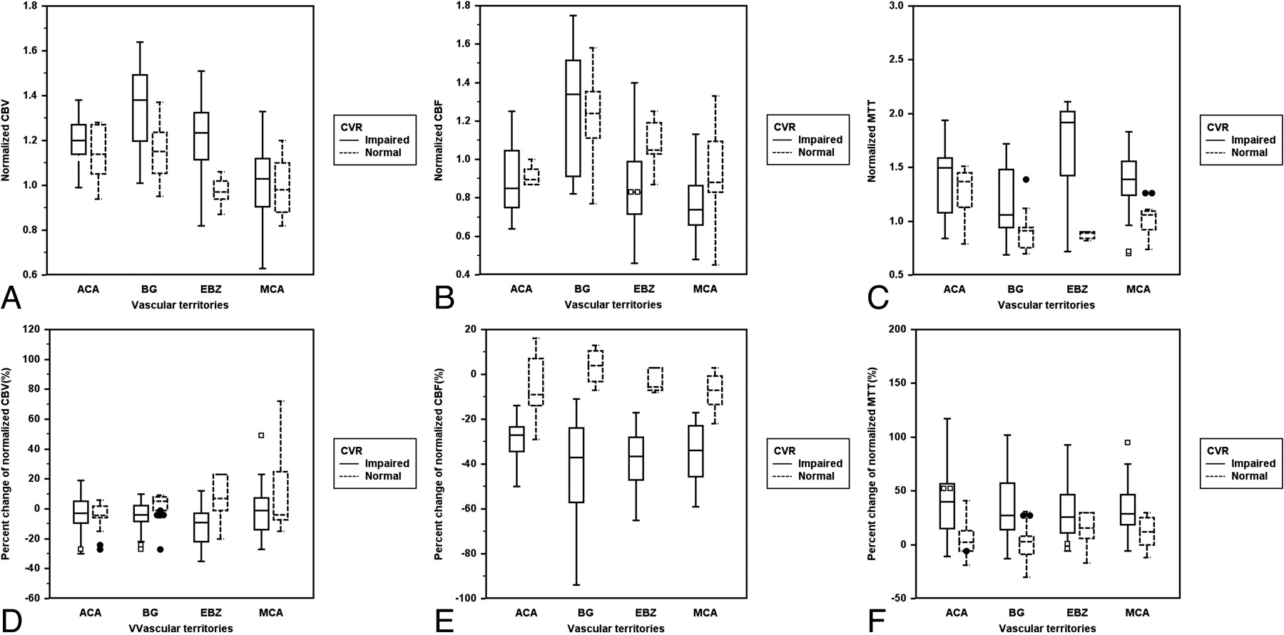
- Fig 2.
Clustered box-and-whisker graphs show the differences in the mean values of baseline and drug-challenged CT perfusion parameters between the normal and impaired cerebrovascular reserve groups in each vascular territory. For analysis of baseline CTP parameters (A to C), the mean values of CBV (A) and MTT (C) in the impaired CVR group were significantly higher than those in normal CVR group in EBZ and BG, and the mean values of CBF (B) were significantly lower than those in normal CVR group only in EBZ. For analysis of the percentage changes of baseline CTP parameters (D to F), pcCBV (D) was significantly different between the normal and impaired CVR groups only in EBZ, and pcCBF (E) and pcMTT (F) were significantly different in all of the vascular territories.
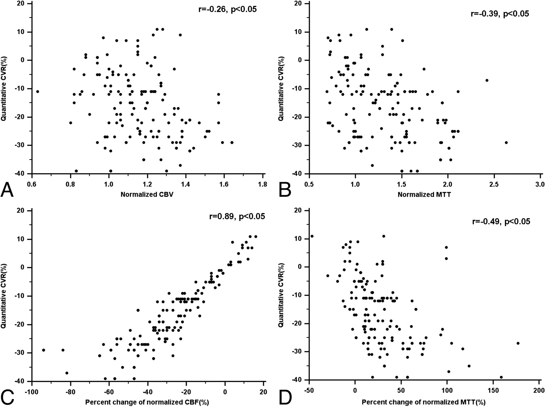
- Fig 3.
Scatter plots show the correlation between the CT perfusion parameters and the quantitative cerebrovascular reserve by using SPECT on the 152 ROIs in all of the vascular territories.
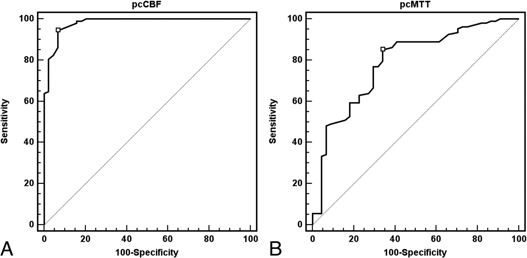
- Fig 4.
ROC curve analysis for each CT perfusion parameter's ability to correctly identify the impaired cerebrovascular reserve. With optimal threshold value (−16%) of pcCBF, the sensitivity, specificity, PPV, and NPV for defining the impaired cerebrovascular reserve are 94.4%, 93.2%, 97.1%, and 87.2%, respectively (the area under the ROC curves = 0.98; A). For defining impaired CVR with pcMTT, statistical analysis gave a threshold value of 10%, with a sensitivity, specificity, PPV, and NPV of 85.2%, 65.9%, 86.0%, and 64.4%, respectively (the area under the ROC curves = 0.79; B).


Tables
- Table 1:
The differences of the mean values of CT perfusion parameters in each vascular territory between the normal and impaired cerebrovascular reserve groups
Variable MCA EBZ ACA BG CBV nCVR 0.99 0.97* 1.14 1.15* iCVR 1.02 1.22* 1.21 1.36* CBF nCVR 0.94 1.07* 0.90 1.24 iCVR 0.77 0.86* 0.90 1.28 MTT nCVR 1.15 0.91* 1.29 0.92* iCVR 1.39 1.67* 1.39 1.16* pcCBV (%) nCVR 12.6 6.5* −6.5 17.3 iCVR −2.2 −11.4* −3.9 −6.1 pcCBF (%) nCVR −7.4* −3.3* −5.9* 3.7* iCVR −35.1* −38.4* −30.0* −44.3* pcMTT (%) nCVR 11.6* 13.3* 2.2* 12.7* iCVR 33.4* 35.6* 43.1* 34.6* Note:—CBV indicates cerebral blood volume; CBF, cerebral blood flow; MTT, mean transit time; pcCBV, percentage of change of CBV; pcCBF, percentage of change of CBF; pcMTT, percentage of change of MTT; nCVR, normal cerebrovascular reserve group; iCVR, impaired cerebrovascular reserve group; MCA, middle cerebral artery; EBZ, anterior external borderzone; ACA, anterior cerebral artery; BG, basal ganglia.
* Values are statistically significant using the Student t test (P < .05).
- Table 2:
The correlation coefficient between the CT perfusion parameters and quantitative cerebrovascular reserve obtained by SPECT
Variable MCA EBZ ACA BG CBV vs CVR −0.04 −0.65* −0.15 −0.40 CBF vs CVR 0.39 0.14 −0.08 0.04 MTT vs CVR −0.43* −0.56* −0.07 −0.45* pcCBV vs CVR 0.32 0.33 −0.26 0.41 pcCBF vs CVR 0.91* 0.92* 0.85* 0.88* pcMTT vs CVR −0.45* −0.52* −0.69* −0.42* Note:—CBV indicates cerebral blood volume; CBF, cerebral blood flow; MTT, mean transit time; pcCBV, percentage of change of CBV; pcCBF, percentage of change of CBF; pcMTT, percentage of change of MTT; CVR, cerebrovascular reserve; MCA, middle cerebral artery; EBZ, anterior external borderzone; ACA, anterior cerebral artery; BG, basal ganglia.
* Values are statistically significant using the Pearson correlation coefficient (P < .05).
In this issue


Jump to section
Related Articles
Cited By...
- Stenotic Transverse Sinus Predisposes to Poststenting Hyperperfusion Syndrome as Evidenced by Quantitative Analysis of Peritherapeutic Cerebral Circulation Time
- Neuroradiologic Correlates of Cognitive Impairment in Adult Moyamoya Disease
- Impact of Extracranial-Intracranial Bypass on Cerebrovascular Reactivity and Clinical Outcome in Patients With Symptomatic Moyamoya Vasculopathy
- Evaluation of CT Perfusion in the Setting of Cerebral Ischemia: Patterns and Pitfalls
- The Acetazolamide Challenge: Techniques and Applications in the Evaluation of Chronic Cerebral Ischemia