
Article Figures & Data
Figures
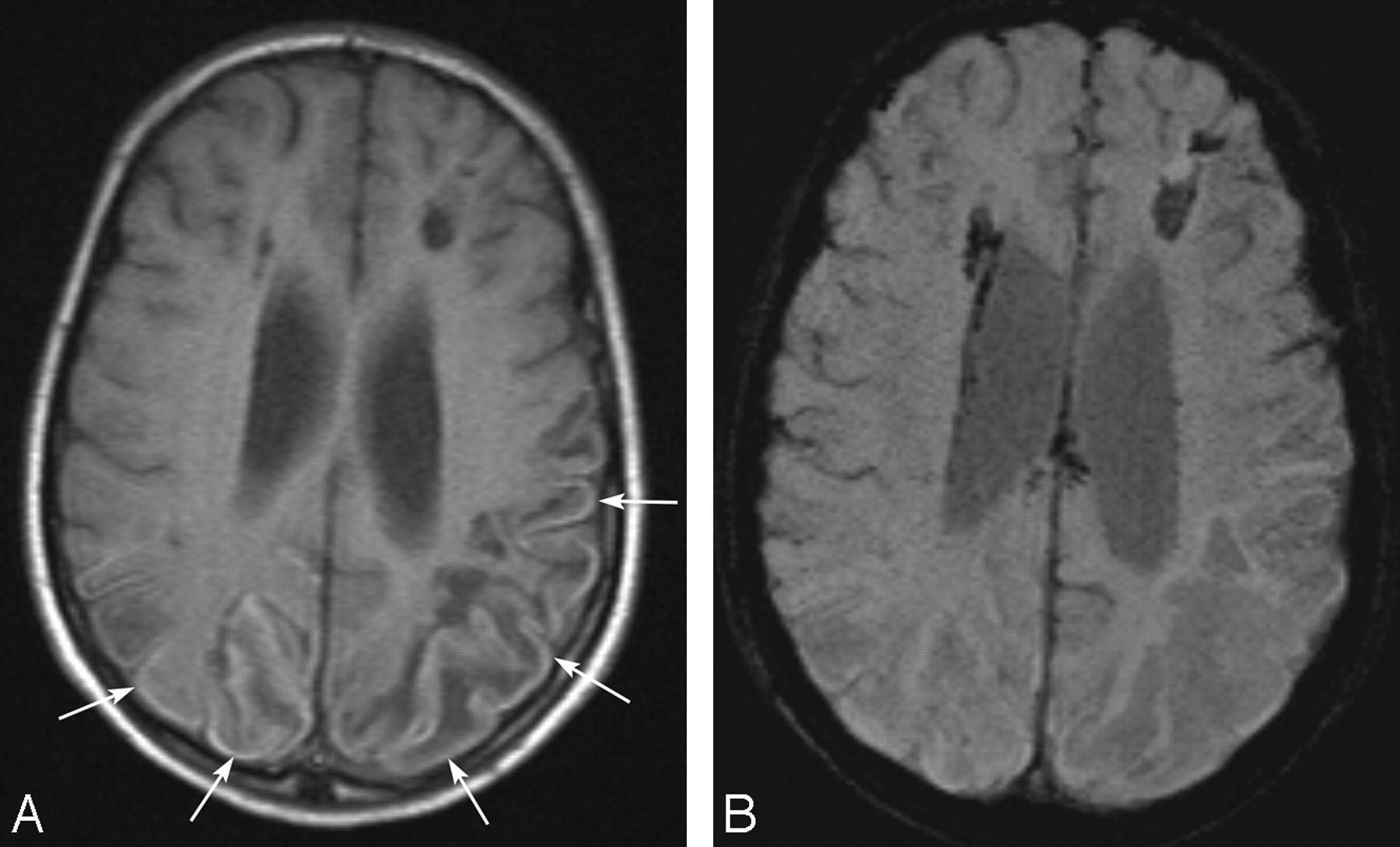
- Fig 1.
A 13-year-old boy with ischemic changes of Moyamoya disease. A, Transverse T1-weighted spin-echo image shows curvilinear hyperintense cortical lesions in the bilateral parietal lobes (arrows). B, Corresponding transverse susceptibility-weighted image shows no signal-intensity loss indicating hemorrhage in the cortical lesions.
- Fig 2.
A 12-month-old girl with chronic infarction in bilateral middle cerebral artery territories. This patient had prior heart surgery. A, Transverse T1-weighted image shows a curvilinear hyperintense cortical lesion (arrows). B, Corresponding transverse susceptibility-weighted image shows dotted signal-intensity loss (arrow), which indicates hemorrhage, in the cortical lesions. Although we consider the signal-intensity loss on the laminar necrosis as hemorrhage because of discontinuity, it is difficult to strictly distinguish it from part of the cortical vein. Note that several small foci of dotted hemorrhage are also found in other parts of the brain parenchyma.
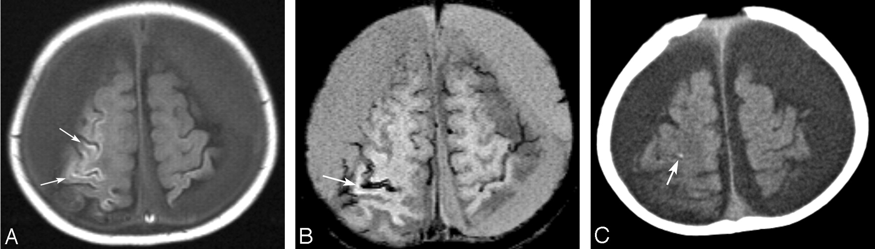
- Fig 3.
An 8-month-old boy with shaken baby syndrome. A, Transverse T1-weighted image shows a curvilinear hyperintense cortical lesion in the damaged area (arrows). B, Susceptibility-weighted image shows laminar signal-intensity loss, which indicates hemorrhage, along with some cortical lesions (arrow). C, CT scan shows dotted calcification (arrow) in part of the area of laminar hemorrhage on SWI.


