Advertisement

Main menu

  • Home
  • Content
    • Current Issue
    • Accepted Manuscripts
    • Article Preview
    • Past Issue Archive
    • AJNR Case Collection
    • Case of the Week Archive
    • Classic Case Archive
    • Case of the Month Archive
  • Special Collections
    • Spinal CSF Leak Articles (Jan 2020-June 2024)
    • 2024 AJNR Journal Awards
    • Most Impactful AJNR Articles
  • Multimedia
    • AJNR Podcast
    • AJNR Scantastics
    • Video Articles
  • For Authors
    • Submit a Manuscript
    • Author Policies
    • Fast publishing of Accepted Manuscripts
    • Graphical Abstract Preparation
    • Manuscript Submission Guidelines
    • Imaging Protocol Submission
    • Submit a Case for the Case Collection
  • About Us
    • About AJNR
    • Editorial Board
  • More
    • Become a Reviewer/Academy of Reviewers
    • Subscribers
    • Permissions
    • Alerts
    • Feedback
    • Advertisers
    • ASNR Home
  • Other Publications
    • ajnr

User menu

  • Alerts
  • Log in
  • Log out

Search

  • Advanced search
American Journal of Neuroradiology

American Journal of Neuroradiology

ASHNR American Society of Functional Neuroradiology ASHNR American Society of Pediatric Neuroradiology ASSR
  • Alerts
  • Log in
  • Log out

Advanced Search

  • Home
  • Content
    • Current Issue
    • Accepted Manuscripts
    • Article Preview
    • Past Issue Archive
    • AJNR Case Collection
    • Case of the Week Archive
    • Classic Case Archive
    • Case of the Month Archive
  • Special Collections
    • Spinal CSF Leak Articles (Jan 2020-June 2024)
    • 2024 AJNR Journal Awards
    • Most Impactful AJNR Articles
  • Multimedia
    • AJNR Podcast
    • AJNR Scantastics
    • Video Articles
  • For Authors
    • Submit a Manuscript
    • Author Policies
    • Fast publishing of Accepted Manuscripts
    • Graphical Abstract Preparation
    • Manuscript Submission Guidelines
    • Imaging Protocol Submission
    • Submit a Case for the Case Collection
  • About Us
    • About AJNR
    • Editorial Board
  • More
    • Become a Reviewer/Academy of Reviewers
    • Subscribers
    • Permissions
    • Alerts
    • Feedback
    • Advertisers
    • ASNR Home
  • Follow AJNR on Twitter
  • Visit AJNR on Facebook
  • Follow AJNR on Instagram
  • Join AJNR on LinkedIn
  • RSS Feeds

Welcome to the new AJNR, Updated Hall of Fame, and more. Read the full announcements.


AJNR is seeking candidates for the position of Associate Section Editor, AJNR Case Collection. Read the full announcement.


LetterLetter

MR Imaging Can Predict the Development of Nonalcoholic Wernicke Encephalopathy

M. Ohira, S. Suzuki, S. Takahashi, K. Takahashi and N. Suzuki
American Journal of Neuroradiology October 2008, 29 (9) e81-e82; DOI: https://doi.org/10.3174/ajnr.A1144
M. Ohira
  • Find this author on Google Scholar
  • Find this author on PubMed
  • Search for this author on this site
S. Suzuki
  • Find this author on Google Scholar
  • Find this author on PubMed
  • Search for this author on this site
S. Takahashi
  • Find this author on Google Scholar
  • Find this author on PubMed
  • Search for this author on this site
K. Takahashi
  • Find this author on Google Scholar
  • Find this author on PubMed
  • Search for this author on this site
N. Suzuki
  • Find this author on Google Scholar
  • Find this author on PubMed
  • Search for this author on this site
  • Article
  • Figures & Data
  • Info & Metrics
  • Responses
  • References
  • PDF
Loading

We read with great interest the article by Fei et al1 in which symmetric paraventricular regions in MR imaging were shown to be useful in the diagnosis of nonalcoholic Wernicke encephalopathy (WE). Among clinical studies, MR imaging is currently considered the most valuable method for confirming a diagnosis of WE, though the typical findings of MR imaging are observed in only 58% of WE patients.2 To advance the clinical facility of MR imaging, we report a case of nonalcoholic WE in which symmetric paraventricular regions could be found before the clinical onset of WE.

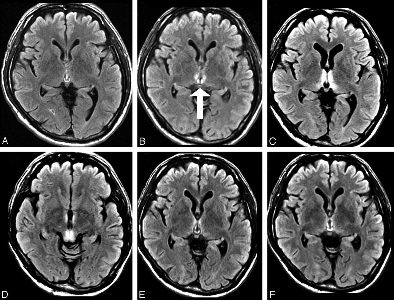
A 61-year-old Japanese man was admitted to our hospital because of impaired attention, gait disturbance, and double vision lasting for 2 days. He had undergone a partial gastrectomy because of gastric cancer 30 months before admission. He had no history of alcoholic abuse or unbalanced nutrition. He noticed transient amnesia and first came to our clinic 24 months before admission. However, there were no abnormal findings in his mental function or neurologic examinations. Brain MR imaging was normal (Fig 1A), and serum thiamine level was 61.1 μg/dL (normal, 2.0–7.2 μg/dL). After that, his symptoms abated; however, the second fluid-attenuated inversion recovery (FLAIR) MR imaging detected small symmetric paraventricular regions 6 months before the admission (Fig 1B).

Neurologic examination on admission revealed a confused state showing sensitivity to sound, oculomotor deficits, horizontal gaze-evoked nystagmus, and truncal ataxia. Routine blood examination and arterial blood analysis were normal. Serum thiamine level was markedly low, at 1.5 μg/dL. MR imaging clearly showed bilateral high-signal intensities over the mammillary bodies and paraventricular lesions in both T2-weighted and FLAIR modalities (Fig 1C, -D). Based on the diagnosis of WE, treatment with intravenous thiamine hydrochloride (500 mg/d) was promptly started. Neurologic manifestations were significantly improved in the following 2 days except for the impairment of recent memory. Thiamine administration for 7 days normalized serum thiamine to 10.9 μg/dL, and repeated brain MR imaging also revealed the diminishing of hyperintense lesions on FLAIR images (Fig 1E). Finally, 15 days after the start of treatment, he achieved normal scores in both the Mini-Mental State Examination and revised Wechsler Adult Intelligence Scale, and his final MR imaging could not detect any abnormal lesions (Fig 1F).

Nonalcoholic WE exhibiting complicated clinical manifestations and an atypical disease progression is prone to be underestimated.1,2 Gastric surgery is known to be one of the predisposing factors of WE, which usually develops weeks or months after the operation.2 The present patient developed WE 30 months after partial gastrectomy without episodes of vomiting or parenteral nutrition. In this regard, Karapanayiotides et al3 reported that a 53-year-old man showing restricted dietary intake and repeated vomiting developed WE 28 years after gastric surgery. In addition, bariatric surgery, commonly performed in the United States, is also a cause of WE, occurring between 4 and 12 weeks postoperatively, especially in young women with vomiting.4 Taken together, it is probable that nonalcoholic WE patients after gastric surgery have usually had antecedent episodes such as dietary restrictions, psychological food refusal, vomiting, and unbalanced total parenteral nutrition.1–4

Approximately 19% of WE patients have none of the typical triad of symptoms, including mental status changes, ocular abnormalities, and ataxic gait disturbance at presentation of WE.2 According to the review of Sechi and Serra,2 early detection of subclinical thiamine deficiency is generally difficult, because symptoms may be vague and nonspecific, such as headache, fatigue, irritability, and abdominal discomfort. In the present patient, however, it was impossible to determine that the transient amnesia at the first examination could really be ascribed to subclinical thiamine deficiency. When the “neuroradiologic” findings in MR imaging were first detected, we could not diagnose WE clinically for the following reasons. First, our patient had no antecedent events of alcohol overdrinking, unbalanced food intake, or vomiting. Second, he had recovered from amnesia, and there were no abnormal neurologic findings. Third, the level of serum thiamine was not low. In fact, he had been well with no treatments for 6 months until he was admitted to our hospital.

In conclusion, we present the chronologic changes of MR imaging before and after the onset of WE. It appears that MR imaging can predict the development of nonalcoholic WE. Thiamine supplementation should be initiated as soon as characteristic findings in MR imaging are seen, even if a clinical diagnosis of WE cannot be established.

  Fig 1.
  • Download figure
  • Open in new tab
  • Download powerpoint
Fig 1.

Serial changes of MR imaging in a patient with nonalcoholic WE. Axial FLAIR images were obtained 24 months (A) and 6 months (B) before the onset of WE, on admission (C and D), and 7 days (E) and 15 days (F) after the start of treatment. Arrow indicates small symmetric paraventricular regions before the clinical onset of WE.

References

  1. ↵
    Fei GQ, Zhong C, Jin L, et al. Clinical characteristics and MR imaging features of nonalcoholic Wernicke encephalopathy. AJNR Am J Neuroradiol 2008;29:164–69
    Abstract/FREE Full Text
  2. ↵
    Sechi G, Serra A. Wernicke's encephalopathy: new clinical settings and recent advances in diagnosis and management. Lancet Neurol 2007;6:442–55
    CrossRefPubMed
  3. ↵
    Karapanayiotides T, Zaloni I, Thomaides T. Delayed development of Wernicke encephalopathy after gastrectomy. Arch Neurol 2006;63:1026–27
    CrossRefPubMed
  4. ↵
    Singh S, Kumar A. Wernicke encephalopathy after obesity surgery: a systematic review. Neurology 2007;68:807–11
    Abstract/FREE Full Text
  • Copyright © American Society of Neuroradiology
PreviousNext
Back to top

In this issue

American Journal of Neuroradiology: 29 (9)
American Journal of Neuroradiology
Vol. 29, Issue 9
October 2008
  • Table of Contents
  • Index by author
Advertisement
Print
Download PDF
Email Article

Thank you for your interest in spreading the word on American Journal of Neuroradiology.

NOTE: We only request your email address so that the person you are recommending the page to knows that you wanted them to see it, and that it is not junk mail. We do not capture any email address.

Enter multiple addresses on separate lines or separate them with commas.
MR Imaging Can Predict the Development of Nonalcoholic Wernicke Encephalopathy
(Your Name) has sent you a message from American Journal of Neuroradiology
(Your Name) thought you would like to see the American Journal of Neuroradiology web site.
CAPTCHA
This question is for testing whether or not you are a human visitor and to prevent automated spam submissions.
Cite this article
M. Ohira, S. Suzuki, S. Takahashi, K. Takahashi, N. Suzuki
MR Imaging Can Predict the Development of Nonalcoholic Wernicke Encephalopathy
American Journal of Neuroradiology Oct 2008, 29 (9) e81-e82; DOI: 10.3174/ajnr.A1144

Citation Manager Formats

  • BibTeX
  • Bookends
  • EasyBib
  • EndNote (tagged)
  • EndNote 8 (xml)
  • Medlars
  • Mendeley
  • Papers
  • RefWorks Tagged
  • Ref Manager
  • RIS
  • Zotero
0 Responses
Respond to this article
Share
Bookmark this article
MR Imaging Can Predict the Development of Nonalcoholic Wernicke Encephalopathy
M. Ohira, S. Suzuki, S. Takahashi, K. Takahashi, N. Suzuki
American Journal of Neuroradiology Oct 2008, 29 (9) e81-e82; DOI: 10.3174/ajnr.A1144
del.icio.us logo Twitter logo Facebook logo Mendeley logo
  • Tweet Widget
  • Facebook Like
  • Google Plus One
Purchase

Jump to section

  • Article
    • References
  • Figures & Data
  • Info & Metrics
  • Responses
  • References
  • PDF

Related Articles

  • No related articles found.
  • PubMed
  • Google Scholar

Cited By...

  • No citing articles found.
  • Crossref (2)
  • Google Scholar

This article has been cited by the following articles in journals that are participating in Crossref Cited-by Linking.

  • Wernicke’s Encephalopathy: Expanding the Diagnostic Toolbox
    Mary E. Lough
    Neuropsychology Review 2012 22 2
  • CT and MRI of Wernicke’s encephalopathy
    A. Cerase, E. Rubenni, A. Rufa, I. Vallone, P. Galluzzi, G. Coratti, F. Franchi, F. Giannini, C. Venturi
    La radiologia medica 2011 116 2

More in this TOC Section

  • Brain AVM’s Nidus: What if We Hadn’t Understood Anything?
  • Letter to the Editor regarding “Automated Volumetric Software in Dementia: Help or Hindrance to the Neuroradiologist?”
  • Reply:
Show more Letters

Similar Articles

Advertisement

Indexed Content

  • Current Issue
  • Accepted Manuscripts
  • Article Preview
  • Past Issues
  • Editorials
  • Editors Choice
  • Fellow Journal Club
  • Letters to the Editor

Cases

  • Case Collection
  • Archive - Case of the Week
  • Archive - Case of the Month
  • Archive - Classic Case

Special Collections

  • Special Collections

Resources

  • News and Updates
  • Turn around Times
  • Submit a Manuscript
  • Author Policies
  • Manuscript Submission Guidelines
  • Evidence-Based Medicine Level Guide
  • Publishing Checklists
  • Graphical Abstract Preparation
  • Imaging Protocol Submission
  • Submit a Case
  • Become a Reviewer/Academy of Reviewers
  • Get Peer Review Credit from Publons

Multimedia

  • AJNR Podcast
  • AJNR SCANtastic
  • Video Articles

About Us

  • About AJNR
  • Editorial Board
  • Not an AJNR Subscriber? Join Now
  • Alerts
  • Feedback
  • Advertise with us
  • Librarian Resources
  • Permissions
  • Terms and Conditions

American Society of Neuroradiology

  • Not an ASNR Member? Join Now

© 2025 by the American Society of Neuroradiology All rights, including for text and data mining, AI training, and similar technologies, are reserved.
Print ISSN: 0195-6108 Online ISSN: 1936-959X

Powered by HighWire