
Article Figures & Data
Figures
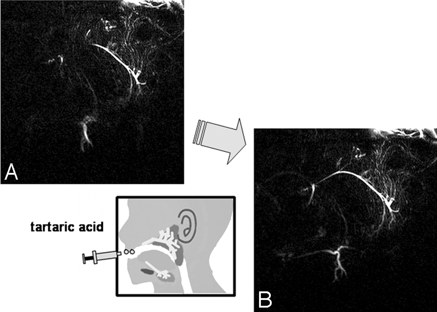
- Fig 1.
MR sialography findings and response to salivary secretion stimulation. A and B, Oblique-sagittal projection MR sialography of a normal (preirradiated) salivary gland before (A) and after (B) secretion stimulation. Salivary secretion stimulation with tartaric acid administration on the tongue improves the depiction of the main duct and distal branches.
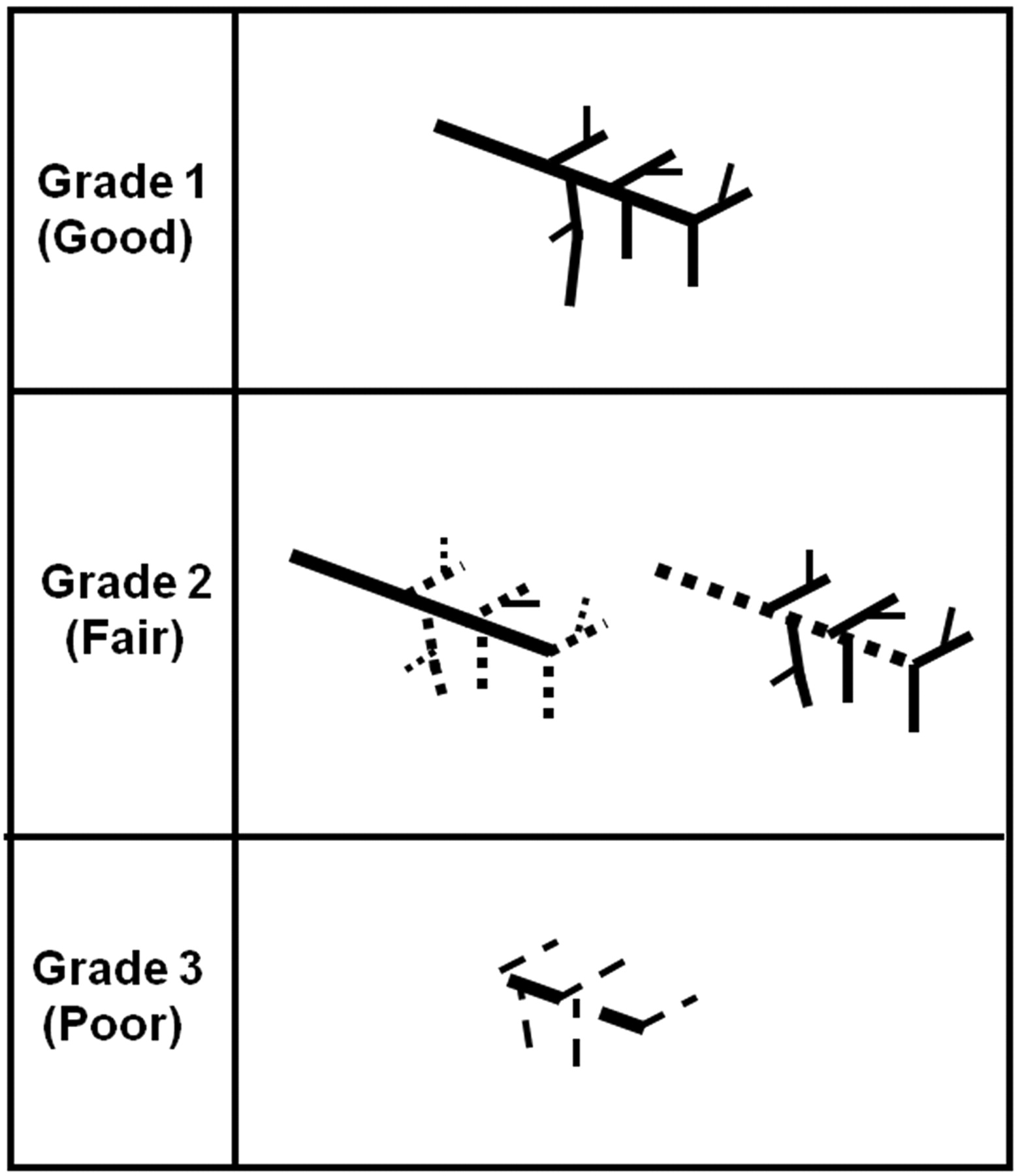
- Fig 2.
Morphologic evaluation (grading) criteria of the salivary gland system on MR sialography. On nonstimulated and stimulated MR sialography, each salivary gland (parotid and submandibular gland) is classified into 3 grades according to the degree of lumen visualization: grade 1, a distinct depiction of both the main trunk and branches; grade 2, a distinct depiction of the main trunk or first- and second-order branches; and grade 3, an indistinct depiction of the main trunk and first- and second-order branches.
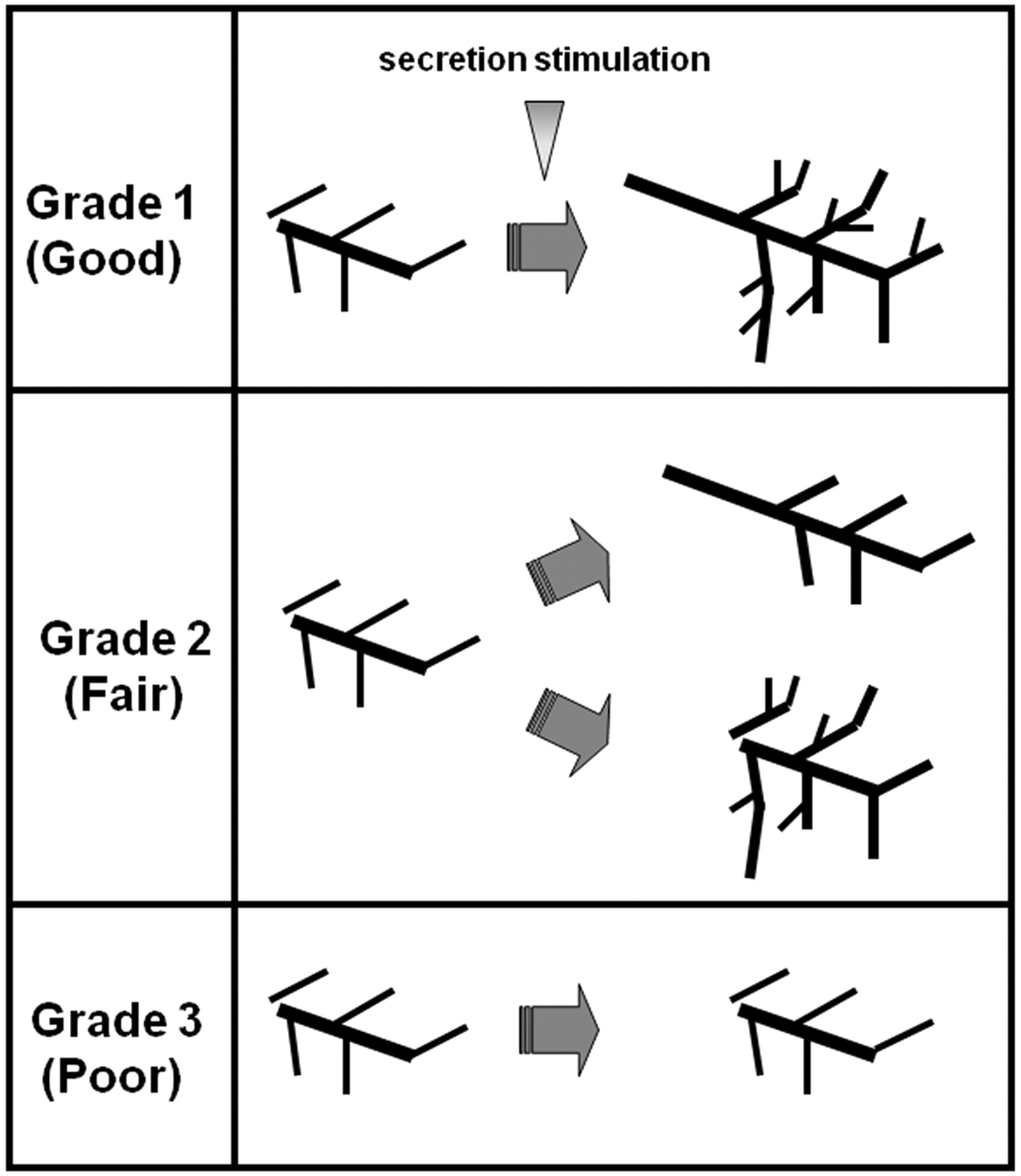
- Fig 3.
Visual evaluation (grading) criteria after salivary secretion stimulation. Grade 1 (good response) is a distinct depiction improvement at the main trunk and each distal branch, grade 2 (fair response) is a distinct improvement at either the main trunk or the distal branches, and grade 3 (poor response) is no distinct response at either the main trunk or distal branches.
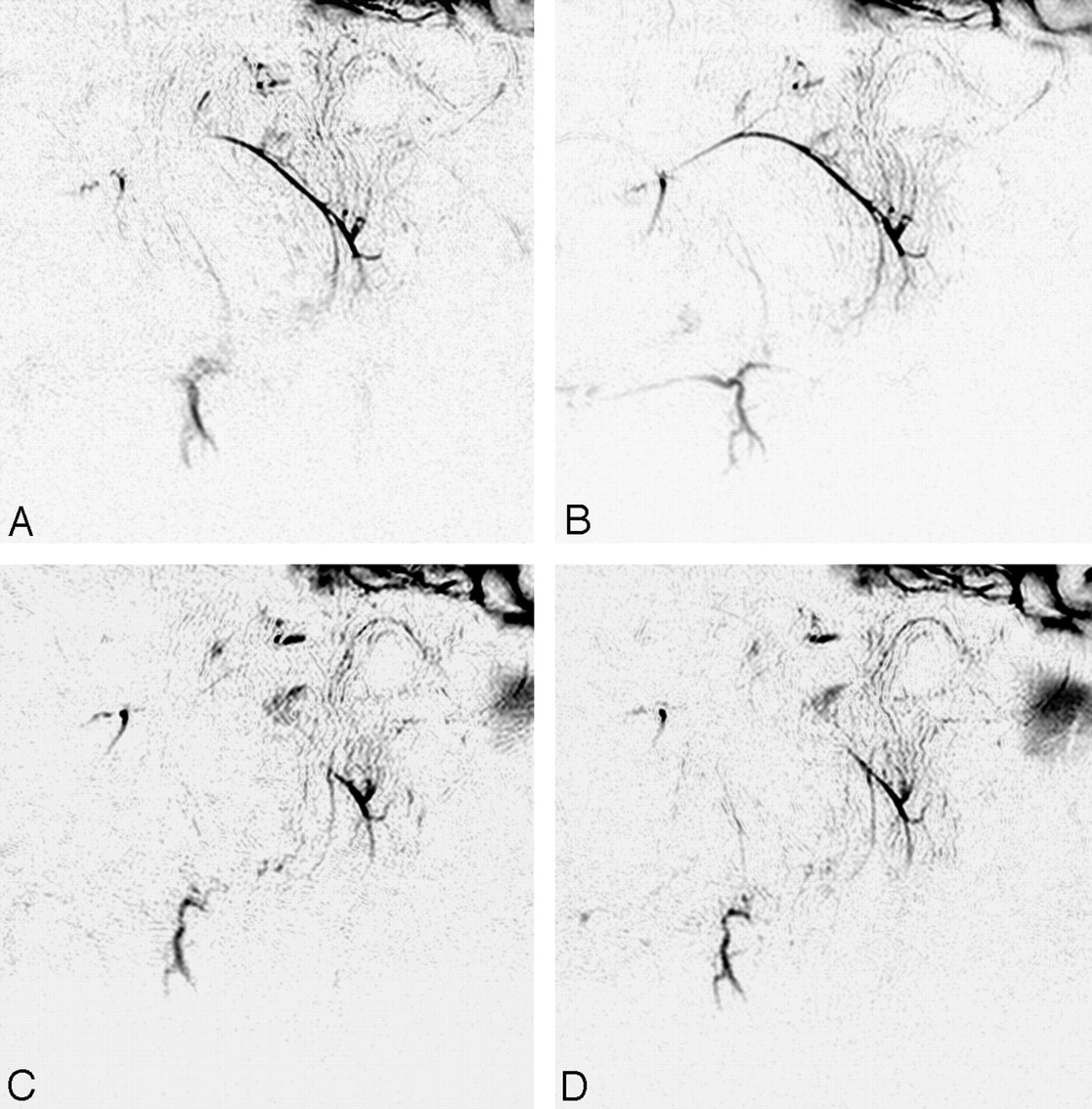
- Fig 4.
MR sialography (inverted images) before (A and B) and after (C and D) 46-Gy irradiation to the salivary gland. (A and C, nonstimulated; B and D; stimulated images). Initial (preirradiation) MR sialography of the right salivary system shows good depiction of parotid and submandibular gland ducts and response to secretion stimulation (A and B). Irradiation to the salivary gland induces insufficient visualization of the main trunk and distal branches and disturbs salivary secretion response (C and D).
- Fig 5.
Nonstimulated (A) and stimulated (B) MR sialography of mild radiation-induced xerostomia. Images show nonstimulated (A) and stimulated (B) MR sialography of the 62-Gy irradiated right salivary system. Both parotid and submandibular glands show good depiction of the salivary duct and secretion response. The clinical xerostomia grade and MR sialography grade for this patient are 1 and 1.08.
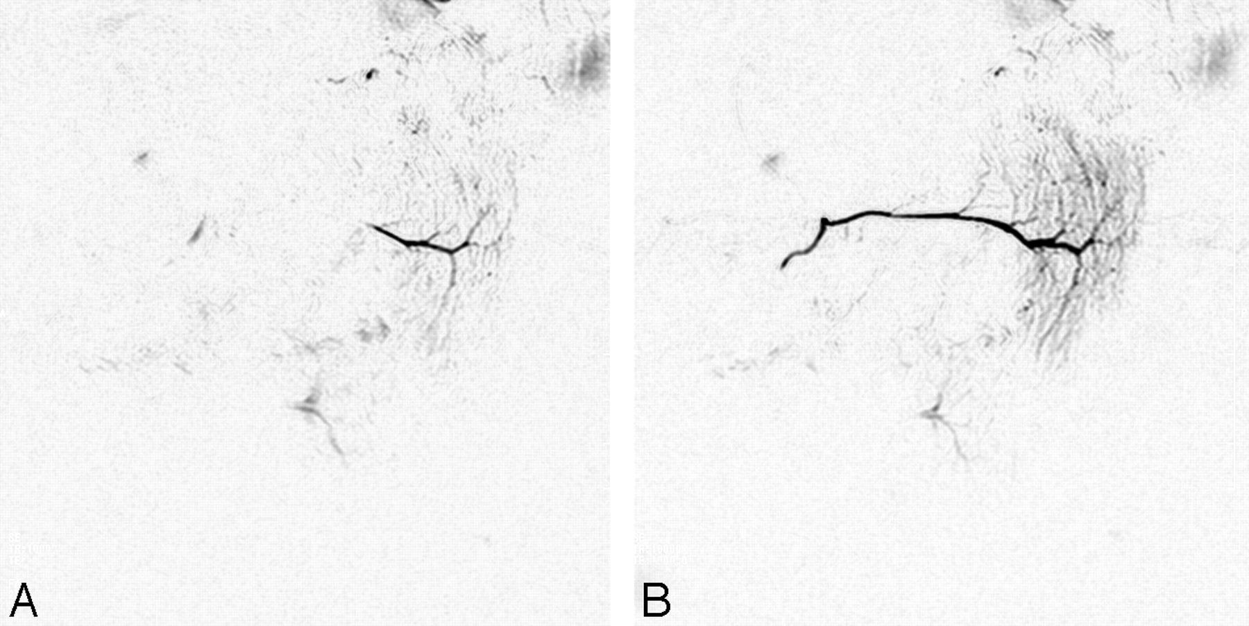
- Fig 6.
Characteristic MR sialography finding of the postirradiated submandibular gland in severe xerostomia. A, Nonstimulated MR sialography of the right salivary system shows poor depiction of both the parotid and submandibular glands. B, After secretion stimulation, the parotid gland shows good response, but the submandibular gland shows no significant response. The clinical grade and MR sialography grade of xerostomia in this patient are 3 and 2.82.
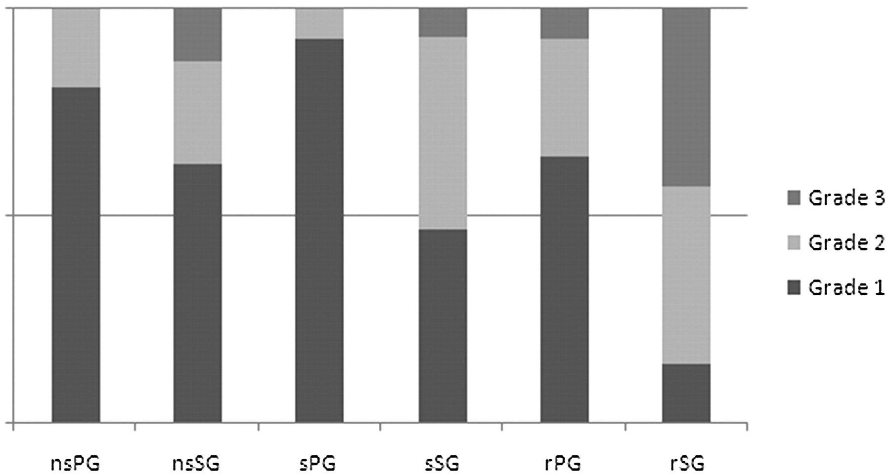
- Fig 7.
MR sialography findings of preirradiation salivary glands and parotid and submandibular glands reveal individual differences in salivary gland visualization and secretion response.
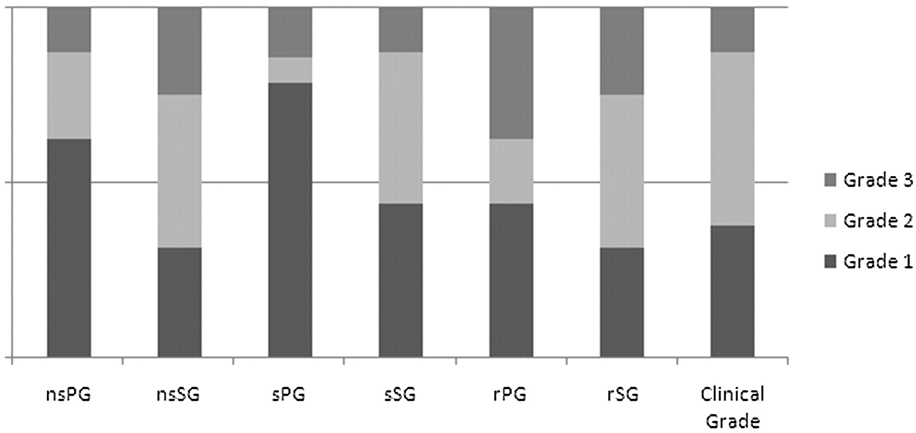
- Fig 8.
MR sialography findings of postirradiation salivary glands and clinical grade of radiation-induced xerostomia. Morphologic findings and secretion response of the submandibular gland seems to be more related to the clinical xerostomia grade than those of the parotid gland.
- Fig 9.
Grading of the radiation-induced xerostomia of our 16 patients: clinical grade and MR sialography-based grade. In 70% of patients with radiation-induced xerostomia, MR sialography-based xerostomia grading with our regression equation is consistent with clinical grade (xerostomia grade = 0.681 + 0.871rSG − 0.471sPG). In patients 3, 5, and 6, the negative coefficient of sPG compensates the MR sialography grading error (overestimation) induced by the high grade of rSG.


Tables
Clinical grading scale of xerostomia*
Grade Characteristics 1, Mild Symptomatic (dry or thick saliva) without significant dietary alteration 2, Moderate Symptomatic and significant oral intake alteration (eg, copious water, other lubricants, diet limited to purees and/or soft moist foods) 3, Severe Symptoms leading to inability to adequately aliment orally; IV fluids, tube feedings, or parenteral nutrition indicated * Clinical severity of radiation-induced xerostomia is classified into 3 grades using CTCAE Version 3.0 modified criteria.


